

Strain Elastography - How To Do It?
- PMID: 29226273
- PMCID: PMC5720889
- DOI: 10.1055/s-0043-119412
Strain Elastography - How To Do It?
Abstract
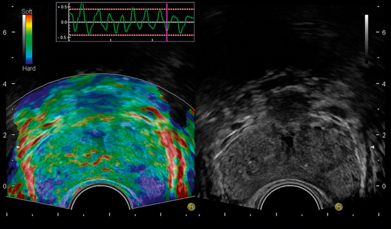
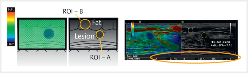
Tissue stiffness assessed by palpation for diagnosing pathology has been used for thousands of years. Ultrasound elastography has been developed more recently to display similar information on tissue stiffness as an image. There are two main types of ultrasound elastography, strain and shear wave. Strain elastography is a qualitative technique and provides information on the relative stiffness between one tissue and another. Shear wave elastography is a quantitative method and provides an estimated value of the tissue stiffness that can be expressed in either the shear wave speed through the tissues in meters/second, or converted to the Young's modulus making some assumptions and expressed in kPa. Each technique has its advantages and disadvantages and they are often complimentary to each other in clinical practice. This article reviews the principles, technique, and interpretation of strain elastography in various organs. It describes how to optimize technique, while pitfalls and artifacts are also discussed.
Keywords: endoscopic ultrasound; real-time; tissue elastography (TE); ultrasound.
Conflict of interest statement
Figures
References
-
- Bamber J, Cosgrove D, Dietrich C F, Fromageau J, Bojunga J, Calliada F, Cantisani V et al.EFSUMB guidelines and recommendations on the clinical use of ultrasound elastography. Part 1: Basic principles and technology. Ultraschall Med. 2013;34:169–184. - PubMed
-
- Shiina T, Nightingale K R, Palmeri M L, Hall T J, Bamber J C, Barr R G, Castera L et al.WFUMB guidelines and recommendations for clinical use of ultrasound elastography: Part 1: basic principles and terminology. Ultrasound Med Biol. 2015;41:1126–1147. - PubMed
-
- Cosgrove D, Piscaglia F, Bamber J, Bojunga J, Correas J M, Gilja O H, Klauser A S et al.EFSUMB guidelines and recommendations on the clinical use of ultrasound elastography. Part 2: Clinical applications. Ultraschall Med. 2013;34:238–253. - PubMed
-
- Ferraioli G, Filice C, Castera L, Choi B I, Sporea I, Wilson S R, Cosgrove D et al.WFUMB guidelines and recommendations for clinical use of ultrasound elastography: Part 3: liver. Ultrasound Med Biol. 2015;41:1161–1179. - PubMed
-
- Dong Y, Sirli R, Ferraioli G, Sporea I, Chiorean L, Cui X, Fan M et al.Shear wave elastography of the liver - review on normal values. Z Gastroenterol. 2017;55:153–166. - PubMed
Publication types
LinkOut - more resources
Full Text Sources
Other Literature Sources