

Differences in the Activity of Endogenous Bone Morphogenetic Protein Signaling Impact on the Ability of Induced Pluripotent Stem Cells to Differentiate to Corneal Epithelial-Like Cells
- PMID: 29226476
- PMCID: PMC5839253
- DOI: 10.1002/stem.2750
Differences in the Activity of Endogenous Bone Morphogenetic Protein Signaling Impact on the Ability of Induced Pluripotent Stem Cells to Differentiate to Corneal Epithelial-Like Cells
Abstract
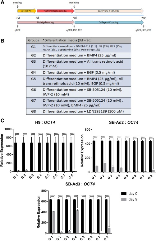
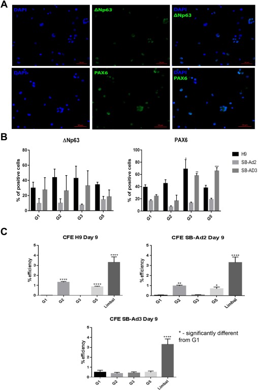
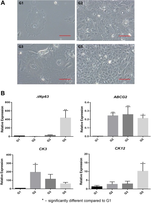
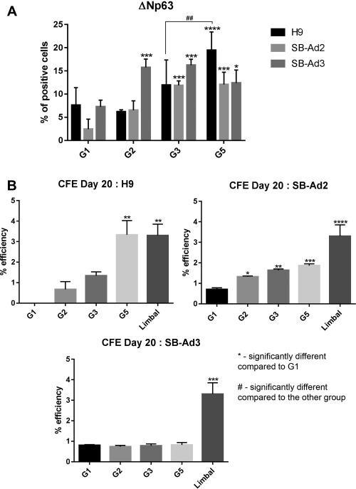
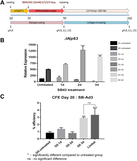
Cornea is a clear outermost layer of the eye which enables transmission of light onto the retina. The transparent corneal epithelium is regenerated by limbal stem cells (LSCs), whose loss/dysfunction results in LSCs deficiency (LSCD). Ex vivo expansion of autologous LSCs obtained from patient's healthy eye followed by transplantation onto the LSCs damaged/deficient eye, has provided a successful treatment for unilateral LSCD. However, this is not applicable to patient with total bilateral LSCD, where LSCs are lost/damaged from both eyes. We investigated the potential of human induced pluripotent stem cell (hiPSC) to differentiate into corneal epithelial-like cells as a source of autologous stem cell treatment for patients with total bilateral LSCD. Our study showed that combined addition of bone morphogenetic protein 4 (BMP4), all trans-retinoic acid and epidermal growth factor for the first 9 days of differentiation followed by cell-replating on collagen-IV-coated surfaces with a corneal-specific-epithelial cell media for an additional 11 days, resulted in step wise differentiation of human embryonic stem cells (hESC) to corneal epithelial progenitors and mature corneal epithelial-like cells. We observed differences in the ability of hiPSC lines to undergo differentiation to corneal epithelial-like cells which were dependent on the level of endogenous BMP signaling and could be restored via the activation of this signaling pathway by a specific transforming growth factor β inhibitor (SB431542). Together our data reveal a differential ability of hiPSC lines to generate corneal epithelial cells which is underlined by the activity of endogenous BMP signaling pathway. Stem Cells 2018;36:337-348.
Keywords: Bone morphogenetic protein 4; Corneal epithelial cells; Corneal epithelial progenitors; Epidermal growth factor; Human embryonic stem cell; Human induced pluripotent stem cell; Retinoic acid.
© 2017 The Authors Stem Cells published by Wiley Periodicals, Inc. on behalf of AlphaMed Press.
Figures

Similar articles
-
Bone Morphogenetic Protein 4 (BMP4) Enhances the Differentiation of Human Induced Pluripotent Stem Cells into Limbal Progenitor Cells.Curr Issues Mol Biol. 2021 Nov 29;43(3):2124-2134. doi: 10.3390/cimb43030147. Curr Issues Mol Biol. 2021. PMID: 34940121 Free PMC article.
-
Limbal stem cell transplantation: an evidence-based analysis.Ont Health Technol Assess Ser. 2008;8(7):1-58. Epub 2008 Oct 1. Ont Health Technol Assess Ser. 2008. PMID: 23074512 Free PMC article.
-
Corneal epithelial differentiation of human pluripotent stem cells generates ABCB5+ and ∆Np63α+ cells with limbal cell characteristics and high wound healing capacity.Stem Cell Res Ther. 2021 Dec 20;12(1):609. doi: 10.1186/s13287-021-02673-3. Stem Cell Res Ther. 2021. PMID: 34930437 Free PMC article.
-
Recent Advances in Stem Cell Therapy for Limbal Stem Cell Deficiency: A Narrative Review.Ophthalmol Ther. 2020 Dec;9(4):809-831. doi: 10.1007/s40123-020-00305-2. Epub 2020 Sep 24. Ophthalmol Ther. 2020. PMID: 32970311 Free PMC article. Review.
-
Characterization, isolation, expansion and clinical therapy of human corneal epithelial stem/progenitor cells.J Stem Cells. 2014;9(2):79-91. J Stem Cells. 2014. PMID: 25158157 Review.
Cited by
-
BMP6 Regulates Corneal Epithelial Cell Stratification by Coordinating Their Proliferation and Differentiation and Is Upregulated in Pterygium.Invest Ophthalmol Vis Sci. 2020 Aug 3;61(10):46. doi: 10.1167/iovs.61.10.46. Invest Ophthalmol Vis Sci. 2020. PMID: 32845956 Free PMC article.
-
Corneal cell therapy: with iPSCs, it is no more a far-sight.Stem Cell Res Ther. 2018 Oct 25;9(1):287. doi: 10.1186/s13287-018-1036-5. Stem Cell Res Ther. 2018. PMID: 30359313 Free PMC article. Review.
-
Differentiation Induction of Human Stem Cells for Corneal Epithelial Regeneration.Int J Mol Sci. 2020 Oct 22;21(21):7834. doi: 10.3390/ijms21217834. Int J Mol Sci. 2020. PMID: 33105778 Free PMC article. Review.
-
Corneal regeneration: insights in epithelial stem cell heterogeneity and dynamics.Curr Opin Genet Dev. 2022 Dec;77:101981. doi: 10.1016/j.gde.2022.101981. Epub 2022 Sep 6. Curr Opin Genet Dev. 2022. PMID: 36084496 Free PMC article. Review.
-
Differentiation of Human Mesenchymal Stem Cells into Corneal Epithelial Cells: Current Progress.Curr Issues Mol Biol. 2024 Nov 21;46(12):13281-13295. doi: 10.3390/cimb46120792. Curr Issues Mol Biol. 2024. PMID: 39727920 Free PMC article. Review.
References
-
- Schlotzer‐Schrehardt U, Kruse FE. Identification and characterization of limbal stem cells. Exp Eye Res 2005;81:247–264. - PubMed
-
- Lu L, Reinach PS, Kao WWY. Corneal epithelial wound healing. Exp Biol Med 2001;226:653–664. - PubMed
-
- Hay ED. Development of the vertebrate cornea. Int Rev Cytol 1980;63:263–322. - PubMed
-
- Amano S, Satoru Y, Tatsuya M et al. Corneal stromal and endothelial cell precursors. Cornea 2006;25:S73–S77. - PubMed
Publication types
MeSH terms
Substances
Grants and funding
- MC_PC_15030/MRC_/Medical Research Council/United Kingdom
- PB-PG-1215-20037/DH_/Department of Health/United Kingdom
- BB/E012841/1/BB_/Biotechnology and Biological Sciences Research Council/United Kingdom
- MR/S035826/1/MRC_/Medical Research Council/United Kingdom
- G0301182/MRC_/Medical Research Council/United Kingdom
LinkOut - more resources
Full Text Sources
Other Literature Sources
