. 2017 Dec;5(6):e00377.
doi: 10.1002/prp2.377.

Identification of an antibody-based immunoassay for measuring direct target binding of RIPK1 inhibitors in cells and tissues

Affiliations

Identification of an antibody-based immunoassay for measuring direct target binding of RIPK1 inhibitors in cells and tissues

Joshua N Finger et al. Pharmacol Res Perspect. 2017 Dec.

Abstract

Therapies that suppress RIPK1 kinase activity are emerging as promising therapeutic agents for the treatment of multiple inflammatory disorders. The ability to directly measure drug binding of a RIPK1 inhibitor to its target is critical for providing insight into pharmacokinetics, pharmacodynamics, safety and clinical efficacy, especially for a first-in-class small-molecule inhibitor where the mechanism has yet to be explored. Here, we report a novel method for measuring drug binding to RIPK1 protein in cells and tissues. This TEAR1 (Target Engagement Assessment for RIPK1) assay is a pair of immunoassays developed on the principle of competition, whereby a first molecule (ie, drug) prevents the binding of a second molecule (ie, antibody) to the target protein. Using the TEAR1 assay, we have validated the direct binding of specific RIPK1 inhibitors in cells, blood and tissues following treatment with benzoxazepinone (BOAz) RIPK1 inhibitors. The TEAR1 assay is a valuable tool for facilitating the clinical development of the lead RIPK1 clinical candidate compound, GSK2982772, as a first-in-class RIPK1 inhibitor for the treatment of inflammatory disease.

Keywords: Benzoxazepinone; RIPK1; TEAR1; TNF; pharmacokinetics/pharmacodynamics; tissue target engagement.


Figures

Figure 1
Figure 1
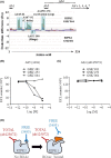
Identification of a target engagement antibody for RIPK1. (A) Comparison of RIPK1 antibodies (Ab1‐Ab7) with changes observed in HDXMS experiment between GSK’481‐bound protein and GSK’064‐bound protein. Ab3 is abcam 125072. Ab5 is Cell Signaling 3493. Ab3, Ab4, Ab6, and Ab7 are shown, though they do not overlap with the RIPK1 (1‐324) truncated protein used in HDXMS. These antibodies recognize a region at the RIPK1 C‐terminus from AA325‐671. The exact epitopes of these antibodies remain unknown. (B) HT‐29 cell lysates (20 μg) were incubated with GSK’481, GSK’253, or GSK’064 at concentrations ranging from 1 μmol·L−1 to 1 pmol·L−1 for 24 hours. Cell lysates were analyzed by immunoassay using Abcam ab72139 mouse monoclonal antibody for RIPK1 capture and Ab5 (Cell Signaling 3493) at 1 μg·mL−1 final concentration. Raw ECL counts were plotted against log molar concentrations of RIPK1 inhibitor. Samples were screened in single well format. (C) HT‐29 cell lysates (20 μg) were incubated with GSK’481, GSK’253, or GSK’064 at concentrations ranging from 1 μmol·L−1 to 1 pmol·L−1 for 24 hours. Cell lysates were analyzed by immunoassay using Abcam ab72139 mouse monoclonal antibody for RIPK1 capture and Ab3 (Abcam ab125072) at 1 μg·mL−1 final concentration. Raw ECL counts were plotted against log molar concentrations of RIPK1 inhibitor. Samples were screened in single well format. (D) Schematic representation of target engagement model demonstrating “competition” of FREERIPK1 antibody by RIPK1 inhibitors of the BOAZ chemical class. ECL, Electrochemiluminescent
Figure 2
Figure 2
RIPK1 target engagement in HT‐29 cells using the TEAR1 assay. (A) HT29 cells were incubated with either GSK’253 or GSK’064 for 24 hours. Cells lysates (20 μg) were analyzed for RIPK1 target engagement using both FREERIPK1 and TOTALRIPK1 immunoassays. FREERIPK1 levels were normalized to TOTALRIPK1 levels. Data are represented as the percent target engagement ±SD; n = 3 replicates per group. *P < .05, **P < .01, and ***P < .001. (B) HT29 cell lysates (10 μg) was analyzed by western blot for RIPK1 (CS3493, ab125072, and ab72139) and normalized to actin
Figure 3
Figure 3
Target engagement of gsk'253 in cynomolgus monkey whole blood. (A) Study design for in vivo evaluation of RIPK1 target engagement. (B) Blood from monkeys (n = 2/group) was collected at various time points following IV administration of 0.12 mg·kg−1 GSK’253 and analyzed by LCMS/MS. ***P < .001. (C) Comparison of predicted and observed RIPK1 target engagement following IV administration of GSK’253. Predicted target engagement was calculated using the known IC50 of 3.1 ng·mL−1 in a monkey whole‐blood challenge assay, assuming a hill slope of 1. Data are represented as the percent target engagement +/‐ SD; n = 2 animals per group. (D) Comparison of GSK’253 PK and in monkey whole blood following IV administration and observed target engagement. Individual measurements are plotted and IC50 was calculated in GraphPad Prism using a nonlinear regression and a 4‐parameter curve fit
Figure 4
Figure 4
Target engagement of GSK’253 in cynomolgus monkey tissues. (A) Skin biopsy‐drug concentrations at each time point following IV administration of 0.12 mg·kg−1 GSK’253. Data are represented mean drug concentration ± SD; n = 2 monkeys per group. *P < .05, **P < .01, and ***P < .001. (B) Comparison of predicted and observed RIPK1 target engagement in skin biopsies following IV administration of GSK’253. Predicted target engagement was calculated using the known IC50 of 3.1 ng·mL−1 in a monkey whole‐blood challenge assay, assuming a hill slope of 1. Data are represented as the percent target engagement ± SD; n = 2‐8 animals per group. *P < .05, **P < .01, and ***P < .001. (C) Comparison of predicted and observed RIPK1 target engagement in terminal colon tissue following IV administration of GSK’253. Predicted target engagement was calculated using the known IC50 of 3.1 ng·mL−1 in a monkey whole‐blood challenge assay, assuming a hill slope of 1. Data are represented as the percent target engagement ±SD; n = 2 animals per group. (D) Comparison of predicted and observed RIPK1 target engagement in terminal synovium tissue from knee joints following IV administration of GSK’253. Predicted target engagement was calculated using the known IC50 of 3.1 ng·mL−1 in a monkey whole‐blood challenge assay, assuming a hill slope of 1. Data are represented as the percent target engagement ±SD; n = 2‐8 animals per group

References

    1. Berger SB, Kasparcova V, Hoffman S, et al. Cutting Edge: RIP1 kinase activity is dispensable for normal development but is a key regulator of inflammation in SHARPIN‐deficient mice. J Immunol. 2014;192:5476‐5480. - PMC - PubMed
    1. Berger SB, Harris P, Kasparcova K, et al. Characterization of GSK′963: a structurally distinct potent and selective inhibitor of RIP1 kinase. Cell Death Disc. 2015;1:15009. - PMC - PubMed
    1. Bonnet MC, Preukschat D, Welz PS, et al. The adaptor protein FADD protects epidermal keratinocytes from necroptosis in vivo and prevents skin inflammation. Immunity. 2011;35:572‐582. - PubMed
    1. Christofferson DE, Yuan J. Necroptosis as an alternative form of programmed cell death. Curr Opin Cell Biol. 2010;22:263‐268. - PMC - PubMed
    1. Christofferson DE, Li Y, Hitomi J, et al. A novel role for RIP1 kinase in mediating TNF[alpha] production. Cell Death Dis. 2012;3:e320. - PMC - PubMed

MeSH terms

Substances

LinkOut - more resources