

CNS high-grade neuroepithelial tumor with BCOR internal tandem duplication: a comparison with its counterparts in the kidney and soft tissue
- PMID: 29226988
- PMCID: PMC8028450
- DOI: 10.1111/bpa.12585
CNS high-grade neuroepithelial tumor with BCOR internal tandem duplication: a comparison with its counterparts in the kidney and soft tissue
Abstract
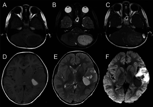
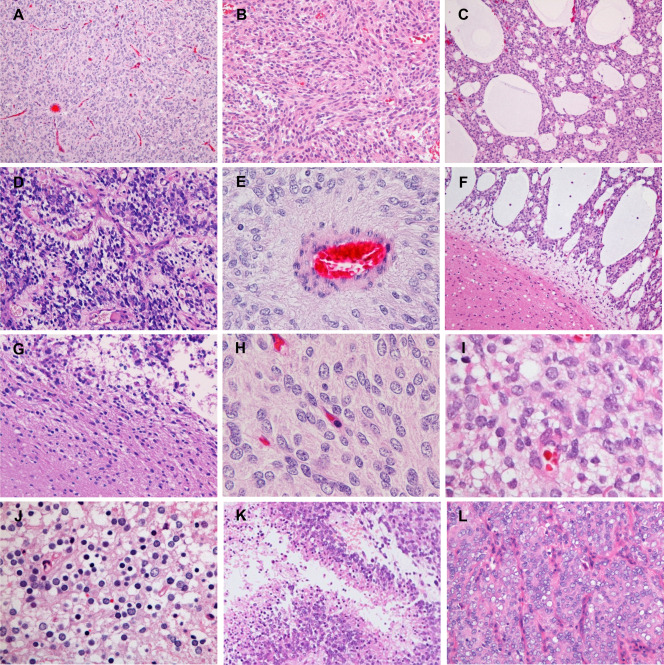
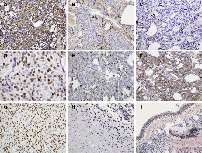
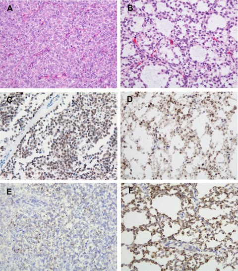
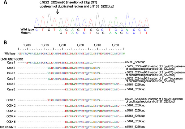
Central nervous system high-grade neuroepithelial tumors with BCOR alteration (CNS HGNET-BCOR) are a recently reported rare entity, identified as a small fraction of tumors previously institutionally diagnosed as so-called CNS primitive neuroectodermal tumors. Their genetic characteristic is a somatic internal tandem duplication in the 3' end of BCOR (BCOR ITD), which has also been found in clear cell sarcomas of the kidney (CCSK) and soft tissue undifferentiated round cell sarcomas/primitive myxoid mesenchymal tumors of infancy (URCS/PMMTI), and these BCOR ITD-positive tumors have been reported to share similar pathological features. In this study, we performed a clinicopathological and molecular analysis of six cases of CNS HGNET-BCOR, and compared them with their counterparts in the kidney and soft tissue. Although these tumors had histologically similar structural patterns and characteristic monotonous nuclei with fine chromatin, CNS HGNET-BCOR exhibited glial cell morphology, ependymoma-like perivascular pseudorosettes and palisading necrosis, whereas these features were not evident in CCSK or URCS/PMMTI. Immunohistochemically, diffuse staining of Olig2 with a mixture of varying degrees of intensity, and only focal staining of GFAP, S-100 protein and synaptophysin were observed in CNS HGNET-BCOR, whereas these common neuroepithelial markers were negative in CCSK and URCS/PMMTI. Therefore, although CNS HGNET-BCOR, CCSK and URCS/PMMTI may constitute a group of BCOR ITD-positive tumors, only CNS HGNET-BCOR has histological features suggestive of glial differentiation. In conclusion, we think CNS HGNET-BCOR are a certain type of neuroepithelial tumor relatively close to glioma, not CCSK or URCS/PMMTI occurring in the CNS.
Keywords: BCOR ITD; clear cell sarcoma of the kidney; high-grade neuroepithelial tumor.
© 2017 International Society of Neuropathology.
Conflict of interest statement
The authors have no conflict of interest.
Figures
Similar articles
-
Pediatric Soft Tissue Tumors With BCOR ITD Express EGFR but Not OLIG2.Pediatr Dev Pathol. 2020 Nov-Dec;23(6):424-430. doi: 10.1177/1093526620945528. Epub 2020 Aug 13. Pediatr Dev Pathol. 2020. PMID: 32790583
-
Recurrent BCOR Internal Tandem Duplication and YWHAE-NUTM2B Fusions in Soft Tissue Undifferentiated Round Cell Sarcoma of Infancy: Overlapping Genetic Features With Clear Cell Sarcoma of Kidney.Am J Surg Pathol. 2016 Aug;40(8):1009-20. doi: 10.1097/PAS.0000000000000629. Am J Surg Pathol. 2016. PMID: 26945340 Free PMC article.
-
High-grade neuroepithelial tumor with BCOR exon 15 internal tandem duplication-a comprehensive clinical, radiographic, pathologic, and genomic analysis.Brain Pathol. 2020 Jan;30(1):46-62. doi: 10.1111/bpa.12747. Epub 2019 Jun 10. Brain Pathol. 2020. PMID: 31104347 Free PMC article.
-
BCOR involvement in cancer.Epigenomics. 2019 May;11(7):835-855. doi: 10.2217/epi-2018-0195. Epub 2019 May 31. Epigenomics. 2019. PMID: 31150281 Free PMC article. Review.
-
CNS tumor with BCOR internal tandem duplication: Clinicopathologic, molecular characteristics and prognosis factors.Pathol Res Pract. 2022 Aug;236:153995. doi: 10.1016/j.prp.2022.153995. Epub 2022 Jun 23. Pathol Res Pract. 2022. PMID: 35809497 Review.
Cited by
-
The spectrum of rare central nervous system (CNS) tumors with EWSR1-non-ETS fusions: experience from three pediatric institutions with review of the literature.Brain Pathol. 2021 Jan;31(1):70-83. doi: 10.1111/bpa.12900. Epub 2020 Nov 6. Brain Pathol. 2021. PMID: 32997853 Free PMC article. Review.
-
Case of the month 1-2019: CNS high-grade neuroepithelial tumor with BCOR alteration.Clin Neuropathol. 2019 Jan/Feb;38(1):4-7. doi: 10.5414/NP301162. Clin Neuropathol. 2019. PMID: 30526817 Free PMC article.
-
Paediatric BCOR-associated sarcomas with a novel long spliced internal tandem duplication of BCOR exon 15.J Pathol Clin Res. 2022 Sep;8(5):470-480. doi: 10.1002/cjp2.287. Epub 2022 Jul 14. J Pathol Clin Res. 2022. PMID: 35836306 Free PMC article.
-
Supratentorial non-RELA, ZFTA-fused ependymomas: a comprehensive phenotype genotype correlation highlighting the number of zinc fingers in ZFTA-NCOA1/2 fusions.Acta Neuropathol Commun. 2021 Aug 13;9(1):135. doi: 10.1186/s40478-021-01238-y. Acta Neuropathol Commun. 2021. PMID: 34389065 Free PMC article.
-
cIMPACT-NOW update 6: new entity and diagnostic principle recommendations of the cIMPACT-Utrecht meeting on future CNS tumor classification and grading.Brain Pathol. 2020 Jul;30(4):844-856. doi: 10.1111/bpa.12832. Epub 2020 Apr 19. Brain Pathol. 2020. PMID: 32307792 Free PMC article.
References
-
- Appay R, Macagno N, Padovani L, Korshunov A, Kool M, André N et al (2017) HGNET‐BCOR tumors of the cerebellum: clinicopathologic and molecular characterization of 3 cases. Am J Surg Pathol 41:1254–1260. - PubMed
-
- Argani P, Perlman EJ, Breslow NE, Browning NG, Green DM, D'Angio GJ et al (2000) Clear cell sarcoma of the kidney: a review of 351 cases from the National Wilms Tumor Study Group Pathology Center. Am J Surg Pathol 24:4–18. - PubMed
-
- Biegel JA (2006) Molecular genetics of atypical teratoid/rhabdoid tumor. Neurosurg Focus 20:E11. - PubMed
-
- Burger PC, Yu IT, Tihan T, Friedman HS, Strother DR, Kepner JL et al (1998) Atypical teratoid/rhabdoid tumor of the central nervous system: a highly malignant tumor of infancy and childhood frequently mistaken for medulloblastoma: a Pediatric Oncology Group study. Am J Surg Pathol 22:1083–1092. - PubMed
Publication types
MeSH terms
Substances
LinkOut - more resources
Full Text Sources
Other Literature Sources
Miscellaneous
