. 2018 Feb;14(2):126-134.
doi: 10.1038/nchembio.2527. Epub 2017 Dec 11.

Structure-inspired design of β-arrestin-biased ligands for aminergic GPCRs

Affiliations

Structure-inspired design of β-arrestin-biased ligands for aminergic GPCRs

John D McCorvy et al. Nat Chem Biol. 2018 Feb.

Abstract

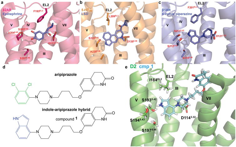
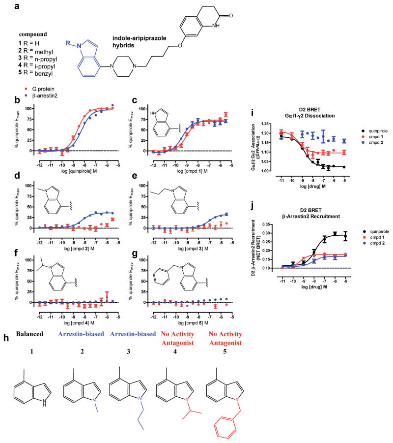
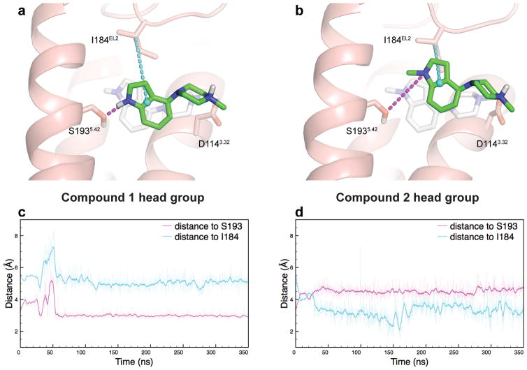
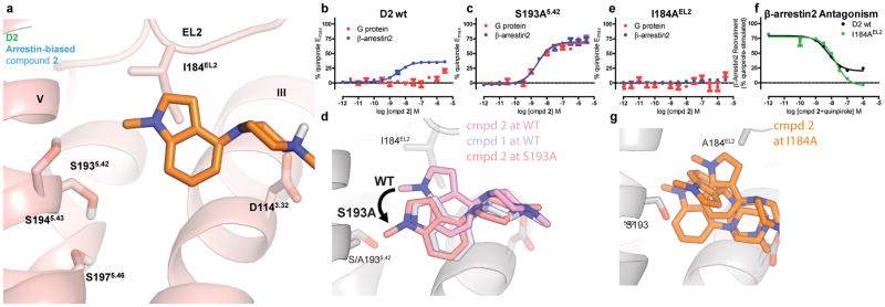
Development of biased ligands targeting G protein-coupled receptors (GPCRs) is a promising approach for current drug discovery. Although structure-based drug design of biased agonists remains challenging even with an abundance of GPCR crystal structures, we present an approach for translating GPCR structural data into β-arrestin-biased ligands for aminergic GPCRs. We identified specific amino acid-ligand contacts at transmembrane helix 5 (TM5) and extracellular loop 2 (EL2) responsible for Gi/o and β-arrestin signaling, respectively, and targeted those residues to develop biased ligands. For these ligands, we found that bias is conserved at other aminergic GPCRs that retain similar residues at TM5 and EL2. Our approach provides a template for generating arrestin-biased ligands by modifying predicted ligand interactions that block TM5 interactions and promote EL2 interactions. This strategy may facilitate the structure-guided design of arrestin-biased ligands at other GPCRs, including polypharmacological biased ligands.


Conflict of interest statement

Competing financial interests

The authors declare no competing financial interests.

Figures

Figure 1
Figure 1. Structure-Inspired Design of Indole-Aripiprazole Hybrid Ligands
D2 ligand design based on comparison of three aminergic crystal structures. a) β2 adrenergic receptor nanobody-stabilized with epinephrine bound (4LDO) indicates the catechol of epinephrine is involved in an extensive hydrogen bond network with transmembrane (TM) 5 serines. b) Structure of the 5-HT2B receptor with LSD bound (5TVN) indicates that EL2.52 Leu209 forms hydrophobic cap over ligand, preventing ligand egress. c) Thermostabolized β1 adrenergic receptor with 4-indole piperazine bound (3ZPQ) shows that the indole N-H interacts with Ser5.42 in a hydrogen bond d) Design of indole-aripiprazole hybrid compounds by addition of 4-indole (blue) replacing the dichlorophenyl (green) of aripiprazole resulting in compound 1. e) Docking of 1 in D2 homology model places the unsubstituted 4-indole moiety of the indole-aripiprazole hybrid 1 in the D2 orthosteric binding pocket making contact with TM5 Ser5.42.
Figure 2
Figure 2. Indole-Aripiprazole Hybrid D2R SFSR
Structure-Functional Selectivity Relationships (SFSRs) of indole N1-substituted analogs of indole-aripiprazole hybrids, which lead to either D2 arrestin-bias or antagonism dependent on substitution. a) Chemical structures of N1-substituted indole-aripiprazole hybrids b-g) Profiling of indole-aripiprazole hybrids measuring D2 G protein activity (Gαi/o-mediated cAMP inhibition; red) and β-arrestin2 recruitment (Tango; blue), normalized to percent quinpirole activity. Data represent n=5 performed in triplicate and in parallel using the same drug dilutions. h) SFSR summary for indole-aripiprazole hybrids. Unsubstituted indole (1) shows weak preference for arrestin with respect to quinpirole (bias factor = 2.5; D2 Gi/o EC50= 0.98 nM, Emax = 66%, D2 β-arrestin2 EC50= 0.71 nM Emax = 69%) comparing Gi/o and arrestin activity but N-methyl (2, D2 β-arrestin2 EC50= 6.3 nM, Emax = 36%) and N-n-propyl (3, D2 β-arrestin2 EC50= 81 nM, Emax = 32%) show arrestin-bias with no measureable Gi/o activity with respect to quinpirole. Larger substitutions such as N-i-propyl (4) and N-benzyl (5) show no activity and instead act as competitive antagonists i) Orthologous assay for D2 G protein activity utilizing D2 Gi1-γ2 dissociation as measured by BRET, showing partial agonism for 1 (EC50 = 0.49 nM, Emax = 55%) and no activity by 2, compared to quinpirole (EC50 = 1.6 nM). Data represent total BRET as calculated using GFP/Rluc ratio j) Orthologous assays for β-arrestin2 recruitment utilizing BRET measuring Venus-tagged-β-arrestin2 and D2long-tagged Rluc association comparing recruitment by 1 (EC50 = 0.52 nM, Emax = 39%) and 2 (EC50 = 11 nM, Emax = 33%) to quinpirole (EC50 = 13 nM). Data are representative and indicate the change in Net BRET with respect to no Venus-β-arrestin2 expressed.
Figure 3
Figure 3. D2R MD Simulations Predict EL2 Engagement for Arrestin-bias
MD simulations of the head groups of compound 1 (a) and compound 2 (b) reveal that β arrestin-biased 2 preferentially interacts with I184 in EL2 over S193 in TM5. By contrast, 1 maintains a stable hydrogen bond with S1935.42 throughout simulation, without interacting substantially with I184EL2. Relative positioning of the head groups to TM5 and EL2 was tracked by the distance from the ligand indole nitrogen to the hydroxyl oxygen of S1935.42 (magenta), and the distance from the center of the indole ring to the β-carbon of I184EL2 (cyan), for compound 1 (c) and 2 (d). The starting pose of the head group simulations, equating to the crystal structure of thermostabilized β1AR (3ZPQ) in complex with indole 4-(piperazin-1-yl)-1H-indole, is shown in light grey, while the green ligand and the protein show a representative snapshot from simulation. In (c) and (d), thin traces are sampled every 100 ps and thick traces are smoothed with a 1 ns moving average.
Figure 4
Figure 4. D2 TM5 and EL2 Mutants Confirm Arrestin-Bias Binding Pose
a) The pose resulting from MD simulation of the head group of arrestin-biased N-methyl indole-aripiprazole hybrid (2) places the N-methyl indole moiety in contact with I184 on EL2, having moved away from S193 on TM5. b) N-methyl indole-aripiprazole hybrid 2 only shows arrestin recruitment activity at D2 wild-type. Data represent mean and standard error of the mean performed in triplicate (Gi/o GloSensor; red, n=3) and β-arrestin2 recruitment (Tango; blue, n=3, EC50 = 3.7 nM, Emax = 36%) c) S193A5.42 transforms arrestin-bias of 2 into balanced signaling with respect to quinpirole. Data represent Gαi/o-mediated cAMP inhibition (Gi/o GloSensor; red, n=3, EC50 = 2.5 nM, Emax = 67%) and β-arrestin2 recruitment (Tango; blue, n=3, EC50 = 2.6 nM, Emax = 69%). d) Representative pose of compound 2 head group from simulation at wild-type (WT) and S193A D2R constructs, and compound 1 head group from wild-type D2R simulation. At S193A, 2 moves to a pose almost identical to 1 at D2 wild-type. e) Mutation of EL2 I184 (I184A) completely abolishes arrestin recruitment for arrestin-biased ligand 2 (Tango; n=5 in triplicate) f) I184A mutation transforms 2 into a D2R β-arrestin2 recruitment antagonist as measured in Tango (n=2, in triplicate), as seen by comparing D2 wild-type (black, IC50 = 6.3 nM) to EL2 I184A (green, IC50 = 13 nM). g) Compound 2 head group is unstable throughout simulation at I184A D2R, sampling many orientations within the ligand-binding pocket. Ligand poses are shown for three points in time during a single simulation.
Figure 5
Figure 5. MD-Assisted Rational Design of Arrestin-biased Compounds
a) Mutagenesis data indicating that 2 requires I184EL2 for β-arrestin recruitment, and MD findings that 2 preferentially interacts with I184 EL2, led to the design of 2-methyl indole derivative 7 to further engage EL2 and enhance β-arrestin recruitment. Compound 6 is the unsubstituted control compound, which can still form a hydrogen bond with S1935.42. Compound 6 shows balanced D2 signaling with respect to quinpirole (bias factor = 1.3, Gi/o EC50 = 0.49 nM, Emax = 86%, β-arrestin2 EC50 = 0.62 nM, Emax = 78%), but compound 7 shows arrestin-bias with respect to quinpirole (bias factor = 20) comparing Gαi/o-mediated cAMP inhibition (GloSensor; red; n=3, EC50 = 23 nM, Emax = 60%) and β-arrestin2 recruitment (Tango; blue; n=3, EC50 = 2.9 nM, Emax = 78%). b) 2-methyl substitution (7, purple, Emax = 78%) shows higher D2 β-arrestin2 recruitment efficacy compared to compound 2 (blue, Emax = 36%) with respect to quinpirole measured by Tango. Data were normalized to quinpirole and represent n=3 in triplicate. c) Interaction with EL2 confirmed with I184AEL2 mutation selectively abolishing β-arrestin2 recruitment (Tango) for biased ligands 2 and 6 and not for balanced 1 (red) and quinpirole (black). Compound 6 (green) shows decreased arrestin recruitment by I184AEL2 mutation but not complete loss of activity (β-arrestin2 EC50 = 2.7 nM, Emax = 40%). Data were normalized to quinpirole and represent n=3 performed in triplicate. d) Structure-function selectivity relationships for indole-aripiprazole hybrid series as outlined using a heat map comparing log log(τ/KA) activities measuring G protein and β-arrestin2 recruitment.
Figure 6
Figure 6. Prediction and Confirmation of Polypharmacological Arrestin-bias
Alignments of D2 TM5 and EL2 residues predict that 2 shows arrestin-bias at D3 (a), D4 (b) with respect to quinpirole and 5-HT7 (c) receptors with respect to 5-HT, where TM5 and EL2 residues in orthosteric sites are well-conserved, except at β2 (d) with respect to isoproterenol, where 2 only shows inverse agonist activity but no arrestin recruitment. G protein-signaling was measured by GloSensor and β-arrestin recruitment was measured by Tango performed in parallel (n=3 in triplicate). e) TM5 and EL2 are key contacts in the orthosteric sites of aminergic GPCRs whereby an arrestin-bias template for ligand design can be used to promote EL2 engagement to enhance β-arrestin recruitment and preclude TM5 engagement to avoid G protein-signaling.

References

    1. Overington JP, Al-Lazikani B, Hopkins AL. How many drug targets are there? Nat Rev Drug Discov. 2006;5:993–996. - PubMed
    1. Rask-Andersen M, Almen MS, Schioth HB. Trends in the exploitation of novel drug targets. Nat Rev Drug Discov. 2011;10:579–590. - PubMed
    1. Wacker D, Stevens RC, Roth BL. How Ligands Illuminate GPCR Molecular Pharmacology. Cell. 2017;170:414–427. - PMC - PubMed
    1. Urban JD, et al. Functional selectivity and classical concepts of quantitative pharmacology. J Pharmacol Exp Ther. 2007;320:1–13. - PubMed
    1. Dewire SM, Ahn S, Lefkowitz RJ, Shenoy SK. Beta-arrestins and cell signaling. Annu Rev Physiol. 2007;69:483–510. - PubMed

Publication types