Comparative Study
. 2017 Dec 1;33(12):834-839.
doi: 10.3928/1081597X-20170921-02.

Efficacy and Safety Comparison Between Suberoylanilide Hydroxamic Acid and Mitomycin C in Reducing the Risk of Corneal Haze After PRK Treatment In Vivo

Comparative Study

Efficacy and Safety Comparison Between Suberoylanilide Hydroxamic Acid and Mitomycin C in Reducing the Risk of Corneal Haze After PRK Treatment In Vivo

Govindaraj Anumanthan et al. J Refract Surg. .

Abstract

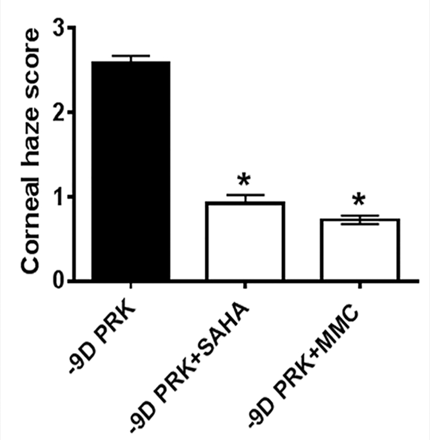
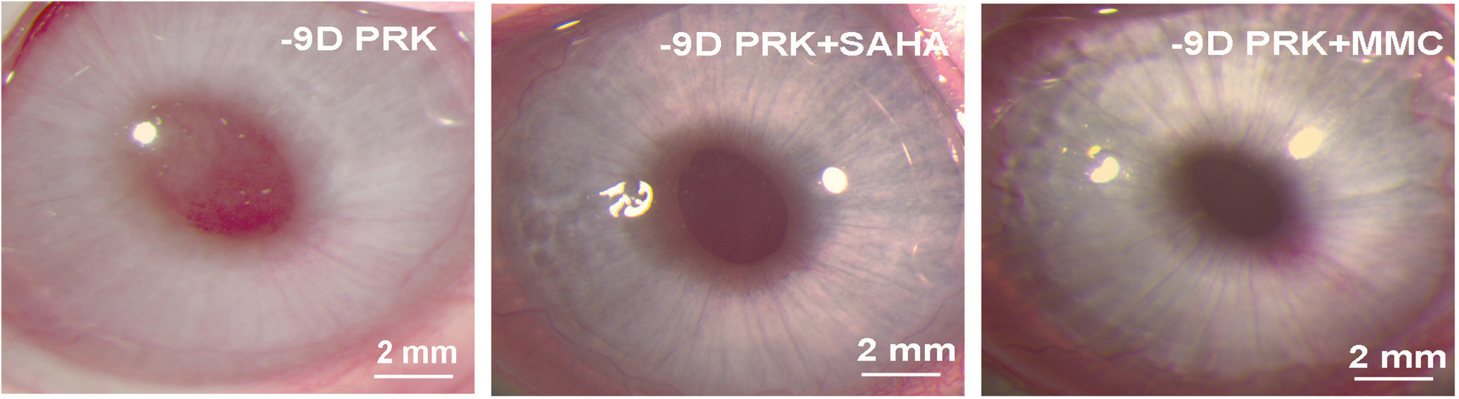
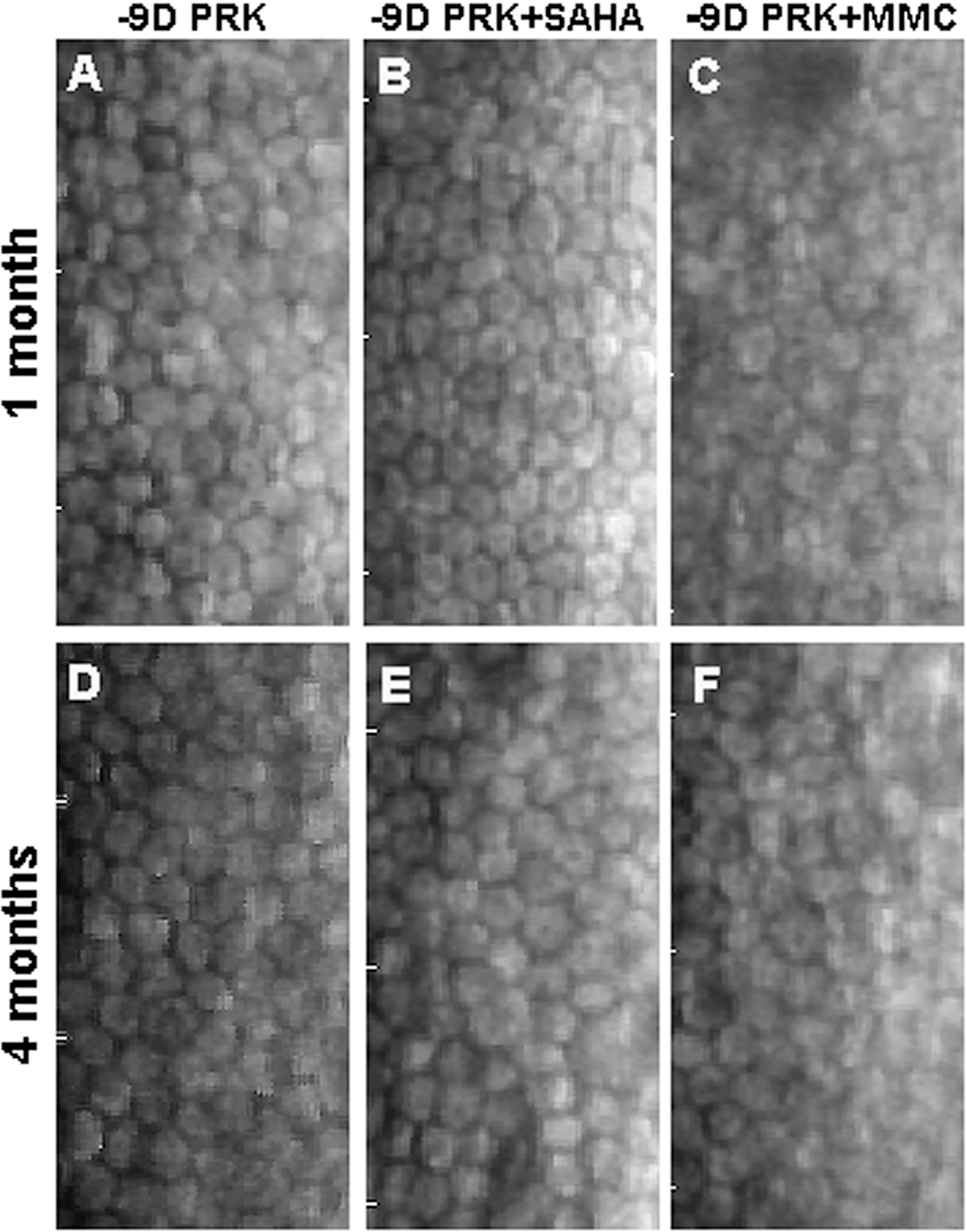
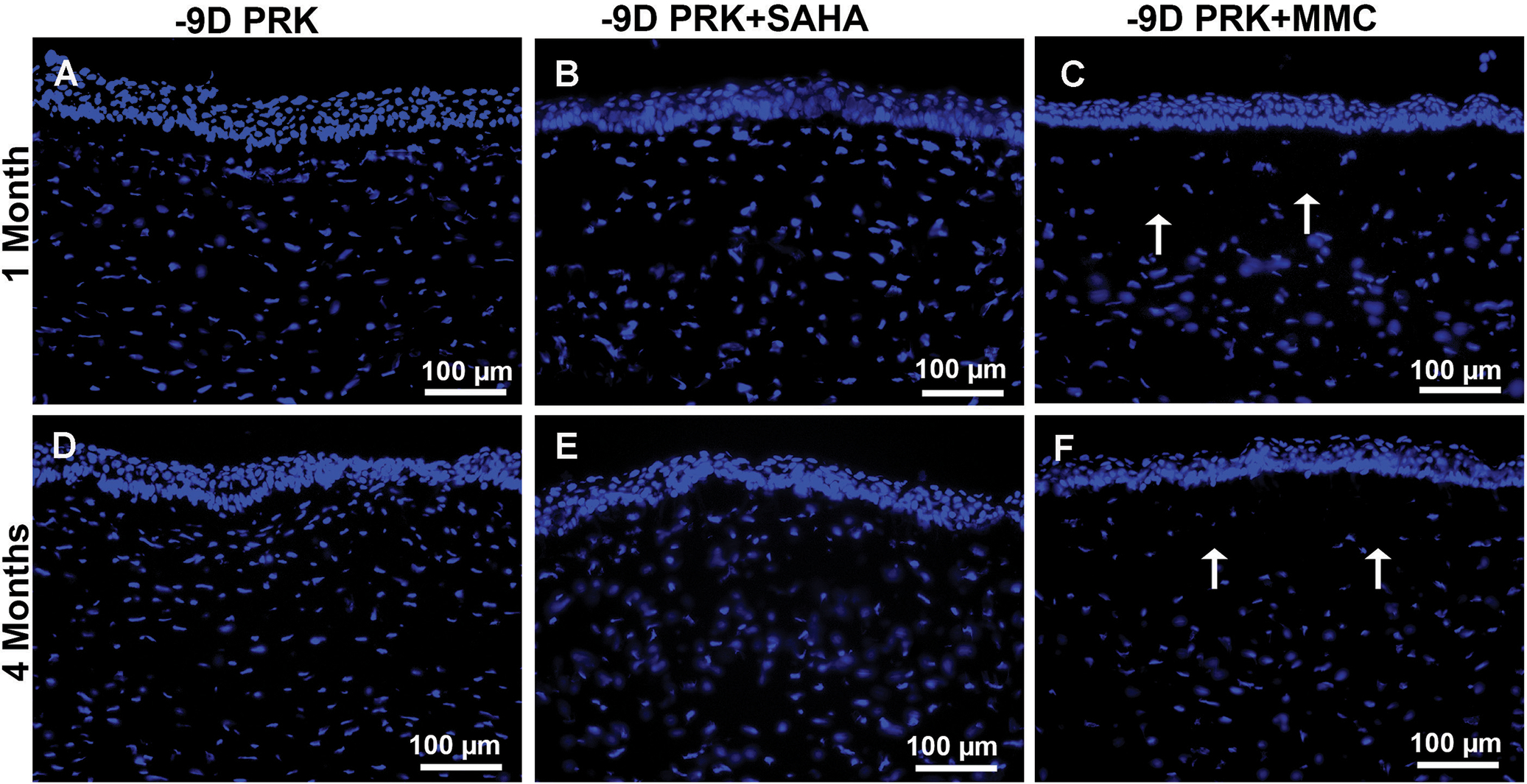
Purpose: This study compared the efficacy and safety of suberoylanilide hydroxamic acid (SAHA) and mitomycin C (MMC) up to 4 months in the prevention of corneal haze induced by photorefractive keratectomy (PRK) in rabbits in vivo.

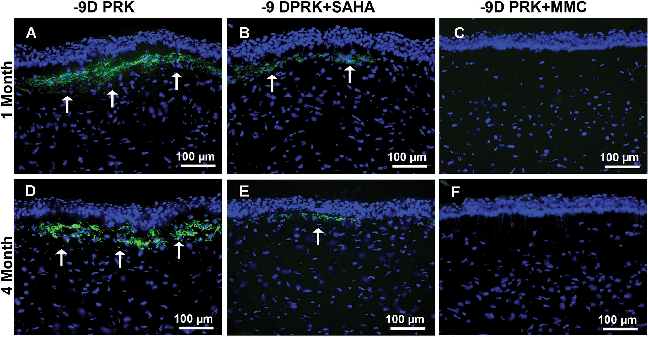
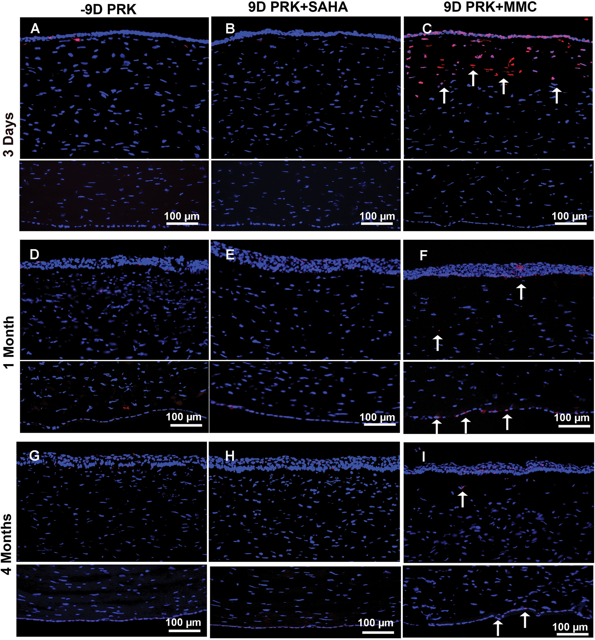
Methods: Corneal haze in rabbits was produced with -9.00 diopter PRK. A single application of SAHA (25 μM) or MMC (0.02%) was applied topically immediately after PRK. Effects of the two drugs were analyzed by slit-lamp microscope, specular microscope, TUNEL assay, and immunofluorescence.

Results: Single topical adjunct use of SAHA (25 μM) or MMC (0.02%) after PRK attenuated more than 95% corneal haze and myofibroblast formation (P < .001). SAHA did not reduce keratocyte density, cause keratocyte apoptosis, or increase immune cell infiltration compared to MMC (P < .01 or .001). Furthermore, SAHA dosing did not compromise corneal endothelial phenotype, density, or function in rabbit eyes, whereas MMC application did (P < .01 or .001).

Conclusions: SAHA and MMC significantly decreased corneal haze after PRK in rabbits in vivo. SAHA exhibited significantly reduced short- and long-term damage to the corneal endothelium compared to MMC in rabbits. SAHA is an effective and potentially safer alternative to MMC for the prevention of corneal haze after PRK. Clinical trials are warranted. [J Refract Surg. 2017;33(12):834-839.].


Conflict of interest statement

The authors have no financial or proprietary interest in the materials presented herein.

Figures

Figure 1.
Figure 1.
Haze quantification showed that a single 5-minute topical application of suberoylanilide hydroxamic acid (SAHA) or mitomycin C (MMC) significantly decreased corneal haze following photorefractive keratectomy (PRK) (P < .001 for both). D = diopters
Figure 2.
Figure 2.
Effect of suberoylanilide hydroxamic acid (SAHA) and mitomycin C (MMC) on the attenuation of myofibroblasts after photorefractive keratectomy (PRK). Representative fluorescence images of rabbit corneas that underwent −9.00 diopter (D) PRK followed by balanced salt solution, or SAHA (25 μM), or MMC (0.02%) treatment at 1 and 4 months after treatment. Myofibroblasts (arrows) for α-smooth muscle actin in the subepithelial stroma (green). SAHA (B and E) and MMC (C and F) treatment significantly decreased α-SMA compared to control corneas (A and D). Scale bar = 100 μm (Blue = DAPI-stained nuclei, Green = α-SMA+ cells)
Figure 3.
Figure 3.
Effect of suberoylanilide hydroxamic acid (SAHA) and mitomycin C (MMC) treatment on keratocyte and endothelial loss. Representative images showing TUNEL staining in rabbit corneas collected 3 days, 1 month, and 4 months after −9.00 diopter (D) photorefractive keratectomy (PRK) and balanced salt solution (BSS), SAHA (25 μM), or MMC (0.02%) single adjunct topical application. BSS (A, D, and G) or SAHA (B, E, and H) treatment did not induce apoptosis in corneal keratocyte or endothelial cells as shown by detection of no TUNEL+ cells. Conversely, MMC treatment (C, F, and I) provoked significant apoptosis in keratocyte and epithelial cells but none in endothelial cells at 3 days (C), with noticeable apoptosis in endothelial cells and some in keratocytes at 1 (F) and 4 (I) months as shown by arrows. These data suggest that MMC caused significant damage to anterior stroma and restricted keratocyte revival in this region lost in the first 3 days of MMC application. Nuclei are stained blue with DAPI. TUNEL+ cells are stained red (arrows). Scale bar = 100 μm
Figure A.
Figure A.
Effect of suberoylanilide hydroxamic acid (SAHA) or mitomycin C (MMC) application on corneal haze in vivo. Representative stereomicroscopy images showing corneal haze levels and its quantification noted at 4 months in −9.00 diopter photorefractive keratectomy (PRK) performed on rabbit corneas that received either balanced salt solution (control), SAHA (25 μM), or MMC (0.02%). Each group had 6 eyes for slit-lamp biomicroscopy. The central scar was considerably reduced and the overall corneal clarity appeared improved in corneas treated with SAHA or MMC.
Figure B.
Figure B.
Effect of suberoylanilide hydroxamic acid (SAHA) and mitomycin C (MMC) on corneal endothelial cells. Representative specular biomicroscopy images of endothelial cells in the rabbit corneas at 1 and 4 months collected from eyes treated with balanced salt solution (control), SAHA (25 μM), or MMC (0.02%) after −9.00 diopter (D) photorefractive keratectomy (PRK). Corneas treated with SAHA (B and E) showed typical hexagonal corneal endothelial morphology and density similar to control (A and D). Conversely, corneas treated with MMC (C and F) showed markedly damaged corneal endothelial with compromised cell size and density at 1 and 4 months.
Figure C.
Figure C.
Effect of suberoylanilide hydroxamic acid (SAHA) and mitomycin C (MMC) on keratocyte number and density. Representative DAPI nuclear staining showing keratocyte population and density at 1 and 4 months in rabbit corneas treated with balanced salt solution (control), SAHA (25 μM), and MMC (0.02%) after photorefractive keratectomy (PRK). Control (A and C) or SAHA (B and D) treatment did not decrease keratocyte number and density in the stroma, whereas MMC treatment (C and F) caused significant keratocyte depletion of keratocyte number and density, especially in the anterior stroma as indicated by the arrows (C and F) at 1 and 4 months. Scale bar = 100 μm (Blue = DAPI-stained nuclei)

Similar articles

Cited by

References

    1. Ljubimov AV, Saghizadeh M. Progress in corneal wound healing. Prog Retin Eye Res. 2015;49:17–45. - PMC - PubMed
    1. Jester JV, Ho-Chang J. Modulation of cultured corneal keratocyte phenotype by growth factors/cytokines control in vitro contractility and extracellular matrix contraction. Exp Eye Res. 2003;77:581–592. - PubMed
    1. Mohan RR, Hutcheon AE, Choi R, et al. Apoptosis, necrosis, proliferation, and myofibroblast generation in the stroma following LASIK and PRK. Exp Eye Res. 2003;76:71–87. - PubMed
    1. Stepp MA, Zieske JD, Trinkaus-Randal V, et al. Wounding the cornea to learn how it heals. Exp Eye Res. 2014;121:178–193. - PMC - PubMed
    1. Tandon A, Tovey JC, Sharma A, Gupta R, Mohan RR. Role of transforming growth factor beta in corneal function, biology and pathology. Curr Mol Med. 2010;10:565–578. - PMC - PubMed

Publication types

MeSH terms

LinkOut - more resources