. 2017 Dec 8;9(12):2529-2543.
doi: 10.18632/aging.101339.

POT1 inhibits the efficiency but promotes the fidelity of nonhomologous end joining at non-telomeric DNA regions

Affiliations

POT1 inhibits the efficiency but promotes the fidelity of nonhomologous end joining at non-telomeric DNA regions

Yang Yu et al. Aging (Albany NY). .

Abstract

Robust DNA double strand break (DSB) repair and stabilized telomeres help maintain genome integrity, preventing the onset of aging or tumorigenesis. POT1 is one of the six factors in the shelterin complex, which protects telomeres from being recognized as DNA damages. TRF1 and TRF2, two other shelterin proteins, have been shown to participate in DNA DSB repair at non-telomeric regions, but whether POT1, which binds to single strand telomeric DNA at chromosomal ends, is involved in DNA DSB repair has not been assessed. Here we found that POT1 arrives at DNA damage sites upon the occurrence of DNA DSBs. It suppresses the efficiency of nonhomologous end joining (NHEJ), the major pathway for fixing DNA DSBs in mammals, but surprisingly promotes NHEJ fidelity. Mechanistic studies indicate that POT1 facilitates the recruitment of Artemis, which is a nuclease and promotes fidelity of NHEJ, to DNA damage sites. In addition, we found that overexpression of POT1 inhibits the protein stability of Lig3, which is the major regulator of alternative NHEJ (alt-NHEJ), therefore suppressing the efficiency of alt-NHEJ. Taken together we propose that POT1 is a key factor regulating the balance between the efficiency and fidelity of NHEJ at non-telomeric DNA regions.

Keywords: Artemis; DNA Lig3; DNA double strand break repair; NHEJ efficiency; NHEJ fidelity; POT1.


Conflict of interest statement

CONFLICTS OF INTEREST

The authors declare that they have no conflicts of interest with the contents of this article.

Figures

Figure 1
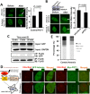
Figure 1. POT1 is rapidly recruited to nontelomeric DNA damage sites
(A) POT1 is recruited to DNA damage sites within 1-second post microirradiation. U2OS cells were microirradiated to generate DSBs in a line pattern using a 405 nm diode laser. (B) Diagram of the site for which ChIP primers were designed (arrows). At different time points post I-SceI transfection, NHEJ-I9a cells were harvested and lysed for ChIP assay with an antibody against POT1, followed by quantitative PCR analysis. The procedure for ChIP is as previously described [12]. (C) Schematic depiction of different domains of POT1 tagged with GFP. (D) Comparable expression of GFP-tagged different domains of POT1. The U2OS cells were transfected with different amounts of vectors encoding OB1 (0.67 μg), OB2 (0.5 μg), OB12 (1 μg), C-terminal (3 μg). At 48 h post transfection, cells were harvested for FACS analysis. (E) Analysis of recruitment of different POT1 domains.
Figure 2
Figure 2. POT1 promotes NHEJ fidelity but inhibits NHEJ efficiency
(A) Schematic picture of NHEJ reporter cassette. The reporter and the cell line harboring it are as previously described [7, 24]. (B) Expression of FLAG-tagged POT1. (C) The effect of POT1 overexpression on NHEJ efficiency. The NHEJ-I9a was transfected with POT1 vector, I-SceI vector and DsRed for normalizing transfection efficiency using Lonza 4D machine. On day 3 post transfection, cells were harvested for FACS analysis. (D) Overexpressing POT1 sensitizes HCA2-hTERT cells to X-Ray. POT1 overexpressing cells were treated with X-Ray at 4 Gy, and then harvested, reseeded to plates at different numbers. On day 14 post IR, cells were stained with Commassie solution and colonies with at least 50 cells were counted. Cell survival was calculated as the ratio of the relative plating efficiencies of X-Ray treated versus control cells. (E) Analysis of NHEJ fidelity. The method is as previously reported [25]. At least forty clones were used for junction sequencing. bp: base pairs.
Figure 3
Figure 3. POT1 stimulates the recruitment of Artemis to DNA damages sites
(A) POT1 overexpression stimulates the recruitment of Artemis to laser induced DNA damage sites. The recruitment of Artemis is quantified using the software of Leica LAS AF Lite. (B) Mildly knocking down POT1 significantly suppresses the recruitment of Artemis to DNA lesions induced by lasers. The recruitment of Artemis is quantified using the software of Leica LAS AF Lite. (C) POT1 interacts with Artemis upon DNA damages. 293FT cells were co-transfected with a plasmid encoding POT1-FLAG and a vector encoding Artemis-GFP. On day 1 post transfection, cells were irradiated with X-Ray at 6 Gy. At different time points, cells were harvested for immunoprecipitation with an antibody against FLAG, followed by Western blot analysis. (D) Both POT1 and Artemis are recruited to a given DNA damage site. The KillerRed (KR) system is as previously described (25). In brief, it is a fluorescent protein derived from hydrozoan. Long exposure of cells expressing the KR protein generates ROS-induced DNA DSBs. The U2OS reporter cell line harboring ~ 200 copies of TRE elements was co-transfected with myc-POT1 or GFP-Artemis and TA (transcription activator) –KR or TA-cherry. Both TA-KR and TA-cherry proteins may recognize the TRE elements. TA-KR causes DNA damages at the given site while TA-cherry is utilized as a negative control. The transfected cells were then exposed to light, followed by fixation and immunostaining for further analysis. (E) The effect of Artemis overexpression on NHEJ fidelity. The analysis of NHEJ fidelity is as described in Figure 2E.
Figure 4
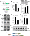
Figure 4. POT1 inhibits alt-NHEJ efficiency and promotes the degradation of Lig3
(A) Schematic diagram of EJ2-GFP for analyzing the alt-NHEJ efficiency. The mechanism of the reporter cassette is as previously described [6]. (B) Overexpression of POT1 inhibits alt-NHEJ efficiency. The reporter construct was digested with I-SceI restriction enzyme in vitro, followed by being transfected to HCA2-hTERT cells together with a control vector or a plasmid encoding POT1. On day 3 post transfection, cells were harvested for FACS analysis. (C) and (D) Mildly knocking down POT1 in HeLa cells significantly stimulates the alt-NHEJ efficiency. HeLa cells were transfected with siRNA against POT1 twice with two days interval, followed by a transfection of I-SceI linearized EJ2-GFP reporter. On day 3 post transfection, cells were harvested for FACS analysis. (E) Expression of important NHEJ factors in the absence or presence of POT1 overexpression. (F) Quantification of Lig3 expression using ImageJ software. The relative expression of Lig3 is calculated as the ratio of Lig3 expression versus TUBULIN. (G) Lig3 expression was not affected at transcriptional level in POT1 overexpressing cells. At 24 h post POT1 transfection, cells were harvested for mRNA extraction. Then Quantitative PCR analysis was performed with primers indicated. The primers used for q-PCR of Lig3 are as follows: Forward: 5′- TATGGCACGGGACCTAG -3′, Reverse: 5′- CTGTTGCTGCTCATCCTC -3′. The primers used for q-PCR of GAPDH are as follows: Forward: 5′ATGACATCAAGAAGGTGGTG3′, Reverse: 5′CATACCAGGAAATGAGCTTG3′. The transcript level of Lig3 was determined using delta CT method [38]. (H) POT1 overexpression promotes Lig3 degradation. 293FT cells with a control vector or a vector encoding POT1 transfected were treated with cycloheximide (CHX) at 50 μg/ml. At different time points post the treatment, cells were harvested for Western blot analysis. (I) The model of POT1 regulating DNA DSB repair at non-telomeric regions.

References

    1. San Filippo J, Sung P, Klein H. Mechanism of eukaryotic homologous recombination. Annu Rev Biochem. 2008;77:229–57. https://doi.org/10.1146/annurev.biochem.77.061306.125255 - DOI - PubMed
    1. Lieber MR. The mechanism of double-strand DNA break repair by the nonhomologous DNA end-joining pathway. Annu Rev Biochem. 2010;79:181–211. https://doi.org/10.1146/annurev.biochem.052308.093131 - DOI - PMC - PubMed
    1. Haber JE. Partners and pathwaysrepairing a double-strand break. Trends Genet. 2000;16:259–64. https://doi.org/10.1016/S0168-9525(00)02022-9 - DOI - PubMed
    1. Yan CT, Boboila C, Souza EK, Franco S, Hickernell TR, Murphy M, Gumaste S, Geyer M, Zarrin AA, Manis JP, Rajewsky K, Alt FW. IgH class switching and translocations use a robust non-classical end-joining pathway. Nature. 2007;449:478–82. https://doi.org/10.1038/nature06020 - DOI - PubMed
    1. Wang M, Wu W, Wu W, Rosidi B, Zhang L, Wang H, Iliakis G. PARP-1 and Ku compete for repair of DNA double strand breaks by distinct NHEJ pathways. Nucleic Acids Res. 2006;34:6170–82. https://doi.org/10.1093/nar/gkl840 - DOI - PMC - PubMed

Publication types

LinkOut - more resources