Occult RV systolic dysfunction detected by CMR derived RV circumferential strain in patients with pectus excavatum
- PMID: 29228013
- PMCID: PMC5724823
- DOI: 10.1371/journal.pone.0189128
Occult RV systolic dysfunction detected by CMR derived RV circumferential strain in patients with pectus excavatum
Abstract
Aims: To investigate the right ventricular (RV) strain in pectus excavatum (PE) patients using cardiac magnetic resonance tissue tracking (CMR TT).
Materials and methods: Fifty consecutive pectus excavatum patients, 10 to 32 years of age (mean age 15 ± 4 years), underwent routine cardiac magnetic resonance imaging (CMR) including standard measures of chest geometry and cardiac size and function. The control group consisted of 20 healthy patients with a mean age of 17 ± 5 years. RV longitudinal and circumferential strain magnitude was assessed by a dedicated RV tissue tracking software.
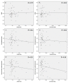
Results: Fifty patients with images of sufficient quality were included in the analysis. The mean right and left ventricular ejection fractions were 55 ± 5% and 59 ± 4%. The RV global longitudinal strain was -21.88 ± 4.63%. The RV circumferential strain at base, mid-cavity and apex were -13.66 ± 3.09%, -11.31 ± 2.79%, -20.73 ± 3.45%, respectively. There was no statistically significant decrease in right ventricular or left ventricular ejection fraction between patients and controls (p > 0.05 for each). There was no significant difference in RV global longitudinal strain between two groups (-21.88 ± 4.63 versus -21.99 ± 3.58; p = 0.93). However, there was significant decrease in mid-cavity circumferential strain magnitude in pectus patients compared with controls (-11.31 ± 2.79 versus -16.19 ± 2.86; p < 0.001). PE patients had a significantly higher basal circumferential strain (-13.66 ± 3.09% versus -9.76 ± 1.79; p < 0.001) as well as apical circumferential strain (-20.73 ± 3.45% versus -12.07 ± 3.38) than control group.
Conclusion: Mid-cavity circumferential strain but not longitudinal strain is reduced in pectus excavatum patients. Basal circumferential strain as well as apical circumferential strain were increased as compensatory mechanism for reduced mid-cavity circumferential strain. Further studies are needed to establish clinical significance of this finding.
Conflict of interest statement
Figures


Similar articles
-
Differences in myocardial strain between pectus excavatum patients and healthy subjects assessed by cardiac MRI: a pilot study.Eur Radiol. 2018 Mar;28(3):1276-1284. doi: 10.1007/s00330-017-5042-2. Epub 2017 Sep 11. Eur Radiol. 2018. PMID: 28894923
-
Cardiovascular magnetic resonance in patients with pectus excavatum compared with normal controls.J Cardiovasc Magn Reson. 2010 Dec 13;12(1):73. doi: 10.1186/1532-429X-12-73. J Cardiovasc Magn Reson. 2010. PMID: 21144053 Free PMC article.
-
Usefulness of strain cardiac magnetic resonance for the exposure of mild left ventricular systolic abnormalities in pectus excavatum.J Pediatr Surg. 2022 Oct;57(10):319-324. doi: 10.1016/j.jpedsurg.2021.09.008. Epub 2021 Sep 17. J Pediatr Surg. 2022. PMID: 34579966
-
The influence of pectus excavatum on cardiac kinetics and function in otherwise healthy individuals: A systematic review.Int J Cardiol. 2023 Jun 15;381:135-144. doi: 10.1016/j.ijcard.2023.03.058. Epub 2023 Mar 31. Int J Cardiol. 2023. PMID: 37003372
-
Value of cardiac magnetic resonance feature-tracking in Arrhythmogenic Cardiomyopathy (ACM): A systematic review and meta-analysis.Int J Cardiol Heart Vasc. 2024 Jul 5;53:101455. doi: 10.1016/j.ijcha.2024.101455. eCollection 2024 Aug. Int J Cardiol Heart Vasc. 2024. PMID: 39228971 Free PMC article. Review.
Cited by
-
Cardiopulmonary function in adolescent patients with pectus excavatum or carinatum.BMJ Open Respir Res. 2021 Jul;8(1):e001020. doi: 10.1136/bmjresp-2021-001020. BMJ Open Respir Res. 2021. PMID: 34326157 Free PMC article.
-
Influence of chest conformation on myocardial strain parameters in healthy subjects with mitral valve prolapse.Int J Cardiovasc Imaging. 2021 Mar;37(3):1009-1022. doi: 10.1007/s10554-020-02085-z. Epub 2020 Oct 30. Int J Cardiovasc Imaging. 2021. PMID: 33128156
-
Transverse and longitudinal right ventricular fractional parameters derived from four-chamber cine MRI are associated with right ventricular dysfunction etiology.Sci Rep. 2023 Mar 30;13(1):5229. doi: 10.1038/s41598-023-32284-2. Sci Rep. 2023. PMID: 36997599 Free PMC article.
-
Description of a new clinical syndrome: thoracic constriction without evidence of the typical funnel-shaped depression-the "invisible" pectus excavatum.Sci Rep. 2023 Jul 25;13(1):12036. doi: 10.1038/s41598-023-38739-w. Sci Rep. 2023. PMID: 37491452 Free PMC article.
-
Evaluation of subclinical myocardial involvement in systemic lupus erythematosus using multiparametric cardiac magnetic resonance imaging.BMC Cardiovasc Disord. 2025 Aug 9;25(1):597. doi: 10.1186/s12872-025-05033-8. BMC Cardiovasc Disord. 2025. PMID: 40783685 Free PMC article.
References
-
- Fokin AA, Steuerwald NM, Ahrens WA, Allen KE. Anatomical, Histologic, and Genetic Characteristics of Congenital Chest Wall Deformities. Seminars in Thoracic and Cardiovascular Surgery. 2009;21(1):44–57. http://dx.doi.org/10.1053/j.semtcvs.2009.03.001. - DOI - PubMed
-
- Jaroszewski D, Notrica D, McMahon L, Steidley DE, Deschamps C. Current Management of Pectus Excavatum: A Review and Update of Therapy and Treatment Recommendations. The Journal of the American Board of Family Medicine. 2010;23(2):230–9. doi: 10.3122/jabfm.2010.02.090234 - DOI - PubMed
-
- Jaroszewski DE, Fonkalsrud EW. Repair of pectus chest deformities in 320 adult patients: 21 year experience. The Annals of thoracic surgery. 2007;84(2):429–33. Epub 2007/07/24. doi: 10.1016/j.athoracsur.2007.03.077 . - DOI - PubMed
-
- Feng J, Hu T, Liu W, Zhang S, Tang Y, Chen R, et al. The biomechanical, morphologic, and histochemical properties of the costal cartilages in children with pectus excavatum. Journal of pediatric surgery. 2001;36(12):1770–6. Epub 2001/12/06. doi: 10.1053/jpsu.2001.28820 . - DOI - PubMed
-
- Oezcan S, Attenhofer Jost CH, Pfyffer M, Kellenberger C, Jenni R, Binggeli C, et al. Pectus excavatum: echocardiography and cardiac MRI reveal frequent pericardial effusion and right-sided heart anomalies. European Heart Journal—Cardiovascular Imaging. 2012;13(8):673–9. doi: 10.1093/ehjci/jer284 - DOI - PubMed
MeSH terms
LinkOut - more resources
Full Text Sources
Other Literature Sources
Miscellaneous

