

In vivo cholinergic basal forebrain atrophy predicts cognitive decline in de novo Parkinson's disease
- PMID: 29228203
- PMCID: PMC5837422
- DOI: 10.1093/brain/awx310
In vivo cholinergic basal forebrain atrophy predicts cognitive decline in de novo Parkinson's disease
Abstract
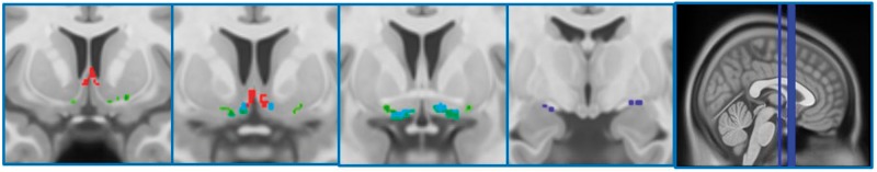
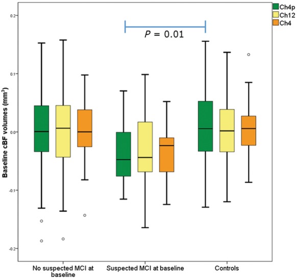
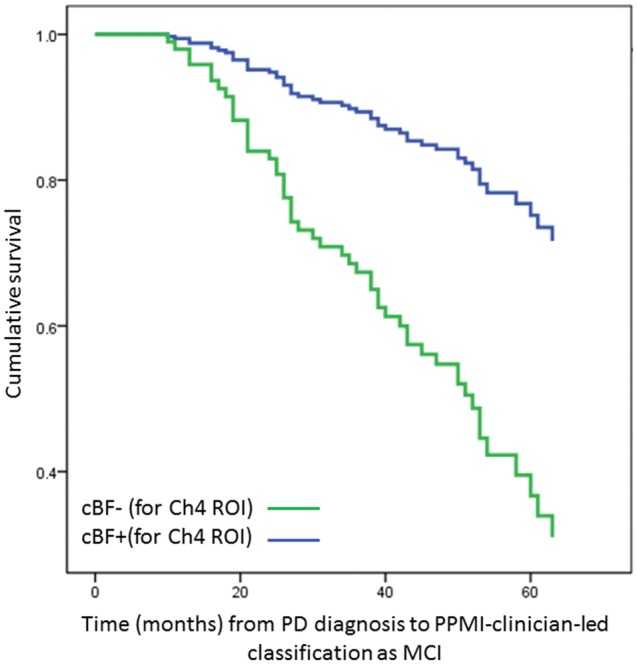
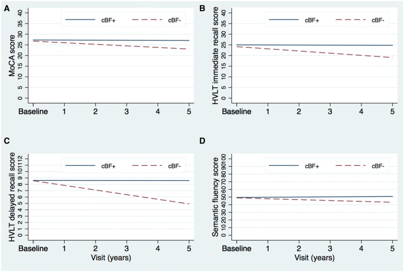
See Gratwicke and Foltynie (doi:10.1093/brain/awx333) for a scientific commentary on this article.Cognitive impairments are a prevalent and disabling non-motor complication of Parkinson's disease, but with variable expression and progression. The onset of serious cognitive decline occurs alongside substantial cholinergic denervation, but imprecision of previously available techniques for in vivo measurement of cholinergic degeneration limit their use as predictive cognitive biomarkers. However, recent developments in stereotactic mapping of the cholinergic basal forebrain have been found useful for predicting cognitive decline in prodromal stages of Alzheimer's disease. These methods have not yet been applied to longitudinal Parkinson's disease data. In a large sample of people with de novo Parkinson's disease (n = 168), retrieved from the Parkinson's Progressive Markers Initiative database, we measured cholinergic basal forebrain volumes, using morphometric analysis of T1-weighted images in combination with a detailed stereotactic atlas of the cholinergic basal forebrain nuclei. Using a binary classification procedure, we defined patients with reduced basal forebrain volumes (relative to age) at baseline, based on volumes measured in a normative sample (n = 76). Additionally, relationships between the basal forebrain volumes at baseline, risk of later cognitive decline, and scores on up to 5 years of annual cognitive assessments were assessed with regression, survival analysis and linear mixed modelling. In patients, smaller volumes in a region corresponding to the nucleus basalis of Meynert were associated with greater change in global cognitive, but not motor scores after 2 years. Using the binary classification procedure, patients classified as having smaller than expected volumes of the nucleus basalis of Meynert had ∼3.5-fold greater risk of being categorized as mildly cognitively impaired over a period of up to 5 years of follow-up (hazard ratio = 3.51). Finally, linear mixed modelling analysis of domain-specific cognitive scores revealed that patients classified as having smaller than expected nucleus basalis volumes showed more severe and rapid decline over up to 5 years on tests of memory and semantic fluency, but not on tests of executive function. Thus, we provide the first evidence that volumetric measurement of the nucleus basalis of Meynert can predict early cognitive decline. Our methods therefore provide the opportunity for multiple-modality biomarker models to include a cholinergic biomarker, which is currently lacking for the prediction of cognitive deterioration in Parkinson's disease. Additionally, finding dissociated relationships between nucleus basalis status and domain-specific cognitive decline has implications for understanding the neural basis of heterogeneity of Parkinson's disease-related cognitive decline.
Keywords: Parkinson’s disease; dementia; mild cognitive impairment; structural MRI.
© The Author (2017). Published by Oxford University Press on behalf of the Guarantors of Brain.
Figures
Comment in
-
Early nucleus basalis of Meynert degeneration predicts cognitive decline in Parkinson's disease.Brain. 2018 Jan 1;141(1):7-10. doi: 10.1093/brain/awx333. Brain. 2018. PMID: 29325047 Free PMC article.
References
Publication types
MeSH terms
Substances
LinkOut - more resources
Full Text Sources
Other Literature Sources
Medical
