. 2018 Jan 1;141(1):132-147.
doi: 10.1093/brain/awx315.

Oestrogen receptor β ligand acts on CD11c+ cells to mediate protection in experimental autoimmune encephalomyelitis

Affiliations

Oestrogen receptor β ligand acts on CD11c+ cells to mediate protection in experimental autoimmune encephalomyelitis

Roy Y Kim et al. Brain. .

Erratum in

  • Corrigendum.
    [No authors listed] [No authors listed] Brain. 2018 Apr 1;141(4):e33. doi: 10.1093/brain/awy043. Brain. 2018. PMID: 29506215 Free PMC article. No abstract available.

Abstract

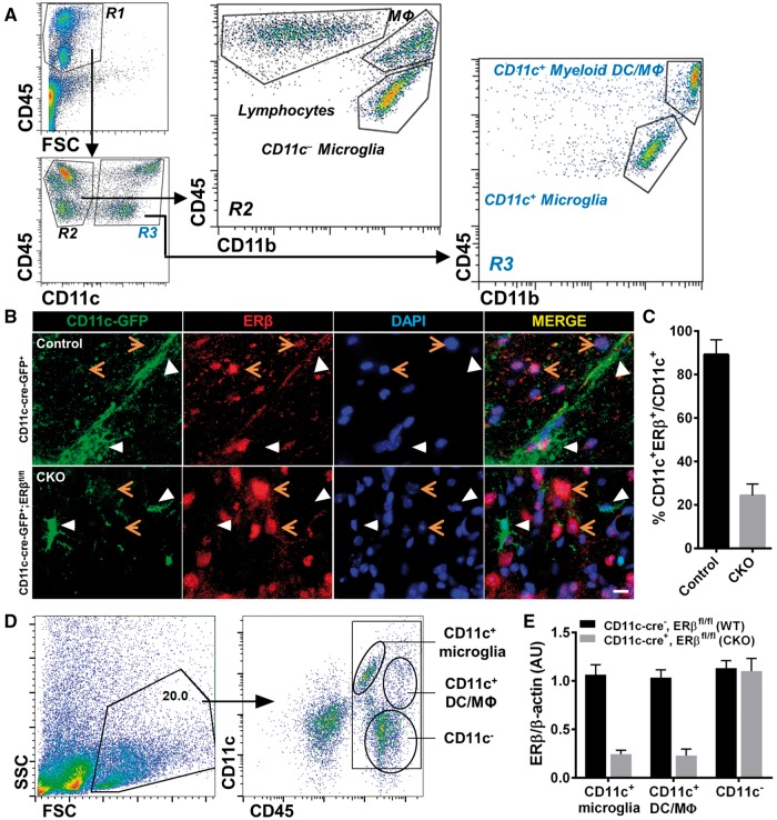
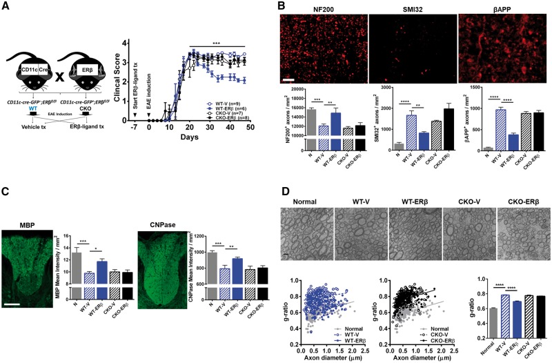
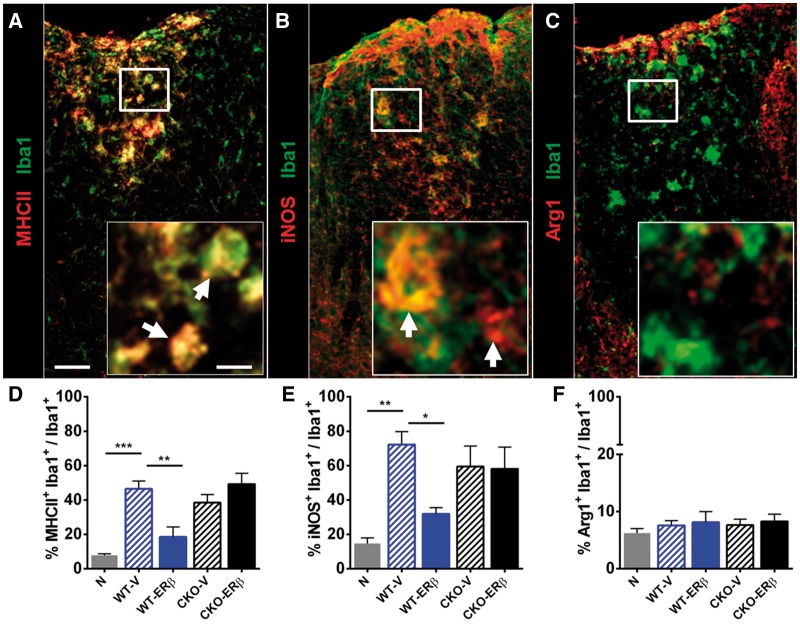
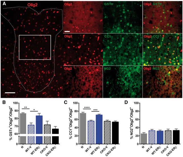
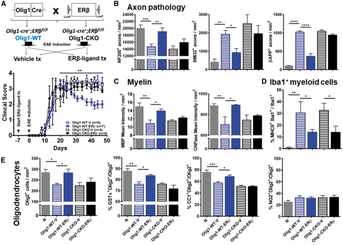
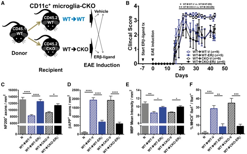
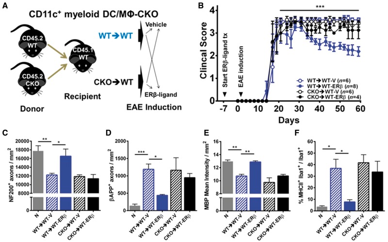
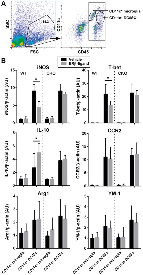
Oestrogen treatments are neuroprotective in a variety of neurodegenerative disease models. Selective oestrogen receptor modifiers are needed to optimize beneficial effects while minimizing adverse effects to achieve neuroprotection in chronic diseases. Oestrogen receptor beta (ERβ) ligands are potential candidates. In the multiple sclerosis model chronic experimental autoimmune encephalomyelitis, ERβ-ligand treatment is neuroprotective, but mechanisms underlying this neuroprotection remain unclear. Specifically, whether there are direct effects of ERβ-ligand on CD11c+ microglia, myeloid dendritic cells or macrophages in vivo during disease is unknown. Here, we generated mice with ERβ deleted from CD11c+ cells to show direct effects of ERβ-ligand treatment in vivo on these cells to mediate neuroprotection during experimental autoimmune encephalomyelitis. Further, we use bone marrow chimeras to show that ERβ in peripherally derived myeloid cells, not resident microglia, are the CD11c+ cells mediating this protection. CD11c+ dendritic cell and macrophages isolated from the central nervous system of wild-type experimental autoimmune encephalomyelitis mice treated with ERβ-ligand expressed less iNOS and T-bet, but more IL-10, and this treatment effect was lost in mice with specific deletion of ERβ in CD11c+ cells. Also, we extend previous reports of ERβ-ligand’s ability to enhance remyelination through a direct effect on oligodendrocytes by showing that the immunomodulatory effect of ERβ-ligand acting on CD11c+ cells is necessary to permit the maturation of oligodendrocytes. Together these results demonstrate that targeting ERβ signalling pathways in CD11c+ myeloid cells is a novel strategy for regulation of the innate immune system in neurodegenerative diseases. To our knowledge, this is the first report showing how direct effects of a candidate neuroprotective treatment on two distinct cell lineages (bone marrow derived myeloid cells and oligodendrocytes) can have complementary neuroprotective effects in vivo.awx315media15688130498001.

Keywords: experimental autoimmune encephalomyelitis; macrophage; multiple sclerosis; neuroprotection; oestrogen receptor beta.


Figures

Figure 1
Figure 1
ERβ-specific deletion in CD11c+ cells during EAE. (A) Flow cytometry dot plots of CNS mononuclear immune cells isolated from brains and spinal cords of EAE mice. CNS mononuclear immune cells were gated based on FSC and CD45 expression. CNS CD45+ gated cells included two separate populations, CD45hi and CD45int, within the R1 gate. CD45+ cells were further gated based on CD11c expression into R2: CD45+CD11c and R3: CD45+CD11c+. CD11b staining revealed that R2 included CD45hiCD11cCD11b lymphocytes, CD45hiCD11cCD11b+ macrophages, and CD45intCD11cCD11b+ resident microglia, and R3 included CD45hiCD11c+CD11b+ peripherally derived myeloid dendritic cells and macrophages (DC/Mφ) and CD45intCD11c+CD11b+ resident microglia. FSC = forward scatter. (B) Immunofluorescence images of spinal cord tissues stained with CD11c-GFP (green), ERβ (red) and nuclear stain DAPI (blue) with merged images for co-localization (yellow) on right. Top row: CD11c-cre-GFP+ control mice showed co-localization (white arrows) of CD11c-GFP and ERβ. Bottom row: CD11c-cre-GFP+;ERβfl/fl CKO mice did not show co-localization (white arrows). Orange arrows represent other cells in the CNS expressing ERβ. Scale bar = 20 μm. (C) Quantitative analysis of CD11c-GFP and ERβ co-localization in immunofluorescence images of CD11c-cre-GFP+ (Control) and CD11c ERβ CKO (CKO) mice with EAE. (D) Representative flow cytometry plots of isolated CNS mononuclear immune cells from a pool of three individual mice. CNS mononuclear immune cells were gated based on SSC and FSC (left), and subpopulations were identified using CD11c and CD45 staining (right). Cell populations labelled as CD11c+ microglia, CD11c+ myeloid dendritic cells and macrophages, and CD11c cells were FACS sorted for mRNA isolation and quantitative PCR analysis. (E) Quantitative analysis of ERβ (Esr2) mRNA expression in sorted CNS CD11c+ microglia, CD11c+ myeloid dendritic cells and macrophages cells, and CD11c cells from mice with ERβ deleted in CD11c+ cells (CKO) and wild-type mice, each with EAE.
Figure 2
Figure 2
ERβ expression in CD11c+ cells is necessary for neuroprotection in EAE. (A) Breeding scheme for creating wild-type (WT) and CKO mice of ERβ in CD11c+ cells. Briefly, CD11c-Cre-GFP+ mice were crossed with mice carrying an ERβ (Esr2) gene flanked by LoxP sites (ERβfl/fl). Homozygous ERβfl/fl mice without (WT) or with (CKO) Cre were generated. Each genotype was separated into two groups and received either vehicle or ERβ-ligand, DPN, treatment (tx). EAE was induced and animals were monitored daily and scored using the standard EAE 0–5 scale. ERβ-ligand treated wild-type EAE mice (WT-ERβ, blue solid) had significantly better clinical scores compared to vehicle treated wild-type EAE mice (WT-V, blue clear), ***P (WT-V versus WT-ERβ) = 0.0002, after Day 20 of EAE. In contrast, ERβ-ligand mediated protection did not occur in ERβ-ligand treated CKO EAE mice (CKO-ERβ, black solid) when compared to vehicle treated CKO EAE mice (CKO-V, black clear), P (CKO-V versus CKO-ERβ) > 0.9999. Detailed EAE statistics are in Supplementary Tables 1 and 2. (B) Representative images and quantitative analyses of NF200+, SMI32+, and βAPP+ axons in dorsal white matter of the spinal cord. Images were taken at 40× magnification. Scale bar = 20 μm. (C) Representative images and quantitative analyses of MBP+ and CNPase+ mean intensity in dorsal white matter of the spinal cord. Scale bar = 100 μm. (D) Representative electron microscopy images of ultraresolution of axons and myelin thickness in the dorsal white matter of spinal cord. Myelin thickness in the wild-type and CKO EAE mice treated with vehicle and ERβ-ligand was measured using the g-ratio (axon diameter) / (axon + outer myelin diameter) and shown in comparison with normal. Quantitative analysis showed that ERβ-ligand treated wild-type EAE mice had a decrease in g-ratio due to increased outer myelin diameter, while CD11c ERβ CKO mice with EAE did not. Scale bar = 1 μm. *P < 0.05; **P < 0.01; ***P < 0.001; ****P < 0.0001. Data are representative of three repeated experiments.
Figure 3
Figure 3
Qualitative effects on inflammatory markers on CNS resident and infiltrated Iba1+ myeloid cells. (A) Representative images of spinal cord tissues stained with MHCII (red) and Iba1 (green), (B) iNOS (red) and Iba1 (green), and (C) ARG1 (red) and Iba1 (green). Scale bar = 50 μm and 10 μm (inset). Inset: white arrows indicate co-localization. (D) MHCII+Iba1+ myeloid cells were increased in vehicle treated wild-type EAE mice (WT-V) compared to healthy controls (N), while ERβ-ligand treated wild-type EAE mice (WT-ERβ) showed a reduction in per cent MHCII+Iba1+ myeloid cells. In contrast, ERβ-ligand treated CKO EAE mice (CKO-ERβ) were no different from vehicle treated CKO EAE mice (CKO-V). (E) iNOS+Iba1+ myeloid cells were also reduced in ERβ-ligand treated wild-type EAE, but not in CKO EAE mice. (F) ARG1+Iba1+ myeloid cells were no different between groups. *P < 0.05; **P < 0.01; ***P < 0.001. Data are representative of two repeated experiments.
Figure 4
Figure 4
ERβ-ligand treatment acts on CD11c+ cells to permit increases in mature oligodendrocytes during EAE. (A) Immunofluorescence images of spinal cord tissues stained with Olig2 (red), and co-stained with GSTπ (green), CC1 (green), and NG2 (green). On the left is a representative image of Olig2+ oligodendrocyte lineage cells (OLC) in dorsal white matter of the spinal cord. On the right are representative images of each co-stain; Olig2-GSTπ (top), Olig2-CC1 (middle) and Olig2-NG2 (bottom). Scale bar = 50 μm (left) and 20 μm (right). White box indicates the area where the co-stains were imaged. Quantitative analyses of (B) Olig2+GSTπ+ mature OLCs, (C) Olig2+CC1+ immature/mature OLCs, and (D) Olig2+NG2+ oligodendrocyte precursor cells, each in dorsal white matter of the spinal cord. *P < 0.05; **P < 0.01; ***P < 0.001; ****P < 0.0001. Data are representative of two repeated experiments.
Figure 5
Figure 5
ERβ expression on Olig1+ cells is necessary for neuroprotection in EAE. (A) Breeding scheme for creating wild-type (WT) and CKO mice of ERβ in Olig1+ cells. Briefly, Olig1-Cre + mice were crossed with mice carrying an ERβ (Esr2) gene flanked by LoxP sites (ERβfl/fl). Homozygous ERβfl/fl mice without (Olig1-WT) or with (Olig1-CKO) Cre were generated. Each genotype was separated into two groups and received either vehicle or ERβ-ligand treatment (tx), EAE was induced and mice were scored for EAE severity as above. ERβ-ligand treated Olig1-WT EAE mice (Olig1-WT-ERβ, blue solid) had significantly better clinical scores compared to vehicle treated Olig1-WT EAE mice (Olig1-WT-V, blue clear), **P (Olig1-WT-V versus Olig1-WT-ERβ) = 0.0095, after Day 20 of EAE. In contrast, ERβ-ligand mediated protection did not occur in ERβ-ligand treated Olig1-CKO EAE mice (Olig1-CKO-ERβ, black solid) when compared to vehicle treated Olig1-CKO EAE mice (Olig1-CKO-V, black clear), P (Olig1-CKO-V versus Olig1-CKO-ERβ) = 0.7967. Detailed EAE statistics are in Supplementary Tables 1 and 2. Quantitative analyses of (B) NF200+, SMI32+, and βAPP+ axonal counts, (C) MBP+ and CNPase+ myelin intensity, and (D) MHCII+Iba1+ myeloid cells in dorsal white matter of the spinal cord. (E) Quantitative analyses of total Olig2+ OLCs, and the percentage of subpopulations: Olig2+NG2+ OPCs, Olig2+CC1+ immature/mature oligodendrocytes, and Olig2+GSTπ+ mature oligodendrocytes in dorsal white matter of the spinal cord. *P < 0.05; **P < 0.01; ***P < 0.001; ****P < 0.0001. Data are representative of three repeated experiments.
Figure 6
Figure 6
CD11c+ microglial ERβ expression is not necessary for neuroprotection in EAE. (A) Diagram for creating CD11c+ microglia-CKO using BMC. Briefly, CD45.1 wild-type mice were used as donors for bone marrow cells, and CD45.2 wild-type (WT→WT) or CKO (WT→CKO) were used as irradiated recipients. Each genotype was separated into two groups and received either vehicle or ERβ-ligand treatment (tx), EAE was induced and mice were scored for EAE severity. (B) ERβ-ligand treated wild-type (WT→WT-ERβ, blue solid) EAE mice had significantly better scores compared to vehicle treated wildtype (WT→WT-V, blue clear) EAE mice, *P (WT→WT-V versus WT→WT-ERβ) = 0.0188. Similarly, ERβ-ligand treated CKO (WT→CKO-ERβ, black solid) EAE mice also had significantly better scores compared to vehicle treated conditional knockout (WT→CKO-V, black clear) EAE mice, **P (WT→CKO-V versus WT→CKO-ERβ) = 0.0077. Detailed EAE statistics are in Supplementary Tables 1 and 2. Quantitative analysis of (C) NF200+ axonal count, (D) βAPP+ axonal count, (E) MBP+ myelin intensity, and (F) MHCII expression on Iba1+ myeloid derived cells (percentage) in dorsal white matter of the spinal cord. *P < 0.05; **P < 0.01; ***P < 0.001; ****P < 0.0001. Data are representative of two repeated experiments.
Figure 7
Figure 7
ERβ expression on CD11c+ myeloid dendritic cells and macrophages is necessary for neuroprotection in EAE. (A) Diagram for creating CD11c+ myeloid dendritic cells and macrophages (DC/MΦ) CKO using bone marrow chimeras. Briefly, CD45.2 wild-type (WT→WT) versus CKO (CKO→WT) mice were used as donors for bone marrow cells, and CD45.1 wild-type mice were used as irradiated recipients. Each genotype was separated into two groups and received either vehicle or ERβ-ligand treatment (tx), EAE was induced and mice were scored for EAE severity. (B) ERβ-ligand treated wild-type (WT→WT-ERβ, blue solid) EAE mice had significantly better EAE scores compared to vehicle treated wild-type (WT→WT-V, blue open) EAE mice, ***P (WT→WT-V versus WT→WT-ERβ) = 0.0005, after Day 20 of EAE, whereas ERβ-ligand mediated protection did not occur in ERβ-ligand treated conditional knockout (CKO→WT-ERβ, black solid) compared to vehicle treated CKO (CKO→WT-V, black clear) EAE mice, P (CKO→WT-V versus CKO→WT-ERβ) > 0.9999. Detailed EAE statistics are in Supplementary Tables 1 and 2. Quantitative analysis of (C) NF200+ axonal count, (D) βAPP+ axonal count, (E) MBP+ myelin intensity, and (F) MHCII expression on Iba1+ myeloid derived cells (percentage) in dorsal white matter of the spinal cord. *P < 0.05; **P < 0.01; ***P < 0.001; ****P < 0.0001.
Figure 8
Figure 8
Gene expression profiles of CD11c+ microglia and CD11c+ dendritic cells and macrophages cells from the CNS of ERβ-ligand or vehicle treated mice with EAE. (A) Representative flow cytometry plots of isolated CNS mononuclear immune cells from a pool of two to four individual mice. CNS mononuclear immune cells were gated based on SSC and FSC (left), and subpopulations were identified using CD11c and CD45 staining (right). Cell populations labelled as CD11c+ microglia and CD11c+ myeloid dendritic cells and macrophages were FACS sorted for mRNA isolation and quantitative PCR analysis. (B) Quantitative analysis of iNOS, T-bet, IL-10, CCR2, ARG1, and YM-1 mRNA expression levels of sorted CD11c+ microglia and CD11c+ myeloid dendritic cells and macrophages from wild type (WT) (left) and CD11c ERβ CKO (right) mice with EAE that were treated with vehicle or ERβ-ligand. Data are from three separate experiments, with error bar representing variation between experiments. *P < 0.05.

Similar articles

Cited by

References

    1. Antal MC, Krust A, Chambon P, Mark M. Sterility and absence of histopathological defects in nonreproductive organs of a mouse ERbeta-null mutant. Proc Natl Acad Sci USA 2008; 105: 2433–8. - PMC - PubMed
    1. Bailey SL, Schreiner B, McMahon EJ, Miller SD. CNS myeloid dendritic cells presenting endogenous myelin peptides ‘preferentially' polarize CD4+ T(H)-17 cells in relapsing EAE. Nat Immunol 2007; 8: 172–80. - PubMed
    1. Bennett ML, Bennett FC, Liddelow SA, Ajami B, Zamanian JL, Fernhoff NB, et al. New tools for studying microglia in the mouse and human CNS. Proc Natl Acad Sci USA 2016; 113: E1738–46. - PMC - PubMed
    1. Bulloch K, Miller MM, Gal-Toth J, Milner TA, Gottfried-Blackmore A, Waters EM, et al. CD11c/EYFP transgene illuminates a discrete network of dendritic cells within the embryonic, neonatal, adult, and injured mouse brain. J Comp Neurol 2008; 508: 687–710. - PubMed
    1. Clarkson BD, Walker A, Harris MG, Rayasam A, Sandor M, Fabry Z. CCR2-dependent dendritic cell accumulation in the central nervous system during early effector experimental autoimmune encephalomyelitis is essential for effector T cell restimulation in situ and disease progression. J Immunol 2015; 194: 531–41. - PMC - PubMed

Publication types

MeSH terms