

Mitochondria Are Critical for BDNF-Mediated Synaptic and Vascular Plasticity of Hippocampus following Repeated Electroconvulsive Seizures
- PMID: 29228215
- PMCID: PMC5838811
- DOI: 10.1093/ijnp/pyx115
Mitochondria Are Critical for BDNF-Mediated Synaptic and Vascular Plasticity of Hippocampus following Repeated Electroconvulsive Seizures
Abstract
Background: Electroconvulsive therapy is a fast-acting and efficient treatment of depression used in the clinic. The underlying mechanism of its therapeutic effect is still unclear. However, recovery of synaptic connections and synaptic remodeling is thought to play a critical role for the clinical efficacy obtained from a rapid antidepressant response. Here, we investigated the relationship between synaptic changes and concomitant nonneuronal changes in microvasculature and mitochondria and its relationship to brain-derived neurotrophic factor level changes after repeated electroconvulsive seizures, an animal model of electroconvulsive therapy.
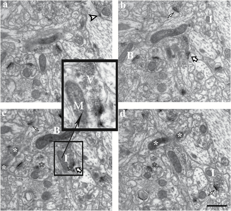
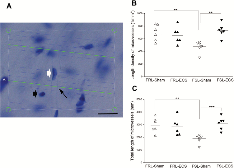
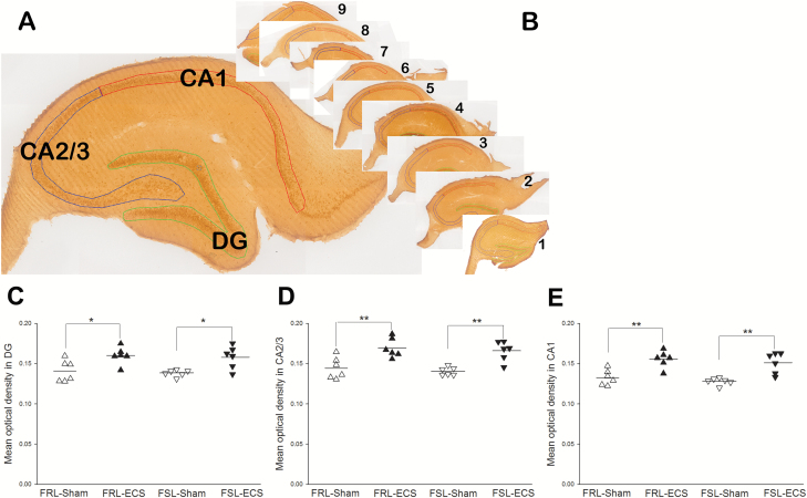
Methods: Electroconvulsive seizures or sham treatment was given daily for 10 days to rats displaying a genetically driven phenotype modelling clinical depression: the Flinders Sensitive and Resistant Line rats. Stereological principles were employed to quantify numbers of synapses and mitochondria, and the length of microvessels in the hippocampus. The brain-derived neurotrophic factor protein levels were quantified with immunohistochemistry.
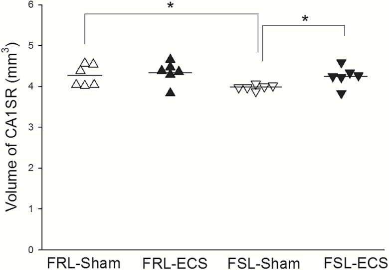
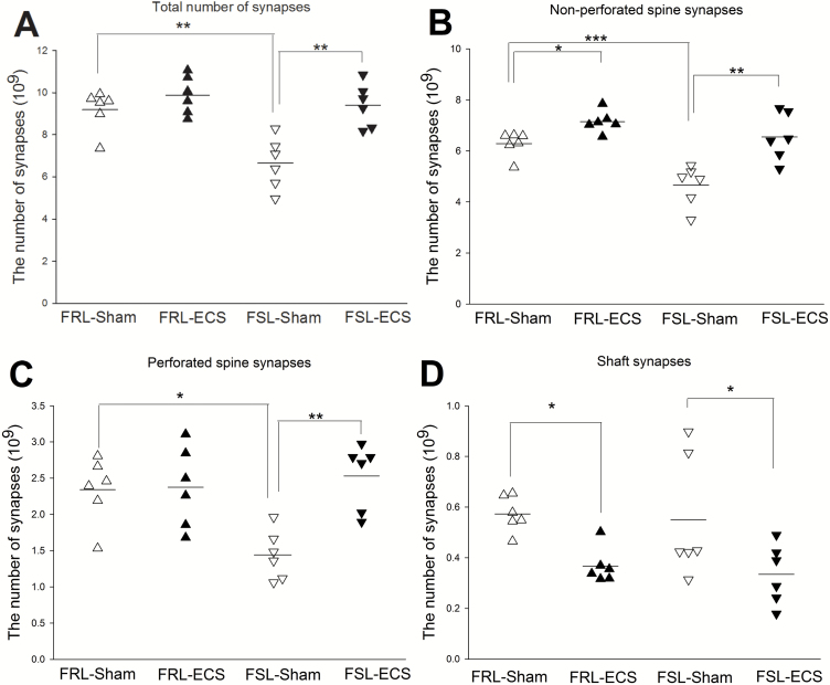
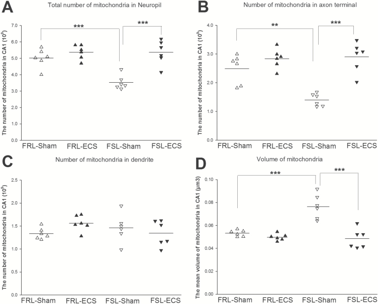
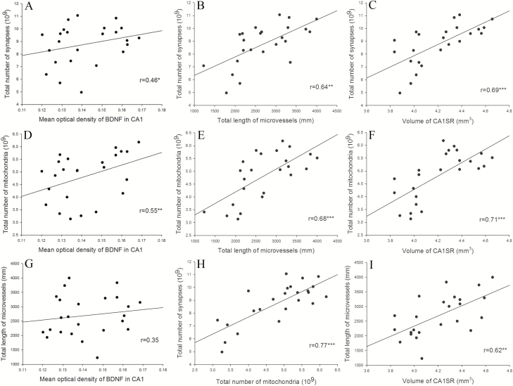
Results: In untreated controls, a lower number of synapses and mitochondria was accompanied by shorter microvessels of the hippocampus in "depressive" phenotype (Flinders Sensitive Line) compared with the "nondepressed" phenotype (Flinders Resistant Line). Electroconvulsive seizure administration significantly increased the number of synapses and mitochondria, and length of microvessels both in Flinders Sensitive Line-electroconvulsive seizures and Flinders Resistant Line-electroconvulsive seizures rats. In addition, the amount of brain-derived neurotrophic factor protein was significantly increased in Flinders Sensitive Line and Flinders Resistant Line rats after electroconvulsive seizures. Furthermore, there was a significant positive correlation between brain-derived neurotrophic factor level and mitochondria/synapses.
Conclusion: Our results indicate that rapid and efficient therapeutic effect of electroconvulsive seizures may be related to synaptic plasticity, accompanied by brain-derived neurotrophic factor protein level elevation and mitochondrial and vascular support.
© The Author(s) 2017. Published by Oxford University Press on behalf of CINP.
Figures
Similar articles
-
Sustained Ultrastructural Changes in Rat Hippocampal Formation After Repeated Electroconvulsive Seizures.Int J Neuropsychopharmacol. 2020 Jul 29;23(7):446-458. doi: 10.1093/ijnp/pyaa021. Int J Neuropsychopharmacol. 2020. PMID: 32215561 Free PMC article.
-
S-Ketamine Rapidly Reverses Synaptic and Vascular Deficits of Hippocampus in Genetic Animal Model of Depression.Int J Neuropsychopharmacol. 2017 Mar 1;20(3):247-256. doi: 10.1093/ijnp/pyw098. Int J Neuropsychopharmacol. 2017. PMID: 27815416 Free PMC article.
-
Quantitative hippocampal structural changes following electroconvulsive seizure treatment in a rat model of depression.Synapse. 2012 Aug;66(8):667-76. doi: 10.1002/syn.21553. Epub 2012 Mar 27. Synapse. 2012. PMID: 22389166
-
Remodeling of axo-spinous synapses in the pathophysiology and treatment of depression.Neuroscience. 2013 Oct 22;251:33-50. doi: 10.1016/j.neuroscience.2012.09.057. Epub 2012 Oct 2. Neuroscience. 2013. PMID: 23036622 Free PMC article. Review.
-
[Strategy to develop a new drug for treatment-resistant depression--role of electroconvulsive stimuli and BDNF].Yakugaku Zasshi. 2007 Apr;127(4):735-42. doi: 10.1248/yakushi.127.735. Yakugaku Zasshi. 2007. PMID: 17409705 Review. Japanese.
Cited by
-
Clozapine: Why Is It So Uniquely Effective in the Treatment of a Range of Neuropsychiatric Disorders?Biomolecules. 2021 Jul 15;11(7):1030. doi: 10.3390/biom11071030. Biomolecules. 2021. PMID: 34356654 Free PMC article. Review.
-
Stereological Changes in Microvascular Parameters in Hippocampus of a Transgenic Rat Model of Alzheimer's Disease.J Alzheimers Dis. 2021;84(1):249-260. doi: 10.3233/JAD-210738. J Alzheimers Dis. 2021. PMID: 34542078 Free PMC article.
-
Microvascular changes associated with epilepsy: A narrative review.J Cereb Blood Flow Metab. 2021 Oct;41(10):2492-2509. doi: 10.1177/0271678X211010388. Epub 2021 Apr 17. J Cereb Blood Flow Metab. 2021. PMID: 33866850 Free PMC article. Review.
-
Upregulation of Wnt2b exerts neuroprotective effect by alleviating mitochondrial dysfunction in Alzheimer's disease.CNS Neurosci Ther. 2023 Jul;29(7):1805-1816. doi: 10.1111/cns.14139. Epub 2023 Feb 27. CNS Neurosci Ther. 2023. PMID: 36852442 Free PMC article.
-
Clinical Improvement in Depression and Cognitive Deficit Following Electroconvulsive Therapy.Diagnostics (Basel). 2023 Apr 28;13(9):1585. doi: 10.3390/diagnostics13091585. Diagnostics (Basel). 2023. PMID: 37174977 Free PMC article. Review.
References
-
- Angelucci F, Aloe L, Vasquez PJ, Mathe AA(2000)Mapping the differences in the brain concentration of brain-derived neurotrophic factor (BDNF) and nerve growth factor (NGF) in an animal model of depression. Neuroreport 11:1369–1373. - PubMed
-
- Angelucci F, Aloe L, Jimenez-Vasquez P, Mathe AA(2003)Electroconvulsive stimuli alter nerve growth factor but not brain-derived neurotrophic factor concentrations in brains of a rat model of depression. Neuropeptides 37:51–56. - PubMed
-
- Ardalan M, Wegener G, Polsinelli B, Madsen TM, Nyengaard JR(2016)Neurovascular plasticity of the hippocampus one week after a single dose of ketamine in genetic rat model of depression. Hippocampus 26:1414–1423. - PubMed
MeSH terms
Substances
LinkOut - more resources
Full Text Sources
Other Literature Sources
Medical
