. 2017 Dec 1;58(14):6232-6240.
doi: 10.1167/iovs.17-22858.

The Magnitude and Time Course of IOP Change in Response to Body Position Change in Nonhuman Primates Measured Using Continuous IOP Telemetry

Affiliations

The Magnitude and Time Course of IOP Change in Response to Body Position Change in Nonhuman Primates Measured Using Continuous IOP Telemetry

Daniel C Turner et al. Invest Ophthalmol Vis Sci. .

Abstract

Purpose: To study the effect and time course of body position changes on IOP in nonhuman primates.

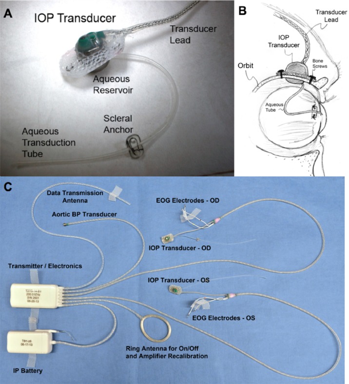
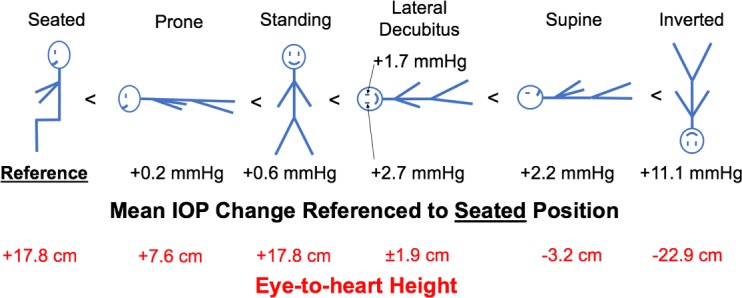
Methods: We recorded continuous bilateral IOP measurements with a wireless telemetry implant in three rhesus macaques in seven different body positions. IOP measurements were acquired in the seated-upright, standing, prone, supine, right and left lateral decubitus positions (LDPs), and head-down inverted positions. Continuous IOP was recorded for 90 seconds in each position before returning to a supine reference position until IOP stabilized; measurements were averaged after IOP stabilized at each position.

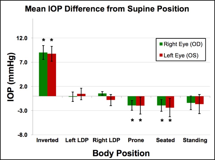
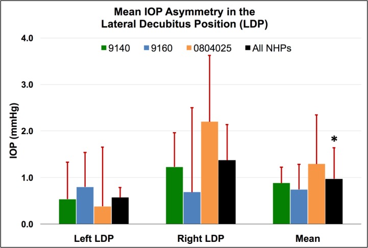
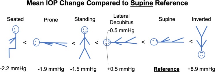
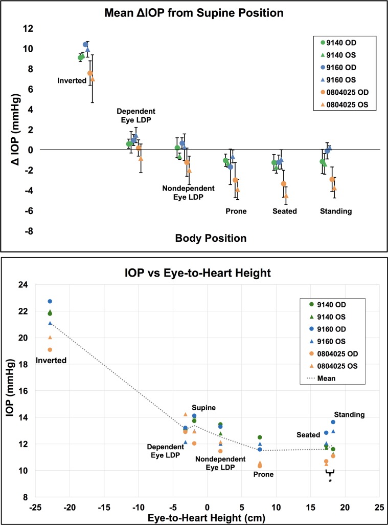
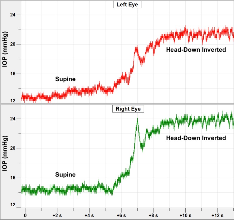
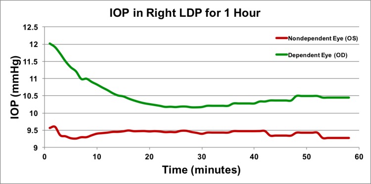
Results: Head-down inversion increased IOP an average of 8.9 mm Hg, compared to the supine reference. In the LDP, IOP decreased an average of 0.5 mm Hg in the nondependent eye (i.e., the higher eye), while the fellow dependent (i.e., lower) eye increased an average of 0.5 mm Hg, compared to supine reference. Standing and seated positions decreased IOP 1.5 and 2.2 mm Hg, respectively, compared with supine reference. IOP changes occurred within 4 to 15 seconds of a body position change, and timing was affected by the speed at which body position was changed. Compared to the IOP in the supine position, the IOP in the inverted, prone, and seated positions was significantly different (P = 0.0313 for all). The IOP in the standing position was not statistically different from the IOP in the supine position (P = 0.094). In addition, the IOP was significantly different between the nondependent eye and the dependent eye in the LDPs compared to the supine position (P = 0.0313).

Conclusions: Body position has a significant effect on IOP and those changes persist over time.


Figures

Figure 1
Figure 1
(A) Photograph of the extraorbital surface of our custom IOP transducer housing that is secured within a ¼-inch hole in the lateral orbital wall with bone screws as shown in (B). A 23-gauge silicone tube delivers aqueous from the anterior chamber to a fluid reservoir on the intraorbital side of the transducer (partially hidden from view in [A]); the tube (with appropriate slack to allow for eye movement) is trimmed, inserted into the anterior chamber, sutured to the sclera by using the integral scleral tube anchor plate, and covered with a scleral patchgraft (not shown). Adapted from Downs et al. (C) Photograph of enhanced Konigsberg Instruments total implant system for continuous monitoring of bilateral IOP, bilateral EOG, aortic blood pressure, and body temperature.
Figure 2
Figure 2
Mean IOP from supine position in all animals (n = 3) across all sessions (n = 5). Error bars represent the standard deviation. *Compared to the IOP in the supine position, the IOP in the inverted, prone, and seated positions were significantly different (P = 0.0313 for all).
Figure 3
Figure 3
Mean IOP asymmetry between dependent and nondependent eyes in the LDP compared to the supine reference position for each NHP, as well as the total mean for all NHPs for all sessions. Error bars represent standard deviation. *The overall change in IOP in the dependent eye was significantly different from the change in IOP in the nondependent eye for all NHPs and both LDP positions combined (P = 0.0313).
Figure 4
Figure 4
Diagram with different body positions and mean IOP change from supine position, ordered from negative change (left) to positive change (right).
Figure 5
Figure 5
Mean IOP changes with body position normalized to the seated position and the eye-to-heart height at each position.
Figure 6
Figure 6
Top: Mean IOP difference from supine position data for each eye (n = 6) by position. Error bars represent standard deviation. Bottom: Mean IOP per eye in each body position versus the approximate eye-to-heart distance for all NHP eyes (n = 6). Legend shows NHP ID and eye. *Seated and standing positions have identical eye-to-heart heights; data points are slightly offset for easier viewing.
Figure 7
Figure 7
Short duration time course of IOP changes in both eyes of one NHP during the process of moving the animal into the inverted position. The IOP transient at ∼7-second mark was due to momentary neck flexion that occurs in the process of picking the NHP up off the table.
Figure 8
Figure 8
Long duration time course of IOP in the lateral decubitus position. Approximately 1 hour of IOP data in the LDP in one animal in one session. Data points are shown as 1-minute averages for first 30 minutes, and as 5-minute averages during the second 30 minutes. To keep the animal sedated for an hour, isoflurane maintenance was performed; this may have generated a temporary rise and fall in the first few minutes of data acquisition.

References

    1. Kim KN, Jeoung JW, Park KH, Kim DM, Ritch R. . Relationship between preferred sleeping position and asymmetric visual field loss in open-angle glaucoma patients. Am J Ophthalmol. 2014; 157: 739– 745. - PubMed
    1. Krieglstein GK, Waller WK, Leydhecker W. . The vascular basis of the positional influence on the intraocular pressure. Albrecht Von Graefes Arch Klin Exp Ophthalmol. 1978; 206: 99– 106. - PubMed
    1. Eklund A, Johannesson G, Johansson E,et al. . The pressure difference between eye and brain changes with posture. Ann Neurol. 2016; 80: 269– 276. - PubMed
    1. Cook JA, Botello AP, Elders A,et al. . Systematic review of the agreement of tonometers with Goldmann applanation tonometry. Ophthalmology. 2012; 119: 1552– 1557. - PubMed
    1. Arora R, Bellamy H, Austin M. . Applanation tonometry: a comparison of the Perkins handheld and Goldmann slit lamp-mounted methods. Clin Ophthalmol. 2014; 8: 605– 610. - PMC - PubMed

Publication types

LinkOut - more resources