. 2018 Jan 25;46(2):840-848.
doi: 10.1093/nar/gkx1191.

The non-specific adenine DNA methyltransferase M.EcoGII

Affiliations

The non-specific adenine DNA methyltransferase M.EcoGII

Iain A Murray et al. Nucleic Acids Res. .

Abstract

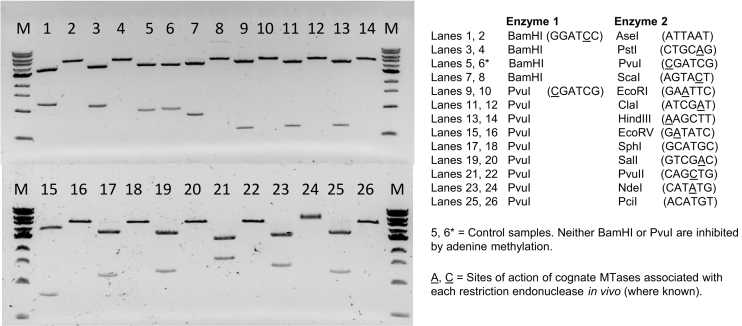
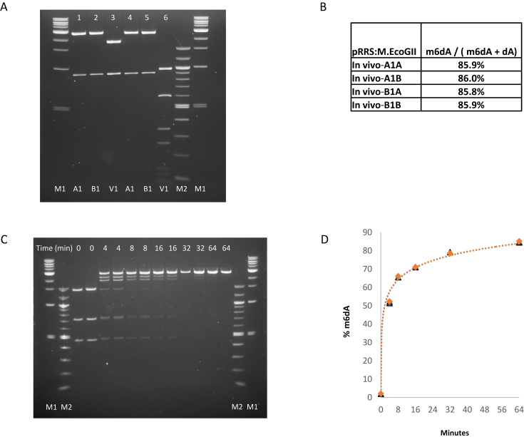
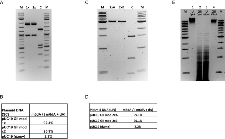
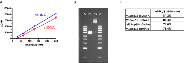
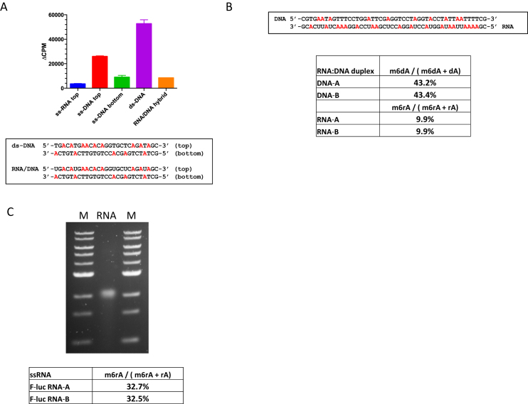
We describe the cloning, expression and characterization of the first truly non-specific adenine DNA methyltransferase, M.EcoGII. It is encoded in the genome of the pathogenic strain Escherichia coli O104:H4 C227-11, where it appears to reside on a cryptic prophage, but is not expressed. However, when the gene encoding M.EcoGII is expressed in vivo - using a high copy pRRS plasmid vector and a methylation-deficient E. coli host-extensive in vivo adenine methylation activity is revealed. M.EcoGII methylates adenine residues in any DNA sequence context and this activity extends to dA and rA bases in either strand of a DNA:RNA-hybrid oligonucleotide duplex and to rA bases in RNAs prepared by in vitro transcription. Using oligonucleotide and bacteriophage M13mp18 virion DNA substrates, we find that M.EcoGII also methylates single-stranded DNA in vitro and that this activity is only slightly less robust than that observed using equivalent double-stranded DNAs. In vitro assays, using purified recombinant M.EcoGII enzyme, demonstrate that up to 99% of dA bases in duplex DNA substrates can be methylated thereby rendering them insensitive to cleavage by multiple restriction endonucleases. These properties suggest that the enzyme could also be used for high resolution mapping of protein binding sites in DNA and RNA substrates.


Figures

Figure 1.
Figure 1.
In vitro methylation of plasmid DNA by M.EcoGII inhibits cleavage activities of multiple restriction endonucleases. pBR322 plasmid DNA was prepared from E. coli ER2796 (a non-methylating strain lacking dam, dcm and M.EcoKI activities) and a 10 μg sample of this DNA was subsequently methylated in vitro using purified M.EcoGII enzyme (1 μM). Unmethylated and M.EcoGII-methylated DNA samples were each incubated with pairs of restriction endonucleases comprising one enzyme that is known to be insensitive to adenine-methylation (either BamHI or PvuI) and a second that is known to be inhibited by this modification. Note that BamHI was only used in the control samples (lanes 5 and 6) and in combination with AseI, PstI and ScaI restriction. The latter three sites are located very close to the PvuI site of pBR322 and would therefore yield products that are not distinguishable from linear pBR322. Lanes 1, 3, 5…..25: unmethylated pBR322 DNA isolated from E. coli ER2796 cells. Lanes 2, 4, 6….26: equivalent DNAs after in vitro methylation with recombinant M.EcoGII. M1 = 1 kb DNA Ladder 0.5–10.0 kb (NEB).
Figure 2.
Figure 2.
Extent of M.EcoGII methylation of plasmid DNA in vivo and in vitro. (A) In vivo assay. Plasmid pRRS:M.EcoGII, a high-copy replicon that expresses M.EcoGII, was introduced into methylation-deficient ER2796 E. coli cells by transformation. Plasmid DNA was recovered from stationary phase cultures of two independent isolates after growth at 37°C for 20 h. pRRS:M.EcoGII and unmethylated pRRS vector control DNAs were restricted with either PvuI alone (lanes 1, 2 and 3) or PvuI plus MboI (lanes 4, 5 and 6). As PvuI activity is insensitive to m6A modification all samples are fully restricted by PvuI (lanes 1–6) but only the unmethylated pRRS vector control DNA is sensitive to MboI restriction (lane 6). M1 = 1 kb DNA Ladder 0.5–10.0 kb (NEB). M2 = 100 bp DNA ladder 0.1–1.5 kb (NEB). (B) LC–MS analysis of pRRS:M.EcoGII plasmid isolates. DNA samples were converted to nucleosides and analyzed in duplicate using LC–MS. In each sample, 86% of the dA bases were methylated in vivo. (C) Analysis of M.EcoGII methylation activity in vitro. Duplicate assays containing 10 μg pUC19 plasmid DNA and 320 μM SAM were set-up on ice and 0.1 ml aliquots of each were removed and snap-frozen in a dry-ice/ethanol bath (as unmethylated control samples). The remainder of each sample was placed in a 37°C water-bath, M.EcoGII enzyme (1 μM) was added and additional 0.1 ml samples were removed and snap-frozen after 4, 8, 16, 32 and 64 minutes of incubation at 37°C. Methylated DNAs were restricted with BamHI and TaqI. M1 = 1 kb DNA Ladder 0.5–10 kb and 100bp DNA Ladder 0.1–1.5 kb (NEB). (D) Time-course of M.EcoGII methylation in vitro. After 4 minutes, 50% of dA is present as m6dA, increasing to over 84% after 64 min. LC–MS data for the individual assays and time points are presented in Supplementary Table S2.
Figure 3.
Figure 3.
M.EcoGII methylates up to 99% of the dA residues in plasmid pUC19 DNA substrates and can be used for genome-wide methylation in vitro. (A) Supercoiled pUC19 plasmid DNA (20 μg) was methylated with M.EcoGII enzyme (1 μM) and 320 μM SAM in vitro, purified by phenol extraction and ethanol precipitation (lane 1x), then an aliquot was re-methylated using the same protocol (Lane 2x). Each experimental sample and the unmodified dam+ control DNA (lane C) were restricted using PvuI and BspHI endonucleases. BspHI cleaves DNA between the first and second nucleotide of TCATGA sequences and its activity is blocked by methylation of either of the dA bases. M = 2-log DNA Ladder 0.1–10.0 kb (NEB). (B) LC–MS data for experimental and control DNA samples. (C) pUC19 plasmid DNA was linearized by SmaI restriction and duplicate samples (2 μg) were subjected to two cycles of methylation using M.EcoGII (1 μM) and 160 μM SAM in vitro. Methylated DNAs were recovered by phenol extraction followed by ethanol precipitation. An aliquot of each sample (lanes 2xA, 2xB) and unmethylated (dam+) control DNA (lane C) were restricted using TaqI endonuclease. (D) LC–MS data for experimental and control DNA samples. (E) Genomic DNA was isolated from E. coli ER2796 cells (which lacks all E. coli DNA MTase activities) and a 20 μg sample was methylated in vitro using M.EcoGII (2 μM) and 320 μM SAM. Lanes 1, 3: unmethylated (ER2796) gDNA samples. Lanes 2, 4: M.EcoGII-methylated (ER2796) gDNA samples. Lanes 1, 2: DpnI (GATC) restricted samples (DpnI requires adenine methylation of both DNA strands for efficient cleavage activity). Lanes 3, 4: MboI (GATC) restricted samples (MboI activity is inhibited by adenine hemi or complete methylation). M = 1 kb-Extend DNA Ladder 0.5–48.5 kb (NEB).
Figure 4.
Figure 4.
M.EcoGII methylates single-stranded and duplex DNA substrates with near equivalent efficiency in vitro. (A) Radioactive assay of M.EcoGII activity using double- and single-stranded oligonucleotide substrates (2μM) containing a single dA nucleotide. Assays used 50mM HEPES buffer, pH 7.0, 0.1 mM EDTA and 3.7 μM [3H-SAM] and were incubated for 10 min at 37°C. (B) and (C) M.EcoGII methylation of single- and double-stranded M13mp18 bacteriophage DNAs in vitro and quantitation of m6dA content using LC–MS. DNA samples (2 μg) were methylated in vitro using M.EcoGII (2 μM). M = 1 kb-Extend DNA Ladder 0.5-to 48.5 kb (NEB).
Figure 5.
Figure 5.
Activity of M.EcoGII on RNA:DNA hybrid duplex and single-stranded RNA substrates in vitro. (A) Radioactive assays of M.EcoGII activity using 27mer ssRNA, ssDNA, dsDNA and RNA:DNA hybrid substrates (2 μM) in vitro. Assays used 50mM HEPES buffer, pH 7.0, 0.1mM EDTA and 3.7 μM [3H-SAM] and were incubated for 10 min at 37°C. (B) Methylation of a synthetic 48mer DNA:RNA hybrid oligonucleotide substrate (0.5 μM) containing 10 dA bases in the DNA strand and 18 rA bases in the RNA strand, Assay used 50 mM HEPES buffer pH 7.0, 1.0 mM EDTA, 320 μM SAM and M.EcoGII (1 μM). After incubation at 37°C for 60 minutes m6dA and m6rA products were quantified using LC–MS. (C) M.EcoGII-methylation of a 1.7 kb in vitro transcribed RNA (5 μg) followed by agarose gel electrophoresis and analysis of m6rA content using LC–MS. Reaction conditions were identical to those used with the DNA:RNA hybrid substrate but incubation at 37°C was extended to 120 min. M = 1 kb-DNA Ladder 0.5-to 10.0 kb (NEB).
Figure 6.
Figure 6.
Predictions of M.EcoGII structure using PHYRE2 analyses. (A) Alignment of predicted structural models for M.EcoGII to the M.EcoP15I:DNA structure (PDB: 4zcf). EcoP15I_ModA (cyan) and the DNA are experimentally determined structures. The various predicted M.EcoGII structure models were derived from threading onto available MTase structures as follows: green - EcoP15I_ModA (pdb:4zcf, CAGCAG); magenta - M1.MboII (pdb:1g60, GAAGA); yellow—M.RsrI (pdb:1nw6, GAATTC); wheat—M.PvuII (pdb:1boo, CAGCTG); gray—M.HpyAVI (pdb:5hfj, GAGG). The AdoMet binding residues (FxGxG motif) and catalytic residues (DPPY motif) of the M.EcoGII models are shown as white, red and blue spheres. Note lack of consensus among the predicted structures for the putative DNA binding/TRD domain (lower left, contacting DNA) versus the high degree of consensus for the methyltransferase structural core (upper middle). (B) Alignment of the EcoP15I_ModA:DNA crystal structure and the predicted M.EcoGII structure derived from threading. The experimentally-determined TRD domain of EcoP15I_ModA (cyan) and the predicted M.EcoGII model (green) appear to be similar with several secondary structural elements in common.

Similar articles

Cited by

References

    1. Jeltsch A. Beyond Watson and Crick: DNA methylation and molecular enzymology of DNA methyltransferases. ChemBioChem. 2002; 3:274–293. - PubMed
    1. Loenen W.A., Dryden D.T., Raleigh E.A., Wilson G.G., Murray N.E.. Highlights of the DNA cutters: a short history of the restriction enzymes. Nucleic Acids Res. 2014; 42:3–19. - PMC - PubMed
    1. Marinus M.G., Casadesus J.. Roles of DNA adenine methylation in host-pathogen interactions: mismatch repair, transcriptional regulation, and more. FEMS Microbiol. Rev. 2009; 33:488–503. - PMC - PubMed
    1. Reisenauer A., Kahng L.S., McCollum S., Shapiro L.. Bacterial DNA methylation: a cell cycle regulator. J. Bacteriol. 1999; 181:5135–5139. - PMC - PubMed
    1. Shell S.S., Prestwich E.G., Baek S-H., Shah R.R., Sassetti C. M., Dedon P.C., Fortune S.M.. DNA methylation impacts gene expression and ensures hypoxic survival of Mycobacterium tuberculosis. PLoS Pathog. 2013; 9:e1003419. - PMC - PubMed

Publication types

MeSH terms

Substances