. 2017 Oct 11;8(58):98371-98383.
doi: 10.18632/oncotarget.21738. eCollection 2017 Nov 17.

MP0250, a VEGF and HGF neutralizing DARPin® molecule shows high anti-tumor efficacy in mouse xenograft and patient-derived tumor models

Affiliations

MP0250, a VEGF and HGF neutralizing DARPin® molecule shows high anti-tumor efficacy in mouse xenograft and patient-derived tumor models

Ulrike Fiedler et al. Oncotarget. .

Abstract

Background: The VEGF/VEGFR and the HGF/cMET pathways are key mediators of the interplay of tumor cells and their microenvironment. However, inhibition of VEGF has been shown to produce only limited clinical benefit and inhibition of the activation of cMET by HGF has not translated into clinical benefit in pivotal trials. MP0250, a DARPin® molecule that specifically inhibits both VEGF and HGF has been developed to explore the clinical potential of dual inhibition of these pathways.

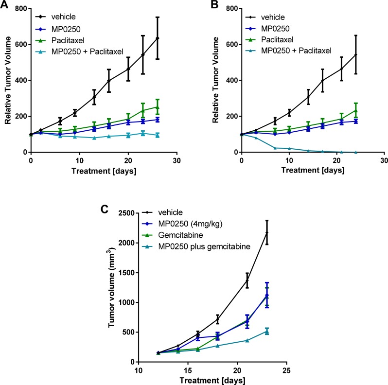
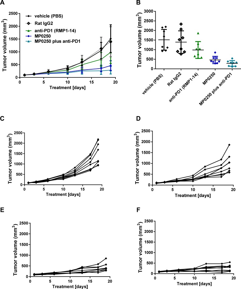
Results: MP0250 binding to VEGF and HGF inhibited downstream signalling through VEGFR2 and cMET resulting in inhibition of proliferation of VEGF- and HGF-dependent cells. Antitumor activity was demonstrated in VEGF- and HGF-dependent xenograft and syngeneic models with activity superior to that of individual VEGF- and HGF-blocking DARPin® molecules. Combination therapy studies showed potentiation of the antitumor activity of chemotherapy and immunotherapy agents, including an anti-PD1 antibody.

Materials and methods: Potency of MP0250 was assessed in cellular models and in a variety of xenograft models as monotherapy or in combination with standard-of-care drugs.

Conclusions: Dual inhibition of VEGF and HGF by MP0250 produced powerful single agent and combination antitumor activity. This, together with increasing understanding of the role of the HGF/cMET pathway in resistance to VEGF (and other agents), supports testing of MP0250 in the clinic.

Keywords: DARPin®; HGF; VEGF; patient-derived xenograft; serum albumin.


Conflict of interest statement

CONFLICTS OF INTEREST The authors declare being employees of Molecular Partners AG or have been employees of Molecular Partners AG at the time MP0250 has been developed.

Figures

Figure 1
Figure 1. MP0250 and its Inhibition of VEGF- and HGF-induced cellular functions
Model of MP0250, which is composed of two human serum albumin (HSA) DARPin® molecules flanking a VEGF and a HGF binding DARPin® molecule (A). The binding of MP0250 to VEGF was determined by quantification of free hVEGF in solution after addition of increasing concentrations of MP0250 (B). Inhibition of VEGF-induced HUVEC proliferation was analyzed in the absence (grey triangle) or presence of increasing concentrations of MP0250. Cell growth was quantified by OD measurement representing BrdU uptake. Error bars indicate the standard deviation of independent duplicates (C). Competition of binding of VEGF-A to sVEGFR2-Fc (D) and sVEGFR1-Fc (E) in the presence of increasing concentrations of MP0250 (filled diamond). As a control, a non-binding DARPin® molecule was titrated (filled grey circle).The HTRF signal was detected. Error bars indicate the standard deviation of independent triplicates (1d, 1e). Inhibition of cMET-phosphorylation in A549 cells by MP0250. Inhibition of phosphorylation as measured by ELISA measurement (OD450-620) versus the concentration of the inhibitor (F). Inhibition of U87MG proliferation by MP0250; error bars indicate the standard deviation of independent duplicates (G). Dashed black lines in the figures indicate IC50s. Data shown in the figure represents one experiment out of independent experiments as outlined in the Materials and Methods section.
Figure 2
Figure 2. Tumor growth inhibition in U87MG and A673 xenograft models
Tumor growth inhibition in the A673 rhabdomyosarcoma xenograft model (A, B) and the U87MG glioblastoma model (C, D). Figures 2A and 2C show the anti-tumor response to MP0250, the anti-HGF DARPin® molecule and the anti-VEGF DARPin® molecule. Figure 2B and 2D show a dose response of MP0250. Tumor growth is plotted as mean +/- SEM.
Figure 3
Figure 3. Tumor growth inhibition in syngeneic models and anti-angiogenic effect of MP0250
Tumor growth inhibition in the orthotopic renal cancer model (RENCA-LN model) (A, B) and the MC38 colorectal cancer model (C, D). Luciferase-transfected RENCA cells were orthotopically implanted into the left kidney of BalbB mice. Tumor growth was monitored by detection of luciferase activity in vivo during the study (Figure 3A) and determination of tumor volume at the end of the study (Figure 3B). MP0250 was compared to sorafenib at doses indicated in the figures. Figure 3C shows the time course of the anti-tumor response to MP0250 and the HGF inhibitor and the VEGF inhibitor. Figure 3D shows the tumor volumes at the end of the study. (E) shows the anti-angiogenic effect of the compounds in the MC38 effect demonstrated by immuno-histochemistry for CD31. Tumor growth is plotted as mean +/− SEM.
Figure 4
Figure 4. Tumor growth inhibition in patient-derived xenograft models
Tumor fragments from two renal cell cancers, RXF2264 (A) and RXF616 (B), two liver cancers, LIXF658 (C) and LIXF575 (D), two lung cancers, LXFS650 (E) and LXFL1121 (F), were implanted into nu/nu mice. MP0250 was dosed at 4 mg/kg 3x weekly (i.v.); sorafenib at 200 mg/kg daily (p.o.), 5-FU at 75 mg/kg 1x weekly (i.p.), and sunitinib at 40 mg/kg daily (p.o).
Figure 5
Figure 5. Tumor growth inhibition by MP0250 in combination with chemotherapy
For combination therapy with paclitaxel tumor fragments from two gastric PDX tumors, GA3002 (A) and GA3027 (B), were implanted into nu/nu mice. For combination therapy with gemcitabine, KP4 tumor cells were implanted into nu/nu mice (C). MP0250 was dosed at 4 mg/kg 3x weekly (i.v.); paclitaxel at 15 mg/kg 1x weekly (i.v.). The combination of MP0250 and paclitaxel was dosed as 4mg/kg MP0250 3x weekly (i.v.) and 15 mg/kg paclitaxel 1x weekly (i.v.). The combination of MP0250 and gemcitabine was dosed as 4 mg/kg MP0250 3x weekly (i.v.) and 100 mg/kg gemcitabine 1x weekly (i.v.). Tumor growth inhibition of PDX models and of KP4 tumors is plotted as mean +/− SEM.
Figure 6
Figure 6. Tumor growth inhibition by MP0250 in combination with anti-PD1
MC38 tumor cells were implanted into C57/Bl6 mice. Mice were randomized into 5 groups of 8 mice. MP0250 was dosed at 4 mg/kg 3x weekly (i.v.); anti-PD1 antibody RMP1-14 was dosed at 5 mg/kg 3x weekly (i.p.). (A) shows the growth curve of the anti-tumor response, (B) shows the tumor volume at the end of the study. Tumor growth is plotted as mean +/- SEM. (C-F) shows the tumor growth curves of the individual animals; (C) shows the vehicle group, (D) the RMP1-14 treatment group, (E) the MP0250 treatment group and (F) the MP0250/RMP1-14 combination group.

References

    1. Hanahan D, Weinberg RA. Hallmarks of Cancer: The Next Generation. Cell. 2011;144:646–74. https://doi.org/10.1016/j.cell.2011.02.013 - DOI - PubMed
    1. Gherardi E, Birchmeier W, Birchmeier C, Vande Woude G. Targeting MET in cancer: rationale and progress. Nat Rev Cancer. 2012;12:89–103. https://doi.org/10.1038/nrc3205 - DOI - PubMed
    1. Carmeliet P. Angiogenesis in life, disease and medicine. Nature. 2005;438:932–6. https://doi.org/10.1038/nature04478 - DOI - PubMed
    1. Jahangiri A, De Lay M, Miller LM, Carbonell WS, Hu YL, Lu K, Tom MW, Paquette J, Tokuyasu TA, Tsao S, Marshall R, Perry A, Bjorgan KM, et al. Gene expression profile identifies tyrosine kinase c-Met as a targetable mediator of antiangiogenic therapy resistance. Clin Cancer Res. 2013;19:1773–83. https://doi.org/10.1158/1078-0432.CCR-12-1281 - DOI - PMC - PubMed
    1. Kerbel RS. Tumor angiogenesis. N Engl J Med. 2008;358:2039–49. https://doi.org/10.1056/NEJMra0706596 - DOI - PMC - PubMed

LinkOut - more resources