. 2017 Oct 24;8(58):98823-98836.
doi: 10.18632/oncotarget.22006. eCollection 2017 Nov 17.

Wisp2 disruption represses Cxcr4 expression and inhibits BMSCs homing to injured liver

Affiliations

Wisp2 disruption represses Cxcr4 expression and inhibits BMSCs homing to injured liver

Dan Qin et al. Oncotarget. .

Abstract

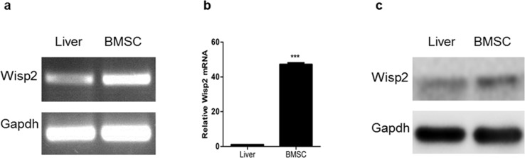
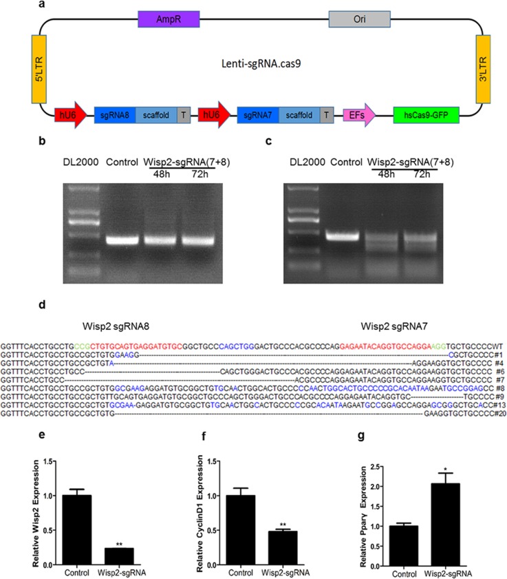
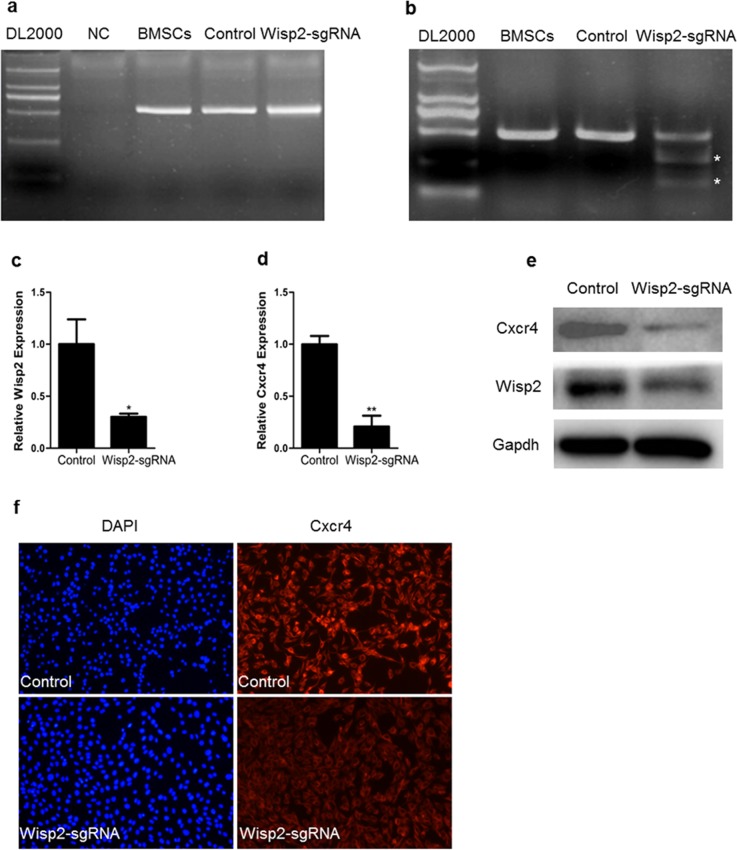
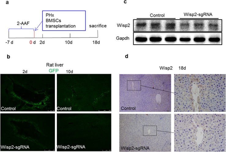
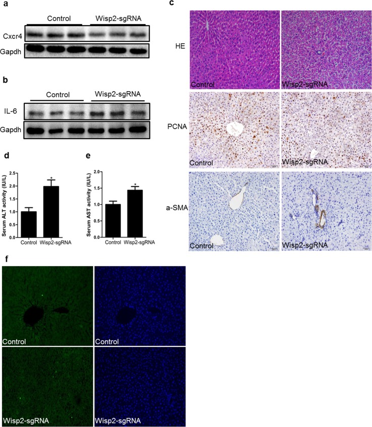
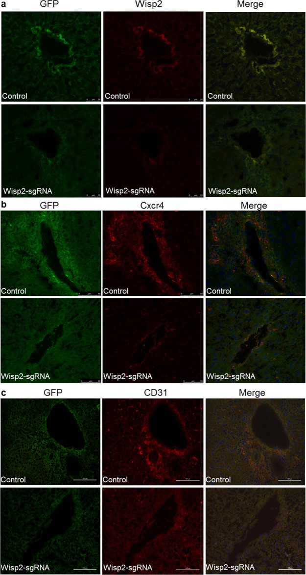
Liver regeneration/repair is a compensatory regrowth following acute liver failure, and bone marrow-derived mesenchyme stem cell (BMSC) transplantation is an effective therapy that promotes liver regeneration/repair. Wnt1 inducible signaling pathway protein 2 (Wisp2) is highly expressed in BMSCs, however, its function remains unclear. In this work, we used clustered regularly interspaced short palindromic repeats (CRISPR)-associated protein -9 nuclease (CRISPR/Cas9) genome editing technology to knockdown Wisp2 in BMSCs, and these modified cells were then transplanted into rats which were induced by the 2-AAF/PH. By linking the expression of Cas9 to green fluorescent protein (GFP), we tracked BMSCs in the rats. Disruption of Wisp2 inhibited the homing of BMSCs to injured liver and aggravated liver damage as indicated by remarkably high levels of ALT and AST. Moreover, the key factor in BMSC transplantation, C-X-C chemokine receptor type 4 (Cxcr4), was down-regulated in the Wisp2 depleted BMSCs and had a lower expression in the livers of the corresponding rats. By tracing the GFP marker, more BMSCs were observed to differentiate into CD31 positive endothelial cells in the functional Wisp2 cells but less in the Wisp2 gene disrupted cells. In summary, Wisp2 promotes the homing of BMSCs through Cxcr4 related signaling during liver repair in rats.

Keywords: BMSCs; Cxcr4; Wisp2; homing; rat.


Conflict of interest statement

CONFLICTS OF INTEREST The authors declare that they have no competing interests.

Figures

Figure 1
Figure 1. High expression of Wisp2 in BMSCs
Total RNA was extracted from rat BMSCs or rat livers, then PCR (a) and qRT-PCR (b) were performed to examine Wisp2 mRNA level. Error bars represent the ± S.E.M. of triplications. “***”, p<0.001. Gene expression level of the samples normalized to Gapdh and the expression of Wisp2 in liver was set as 1. (c) The protein level of Wisp2 was measured in BMSCs and livers with western blot analysis. 20μg total protein per lane and Gapdh was used as an internal control.
Figure 2
Figure 2. Establishment of Wisp2 knockdown cells by Crispr/Cas9 system
(a) Sketch of the “all in one” lentivirus expression vector containing Cas9 and sgRNAs (Lenti.sgRNAs.Cas9). SgRNAs insertion sites are highlighted in blue. (b and c) High cutting effiency of lentivirus against Wisp2 in C3H10T1/2 cells. PCR products were amplified and subjected to T7EN1. Control, C3H10T1/2 infected with basic vector; sgW(7+8), cells infected with lentivirus against Wisp2 containing sgW(7+8); supernatants containing lentivirus were collected 48 hours and 72 hours after the transfection. (d) Sequences of modified Wisp2 detected in C3H10T1/2 cells. 8 of 15 TA clones from the PCR products were analyzed by DNA sequencing and displayed in the images. The PAM sequences are highlighted in green; the targeting sequences are highlighted in red; the mutations are highlighted in blue; deletion (-); insertions (^), lower case; #: clone number. (e-g) QRT-PCR analysis of the expression of Wisp2, CyclinD1 and Pparγ in C3H10T1/2 cells infected with lentivirus against Wisp2. CyclinD1 and Pparγ were used as positive controls. Gene expression level of the control samples normalized to 36B4 was set as 1. Results are shown as mean ± S.E.M. of 3 independent experiments. “**”, p<0.01, “*”, P<0.05.
Figure 3
Figure 3. Down-regulation of Cxcr4 in Wisp2 genetically modified cells
(a) PCR products of the targeted Wisp2 from BMSCs infected with lentivirus against Wisp2 (Wisp2-sgRNA) or lentivirus without sgRNAs (Control). (b) Cas9 mediated on-target cleavage of Wisp2 by T7EN1. PCR products were amplified and subjected to T7EN1. Samples with cleavage bands were marked with an asterisk“*”. (c and d) QRT-PCR analysis of the expression of Wisp2 and Cxcr4 in BMSCs infected with lentivirus against Wisp2. The error bars indicated the ± S.E.M. of at least three independent experiments, “*”, p<0.05. Gene expression level of the control samples normalized to Gapdh was set as 1. (e) Wisp2 and Cxcr4 protein expression levels were depressed in the Wisp2-sgRNA-BMSCs. Total cellular protein was analyzed by immunoblotting for Gapdh. 20μg of total protein per lane. (f) Immunofluorescence showed that Cxcr4 was reduced in the Wisp2-sgRNA-BMSCs. Blue, DAPI; Red, Cxcr4.
Figure 4
Figure 4. Wisp2 was disrupted in rat livers after 2-AAF/PH
(a) Seven days after the intragastric administration of 2-AAF, the rats underwent PH and were transplanted with 1.8×10⁶ Wisp2-sgRNA-BMSCs or equal amounts of control BMSCs and then sacrificed and livers collected as indicated in the scheme. (b) Representative images showing homing BMSCs to the injured liver. (c and d) Wisp2 expressed in rat livers. Expression of Wisp2 was detected in rat livers 18 days post-transplantation by Immunoblot analysis (c). 40μg of total protein per lane. (d) Immunostaining was performed. Original magnification: 10× (Left panel) and 40× (right panel).
Figure 5
Figure 5. Cxcr4 was decreased and repair capability was reduced in lentiviral Wisp2 disrupted BMSC transplantation rats
Liver samples treated with Wisp2-sgRNA or not were examined for the expression of Cxcr4 (a) and IL-6 (b). 40μg of total protein per lane, as indicated by western analysis 18 days after transplantation. (c) Paraffin- embedded sections from livers obtained 18 days after the transplantation were stained with H&E and PCNA or α-SMA. The Wisp2-sgRNA group showed more extensive liver damage and cellular swelling (20×). Serum ALT (d) and AST (e) levels were assessed to determine the degree of liver injury. The control group was set as 1. Asterisks indicate significant differences, “*”, P< 0.05. (f) Representative micrographs of TUNEL staining of liver tissue. Liver sections collected 18 days after BMSCs transplantation were stained with FITC-conjugated TUNEL to identify apoptotic cells. The results showed that disrupted Wisp2 incresed the levels of apoptosis caused by 2-AAF/PH. Original magnification: 20×.
Figure 6
Figure 6. Wisp2 was required for BMSC migration and differentiation to endothelial cells
Significantly decreased expression of Wisp2 and Cxcr4 in rat livers after the Wisp2-sgRNA-BMSC transplanted 18 days, the expression of Wisp2 (a) and Cxcr4 (b) were determined in liver sections. Liver cyrosections were stained for GFP (Green fluorescence) and Wisp2/Cxcr4 (Red fluorescence). Images were merged with DAPI (Blue fluorescence) staining reveal the nuclei. (c) Expression of the endothelial cell marker CD31 (Red fluorescence) was also examined in the liver sections by immunostaining. The expression of CD31 was reduced in the Wisp2-sgRNA BMSCs transplanted rat livers.

Similar articles

Cited by

References

    1. Jemal A, Bray F, Center MM, Ferlay J, Ward E, Forman D. Global cancer statistics. CA Cancer Journal for Clinicians. 2011;61:69–90. - PubMed
    1. Torre LA, Bray F, Siegel RL, Ferlay J, Lortet-Tieulent J, Jemal A. Global cancer statistics, 2012. CA Cancer Journal for Clinicians. 2015;65:87–108. - PubMed
    1. McDowell TD, Stevens RD, Gurakar A. Acute liver failure: a management challenge for the practicing gastroenterologist. Gastroenterology & Hepatology. 2010;6:444–450. - PMC - PubMed
    1. Oertel M, Shafritz DA. Stem cells, cell transplantation and liver repopulation. Biochimica Et Biophysica Acta. 2008;1782:61–74. - PMC - PubMed
    1. Oh SH, Witek RP, Bae SH, Zheng D, Jung Y, Piscaglia AC, Petersen BE. Bone marrow-derived hepatic oval cells differentiate into hepatocytes in 2-acetylaminofluorene/partial hepatectomy induced liver regeneration. Gastroenterology. 2007;132:1077–1087. - PubMed

LinkOut - more resources