

Comprehensive analysis of ectopic mandibular third molar: a rare clinical entity revisited
- PMID: 29229002
- PMCID: PMC5725881
- DOI: 10.1186/s13005-017-0157-x
Comprehensive analysis of ectopic mandibular third molar: a rare clinical entity revisited
Abstract
Background: Ectopic mandibular third molar is a rare clinical entity with incompletely known etiology. Here, we sought to delineate its epidemiological, clinical and radiographic characteristics, and therapy by integrating and analyzing the cases treated in our institution together with previously reported cases.
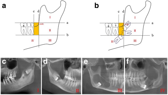
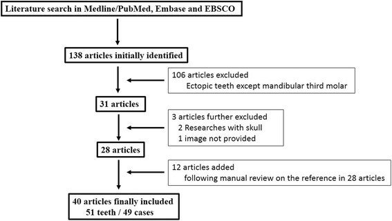
Method: A new definition and classification for ectopic mandibular third molar was proposed based on its anatomic location on panoramic images. Thirty-eight ectopic mandibular third molars in 37 patients and 51 teeth in 49 patients were identified in our disease registry and from literature (1990-2016), respectively. These cases were further categorized and compared according to our classification protocol. The demographic, clinicopathological and radiographic data were collected and analyzed.
Results: These ectopic teeth were categorized into four levels, 33 in level I(upper ramus), 32 in level II (middle ramus), 15 in level III (mandibular angle) and 9 in level IV (mandibular body). The common clinical presentations included pain, swelling and limited mouth opening, although sometimes asymptomatic. Most teeth were associated with pathological lesions. Treatments included clinical monitor and surgical removal by intra- or extraoral approach with favorable outcomes. Clinical presentations and treatment options for these teeth were significantly associated with their ectopic locations as we classified.
Conclusions: Ectopic mandibular third molars are usually found in patients with middle ages and in upper and middle ramus of mandible. Surgery is preferred to remove these ectopic teeth and associated pathologies when possible.
Keywords: Ectopic tooth; Impacted third molar; Mandibular third molar; Panoramic radiography.
Conflict of interest statement
Ethics approval and consent to participate
The research protocol (2016–256) was reviewed and approved by the Ethics and Research Committee of Nanjing Medical University. Written informed consent was obtained from all enrolled patients before treatment. The methods and protocols during the whole study were performed in accordance with the tenets of the Declaration of Helsinki for research involving human subjects as well as our institutional guidelines.
Consent for publication
Not applicable.
Competing interests
The authors declare that they have no competing interests.
Publisher’s Note
Springer Nature remains neutral with regard to jurisdictional claims in published maps and institutional affiliations.
Figures

Similar articles
-
Ectopic mandibular third molar in subcondylar region: report of a rare case.Oral Maxillofac Surg. 2012 Mar;16(1):153-5. doi: 10.1007/s10006-011-0284-7. Epub 2011 Jun 24. Oral Maxillofac Surg. 2012. PMID: 21701799
-
Surgical approach to impacted mandibular third molars--operative classification.J Oral Maxillofac Surg. 2010 Mar;68(3):628-33. doi: 10.1016/j.joms.2009.07.072. Epub 2009 Dec 23. J Oral Maxillofac Surg. 2010. PMID: 20031288
-
Ectopic mandibular third molar in the ramus region: report of a case and literature review.Oral Surg Oral Med Oral Pathol Oral Radiol Endod. 2008 Feb;105(2):155-61. doi: 10.1016/j.tripleo.2007.04.009. Epub 2007 Aug 30. Oral Surg Oral Med Oral Pathol Oral Radiol Endod. 2008. PMID: 17764987 Review.
-
Intraoral extraction of an ectopic mandibular third molar detected in the subcondylar region without a pathological cause: A case report and literature review.Cranio. 2017 Sep;35(5):327-331. doi: 10.1080/08869634.2016.1240466. Epub 2016 Oct 3. Cranio. 2017. PMID: 27690832 Review.
-
A comparative evaluation of film and digital panoramic radiographs in the assessment of position and morphology of impacted mandibular third molars.Indian J Dent Res. 2011 Mar-Apr;22(2):219-24. doi: 10.4103/0970-9290.84290. Indian J Dent Res. 2011. PMID: 21891889
Cited by
-
Piezo-surgery technique and intramuscular dexamethasone injection to reduce postoperative pain after impacted mandibular third molar surgery: a randomized clinical trial.BMC Oral Health. 2021 Aug 11;21(1):393. doi: 10.1186/s12903-021-01759-x. BMC Oral Health. 2021. PMID: 34380473 Free PMC article. Clinical Trial.
-
Analysis of the unpredictable migration of impacted mandibular third molars: A pilot study.J Clin Exp Dent. 2020 Dec 1;12(12):e1145-e1149. doi: 10.4317/jced.56891. eCollection 2020 Dec. J Clin Exp Dent. 2020. PMID: 33282135 Free PMC article.
-
Ectopic third mandibular molar: evaluation of surgical practices and meta-analysis.Clin Oral Investig. 2021 Aug;25(8):4781-4799. doi: 10.1007/s00784-021-04018-z. Epub 2021 Jun 17. Clin Oral Investig. 2021. PMID: 34137925
-
Minimum size and positioning of imaging field for CBCT-scans of impacted lower third molars: a retrospective study.BMC Oral Health. 2021 Dec 29;21(1):670. doi: 10.1186/s12903-021-02029-6. BMC Oral Health. 2021. PMID: 34965859 Free PMC article.
-
Extraoral surgical removal of an ectopic impacted third molar of the mandible. Report of a case.J Clin Exp Dent. 2020 Jun 1;12(6):e615-e619. doi: 10.4317/jced.56602. eCollection 2020 Jun. J Clin Exp Dent. 2020. PMID: 32665824 Free PMC article.
References
-
- Ahmed NM, Speculand B. Removal of ectopic mandibular third molar teeth: literature review and a report of three cases. Oral Surg. 2012;5:39–44. doi: 10.1111/j.1752-248X.2011.01145.x. - DOI
MeSH terms
Grants and funding
LinkOut - more resources
Full Text Sources
Other Literature Sources
