Review
. 2018 May:64:96-130.
doi: 10.1016/j.preteyeres.2017.12.001. Epub 2017 Dec 8.

Neural control of choroidal blood flow

Affiliations
Review

Neural control of choroidal blood flow

Anton Reiner et al. Prog Retin Eye Res. 2018 May.

Abstract

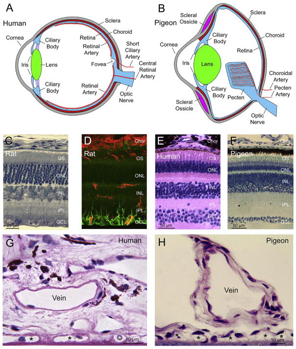
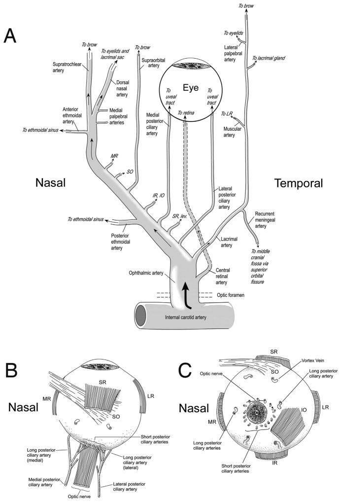
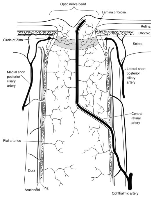
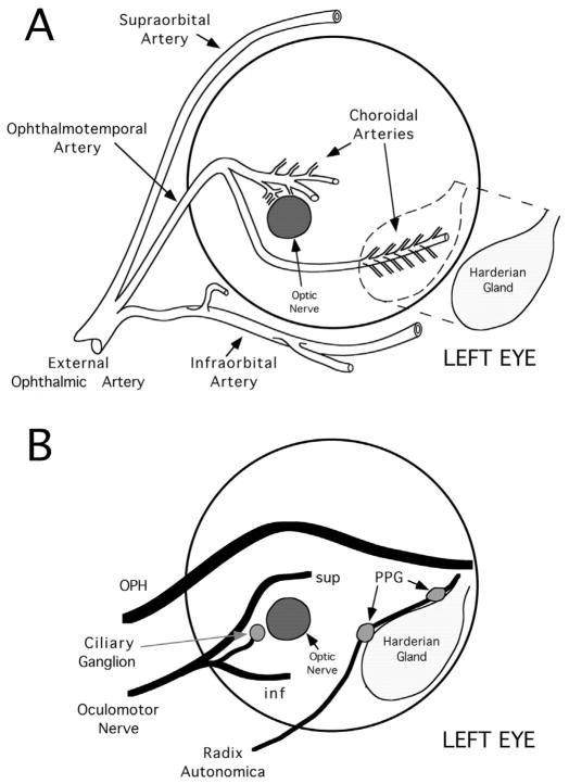
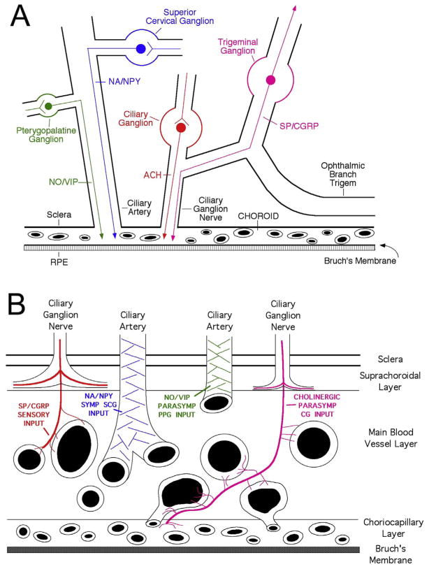
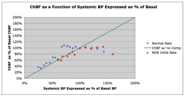
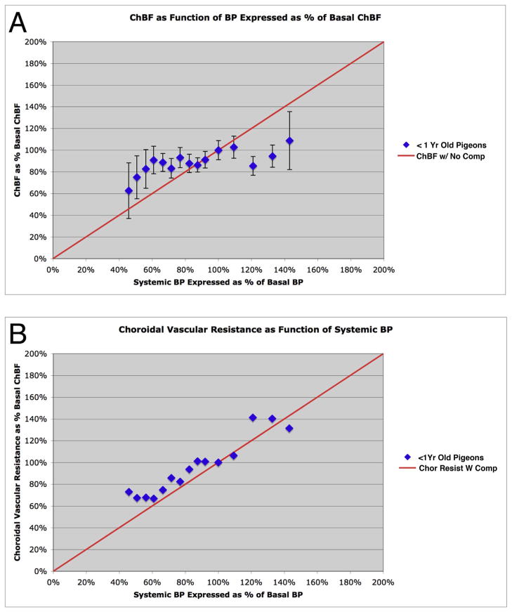
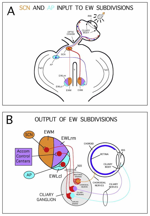
The choroid is richly innervated by parasympathetic, sympathetic and trigeminal sensory nerve fibers that regulate choroidal blood flow in birds and mammals, and presumably other vertebrate classes as well. The parasympathetic innervation has been shown to vasodilate and increase choroidal blood flow, the sympathetic input has been shown to vasoconstrict and decrease choroidal blood flow, and the sensory input has been shown to both convey pain and thermal information centrally and act locally to vasodilate and increase choroidal blood flow. As the choroid lies behind the retina and cannot respond readily to retinal metabolic signals, its innervation is important for adjustments in flow required by either retinal activity, by fluctuations in the systemic blood pressure driving choroidal perfusion, and possibly by retinal temperature. The former two appear to be mediated by the sympathetic and parasympathetic nervous systems, via central circuits responsive to retinal activity and systemic blood pressure, but adjustments for ocular perfusion pressure also appear to be influenced by local autoregulatory myogenic mechanisms. Adaptive choroidal responses to temperature may be mediated by trigeminal sensory fibers. Impairments in the neural control of choroidal blood flow occur with aging, and various ocular or systemic diseases such as glaucoma, age-related macular degeneration (AMD), hypertension, and diabetes, and may contribute to retinal pathology and dysfunction in these conditions, or in the case of AMD be a precondition. The present manuscript reviews findings in birds and mammals that contribute to the above-summarized understanding of the roles of the autonomic and sensory innervation of the choroid in controlling choroidal blood flow, and in the importance of such regulation for maintaining retinal health.

Keywords: Choroidal blood flow; Ciliary ganglion; Ocular blood flow; Parasympathetic; Pterygopalatine ganglion; Superior cervical ganglion; Sympathetic; Uvea.


Figures

Fig. 1
Fig. 1
Schematics and images illustrating similarities and differences between mammalian and avian eye, using as examples of mammals the human eye (A, E, G) and the rat eye (C, D) and as an example of mammals the pigeon eye (B, F, H). Images A and B show schematics of human (A) and pigeon eye (B). The major differences are the more flattened shape of the avian eye, the presence of scleral ossicles ringing the cornea in birds, and presence of the pecten and its vasculature in birds instead of the retinal vasculature in mammals. In A and B, red is used to illustrate blood vessels or vascular layers of the eye, with small branches from the retinal arteries penetrating into the retina in the human eye. Images C–F present comparable cross-sections through rat (C, D), human (E) and pigeon (F) retina. The two images of rat retina show a transmitted light micrograph of a toluidine blue-stained plastic-embedded section showing the retinal layers (C) juxtaposed to a confocal laser scanning microscope image of a comparable view of a section through rat retina in which blood vessels are labeled red with the lectin IB4 and Müller cell endfeet and processes in the inner retina are immunolabeled green for GFAP (D). The two images show the location of the retinal vessels, fragments of which are seen in the nerve fiber layer and ganglion cell layer (GCL), in the inner plexiform layer (IPL), and in the outer plexiform layer between the INL and outer nuclear layer (ONL). Choroid (Chor) and outer segments (OS) are also present in the field of view. Note that some outer segments label with IB4. Note also that the GCL and INL are relatively thicker and the ONL relatively thinner in human (E) and pigeon (F) retina than in rat retina (C), but lamination is otherwise comparable. The human retinal image (E) is from the histology image bank at the online Yale University Medical School website (http://medcell.med.yale.edu/histology/). Images G and H show that human and pigeon choriocapillaris (asterisks) show close resemblance, but human (and in general mammalian choroid) is richer in connective tissue surrounding the choroidal vessels. Avian choroid by contrast is characterized by lucunar spaces around blood vessels. Pigeon choroid is from a 1-μm plastic embedded section of inferior retina. The human image shows submacular choroid in a paraffin embedded section from a 71-year old normal, caucasian male, and is stained with PAS and hematoxylin. The human section and image is unpublished and was prepared and kindly provided by D. Scott McLeod and Dr. Gerard Lutty of Johns Hopkins University (MD).
Fig. 2
Fig. 2
Schematic of ophthalmic artery in human and its arterial branches for the right eye. Schematic A is adapted from Oyster Fig. 6.8 (1999) and shows the various orbital branches of the ophthalmic artery. Blood is supplied to the eye by the central retinal artery, the posterior ciliary arteries, and the rectus muscle branches that give rise to the ciliary arteries. Other orbital arteries pass through the orbit to supply structures outside the orbit, some of which also give rise to intraorbital branches in passing (such as glandular branches). Schematics B and C are redrawn from Oyster Fig. 6.15 (1999) by N. Guley and show the posterior ciliary artery and its branches, as viewed from the top of the eye (B) and back of the eye (C). A medial and a lateral posterior ciliary artery typically arise from the ophthalmic artery. The small branches from the posterior ciliary arteries that penetrate the sclera around the optic nerve are the short posterior ciliary arteries, while the main posterior ciliary arteries continue as the medial and lateral long posterior ciliary arteries. Abbreviations: MR-medial rectus; SO-superior oblique; IR-inferior rectus; IO-inferior oblique; SR-superior rectus; LR-lateral rectus.
Fig. 3
Fig. 3
Schematic of the central retinal artery, and the optic nerve and nerve head blood supply in human, redrawn from Oyster Fig. 6.23 (1999) by N. Guley. The central core of the optic nerve is supplied and drained by the central retinal artery and vein, respectively. The more peripheral portions of the nerve and nerve head are supplied by the short posterior ciliary arteries and their branches. The laminar part of the optic nerve is also supplied from the central retinal artery and the short posterior ciliary arteries. By contrast, the prelaminar part of the optic nerve head is supplied from the central retinal artery and the choroid.
Fig. 4
Fig. 4
Schematic llustrations of the major ocular vessels (A) and nerves (B) and their relationship to the Harderian gland in birds, as viewed from the posterior aspect of the left eye. Schematic A illustrates the origin of the ophthalmotemporal artery from the external ophthalmic artery (which is itself a branch of the internal carotid) and its ocular course along the left eye. Note the course of the ophthalmotemporal artery along the temporal, posterior, and nasal poles of the eye, and note that it gives rise to choroidal arteries throughout its course. It also gives rise to additional muscular and glandular branches. The ophthalmotemporal artery is accompanied by a vein of the same name whose major branches are somewhat different from those of the artery. Schematic B shows the course and relative locations of several major orbital nerves, as well as the locations of the ciliary ganglion (CG) and a simplified version of the PPG system of microganglia. Superior is to the top and nasal to the right in both schematics. Abbreviations: inf - inferior branch of oculomotor nerve; OPH - ophthalmic nerve; sup - superior branch of oculomotor nerve.
Fig. 5
Fig. 5
Schematics illustrating the ganglia innervating choroid (A) and the route fibers from each ganglion take to enter the choroid (B). This organization and neurochemistry for the choroidal innervation shown is true of both birds and mammals.
Fig. 6
Fig. 6
Series of images of fluorescent labeling showing that neurons of the rat PPG that project to the choroid contain NOS, VIP, and ChAT. Image A shows neurons of the PPG that had been retrogradely labeled by intrachoroidal fluorogold (FG) injection into the temporal sector of the choroid, and image D shows NOS immunolabeling in this same field of view. NOS is present in most of the FG-labeled PPG neurons, as indicated by arrows for some of the double-labeled neurons. Image B shows neurons of the PPG that were retrogradely labeled by the same intrachoroidal FG injection, and image E shows VIP + neurons in this same field. Note that VIP is also present in most of the FG-labeled PPG neurons (some indicated by arrows). Image C shows neurons of the PPG that had been retrogradely labeled by the same intrachoroidal FG injection as in A and B, while image F shows ChAT immunolabeling in the same field. Note that ChAT is also present in most of the FG-labeled PPG neurons (some indicated by arrows). All images are at the same magnification.
Fig. 7
Fig. 7
Images showing VIP + nerve fibers of presumptive PPG origin in choroid in rats (A) and rhesus monkey (B) (colorized from Stone, 1986). Arrows indicate nerve fibers in monkey choroid.
Fig. 8
Fig. 8
A pair of images of transverse sections of the SSN at its rostral level (A, B), showing neurons in rat SSN that are preganglionic to choroidal neurons of the PPG. Image A shows a section from a normal rat immunolabeled for choline acetyltransferase (ChAT), a marker of cholinergic neurons, while image B shows neurons in SSN transneuronally labeled 63 h after a pseudorabies injection into the choroid. The pair of images shows that SSN and facial motor nucleus (n7) neurons are ChAT+, and neurons regulating choroid are restricted to more ventromedial SSN. The magnification is the same in A and B. Images C and D show a single field of view of the SSN from tissue double-labeled by immunofluorescence for pseudorabies (A) and ChAT (B), from an animal that survived 65 h after ipsilateral virus injection into the choroid. The arrows indicate neurons within the SSN that were labeled for PRV from choroid and were cholinergic. These results show that the PRV + neurons within the SSN transneuronally labeled from the choroid were cholinergic preganglionic neurons, which represent a subset of SSN neurons. Images C and D are at the same magnification. All four fields are of the right side of the brain, with medial to the left and dorsal to the top.
Fig. 9
Fig. 9
Schematic distribution of PRV + neurons in PVN, representing a composite of sections from two rats with minute intrachoroidal injection of PRV (A–D). Note that the majority of PRV + neurons are localized in the parvocellular subdivisions of PVN. Schematic distribution of PRV + neurons in NTS (E–H). Note that the majority of PRV + neurons are localized to the dorsal, intermediate and solitary tract subdivisions of NTS. PVN Abbreviations: DP - dorsal parvocellular subdivision of PVN; FA - fornical area of PVN; LP - lateral parvocellular subdivision of PVN; LPL - lateral part of the lateral parvocellular subdivision of PVN; MPdd - dorsal part of the dorsal medial parvocellular subdivision of PVN; MPdv - ventral part of the dorsal medial parvocellular subdivision of PVN; PM - posterior magnocellular division of PVN; PMv - ventral part of the posterior magnocellular division of PVN; PV - periventricular region of hypothalamus. NTS Abbreviations: AP – area postrema; C - central subnucleus of NTS; Com - commissural subnucleus of NTS; Cu - cuneate nucleus; D - dorsal subnucleus of NTS; G - gelatinous subnucleus of NTS; Gr – gracile nucleus; IM - intermediate subnucleus of NTS; M - medial subnucleus of NTS; M10 - dorsal vagal motor nucleus; sol - solitary tract and subnucleus of NTS; V - ventral subnucleus of NTS; VL - ventrolateral subnucleus of NTS.
Fig. 10
Fig. 10
Schematic summarizing the major inputs to SSN and their neurotransmitters, as discussed in Li et al. (2015a). Abbreviations: Glu – glutamate; NA – noradrenaline; OT – oxytocin; 5HT – serotonin.
Fig. 11
Fig. 11
Graph showing the time course of the mean ChBF, ChBVol, ChBVel and ABP responses to stimulation at effective anodal NTS sites. The blue bar marks the stimulation period. Each data point is the mean for a 333 ms interval, and ChBF, ChBVol, ChBVel and ABP responses are all expressed as percent of basal. The rapid ChBF increases are driven by rapid increases in both ChBVel and ChBVol.
Fig. 12
Fig. 12
Schematic illustrating the hypothetical circuit by which baroreceptive input to the NTS may mediate disinhibitory control of choroidal vasodilation via the SSN.
Fig. 13
Fig. 13
Baroregulation in rat choroid in which arterial BP was allowed to fluctuate spontaneously. Over a range of about 35% above and below basal blood pressure (BP), ChBF remains stable at about 100% of basal ChBF. Inhibition of neuronal NOS prevents ChBF baroregulation to low BP but not to high BP. Since the parasympathetic input to choroid in mammals from the PPG uses NO as a vasodilator, these data suggest that compensatory vasodilation of choroid to low BP is mediated by parasympathetic circuitry.
Fig. 14
Fig. 14
The effects of choroidal SSN destruction on retinal function and structure. At 8 weeks post-lesion, the flash-evoked scotopic b-wave ERG peak was significantly reduced for the right eye in right SSN-Lx rats (n = 10) compared to control rats (n = 9) for all light intensities (A). Intense GFAP immunolabeling was seen in Müller cell processes in the IPL of the right eye after right choroidal SSN destruction (D, F), but not in control retinas, which included SSN-miss cases (E) and normal control rats. GFAP upregulation occurred throughout the topographic extent of the retina (above images show superior retina). Magnification the same in B–F.
Fig. 15
Fig. 15
Graphs showing baroregulation in normal young pigeons. The graphs show mean ChBF (A) and choroidal resistance (B) (±SEM) plotted as a function of the corresponding ABP, over an ABP range of 40 mmHg–135 mmHg (45%–145% of basal ABP) in 50 normal young pigeons under a year of age. The mean ChBF and ABP are graphed as a percent of basal ChBF and ABP for the 50 < 1 year old pigeons. The red line in A shows ChBF as it would be if it linearly followed ABP, that is, with no compensation. Note that from about 30% above and 40% below basal ABP, ChBF remains between 80% and 100% of basal. The red line in B shows choroidal resistance as it would be if it linearly followed ABP, that is, with baroregulatory compensation. Note that choroidal resistance in the young pigeons decreased linearly as ABP declined from 40% above to 50% below basal ABP.
Fig. 16
Fig. 16
Schematized horizontal views of the midbrain and the eye (A) and of the nucleus of Edinger-Westphal (EW), the ciliary ganglion and the eye (B) showing the circuitry in pigeon for the bisynaptic retinal pathways to the nucleus of EW that drive ChBF increases and pupil constriction. The pathway shown in A with red lines depicts the crossed projection from retinal ganglion cells to the suprachiasmatic nucleus (SCN) that, in turn, has a bilateral (but greater contralateral than ipsilateral) projection to medial EW (EWM), which controls ChBF via its ipsilateral projection to choroidal neurons of the ciliary ganglion, as depicted in (B). The pathway depicted with blue lines in (A) shows a crossed projection from retinal ganglion cells to area pretectalis (AP), which then projects to the contralateral caudolateral part of lateral EW (EWLcl), which controls the pupillary light reflex (PLR) via an ipsilateral projection to pupilloconstrictive neurons of the ciliary ganglion, as depicted in B. The lower schematic (B) details the peripheral circuitry controlling ChBF and PLR, with EW, the ciliary ganglion (CG), and the eye, all in horizontal view. The EW projects ipsilaterally via the oculomotor nerve to the CG, where EWM input terminates with boutonal endings on choroidal neurons that project to choroidal blood vessels. Projections from both the rostromedial part of lateral EW (EWLrm) and from EWLcl terminate with cap-like endings on ciliary neurons that project to the ciliary body and the iris, and control accommodation and the PLR, respectively. The subdivisions of EW are color-coded in (A) and (B), and the projections of each to the eye via the ciliary ganglion in B follow the same color code. Other abbreviations: EWL - lateral subdivision of the nucleus of Edinger-Westphal; TeO - optic tectum.
Fig. 17
Fig. 17
Images of representative GFAP-immunolabeled sections through the superior - central retina of normal pigeons (B, C), a pigeon with a left area pretectalis (AP) lesion (D, E), a pigeon that survived for 3 weeks with a complete right EW lesion (G), a pigeon that survived for 9 weeks with a complete right EW lesion (H), a pigeon that survived for 22 weeks with a complete right EW lesion (I), and a pigeon that survived for 40 weeks with a complete right EW lesion (J), all housed in a 12 h moderate light/12 h dark cycle. Images (A) and (F) show a 1 mm thick toluidine blue-stained plastic-embedded section of pigeon retina, with the different retinal layers delimited by hash marks, and the hash mark between the outer nuclear layer and the inner segment layer located at the outer limiting membrane. Retinal sections from a normal pigeon never housed in an individual cage, show no GFAP immunolabeling (B), while the retinal section from a normal pigeon housed for 3 weeks in an individual cage shows slight GFAP-immunostaining of the nerve fiber layer (NFL) and ganglion cell layer (GCL) (C). The lesion of left AP eliminated the pupil light reflex and chronically dilated the pupil of the right eye. In the left eye, GFAP-immunolabeling is weak and does not extend beyond the NFL (D), while GFAP-immunolabeling in the right eye fills Müller cell processes into the GCL (E). The image of the right eye (G) of a bird 3 weeks after a complete lesion of both the right EWM and EWL shows GFAP immunolabeling fills Müller cell processes into the inner nuclear layer (INL). The image of the right eye (H) of a bird 9 weeks after a complete lesion of right EWM and EWL shows GFAP-immunolabeling extends through the outer plexiform layer (OPL). The image of the right (I) eye of a bird 22 weeks after a complete lesion of EWM and EWL shows GFAP-immunolabeling extends through the outer plexiform layer to the outer limiting membrane (OLM). Finally, the image of the right eye (J) of a pigeon 40 weeks after a complete lesion of EWM and EWL shows GFAP-immunolabeling filled the Müller cell processes through the INL. Abbreviations: GCL - ganglion cell layer; INL - inner nuclear layer; IPL, inner plexiform layer; IS - inner segment layer; NFL - nerve fiber layer; OLM - outer limiting membrane; OS - outer segment; and RPE - retinal pigment epithelium. Magnification the same in all images.
Fig. 18
Fig. 18
Images A and B show sympathetic nerve fibers with varicosities in rat central choroid immunolabeled for dopamine beta-hydroxylase (DBH) and vesicular monoamine transporter-2 (VMAT2). Both images are at the same magnification.
Fig. 19
Fig. 19
At 2–3 months post-SCGx, choroidal baroregulation during high arterial BP was impaired. Image A shows a plot of ChBF as function of arterial BP for sham rat eyes (n = 14) and SCGx rat eyes (n = 14). As ABP rapidly rose above baseline after LNAME administration, ChBF in sham eyes remained relatively stable but followed ABP in SCGx eyes. After ABP had stabilized at an elevated level, ChBF in SCGx eyes declined toward baseline but remained elevated compared to sham eyes. The impairment in ChBF baroregulation was associated with a deficit in the flash-evoked scotopic b-wave ERG peak, which was significantly reduced for SCGx eyes compared to control eyes (B). Additionally, GFAP immunolabeling of Müller cells was increased in retina by 3 weeks after SCGx (C, D). The immunolabeled Müller cell processes in SCGx eyes traversed the IPL and some extended into the INL (C). By contrast, in control retinas, GFAP labeling of Müller cell processes did not extend much beyond the GCL (D).
Fig. 20
Fig. 20
Examples of VIP immunolabeling of nerve fibers on macular choroidal vessels (V) in humans of differing age. The abundant beaded striae running across the vessel lumens are the VIP-positive fibers. Note the generally lesser amount of VIP-positive fibers in elderly compared with young eyes. Note also the VIP + intrachoroidal neuron in C. Magnification is the same in all images.
Fig. 21
Fig. 21
A time line summarizing the ages by which or at which significant change in visual and choroidal parameters occurred in our sample of pigeons. The age by which 50% loss had occurred for the retinal illumination and EWM-evoked ChBF responses was around three years (2.9 and 3.16, respectively). Despite the early decline in the function of the EWM-ciliary ganglion circuit, the age by which 50% of the loss in the ciliary ganglion innervation of the choroid was not until 10 years. These results suggest that the functional decline in this circuit precedes the actual intrachoroidal fiber loss. These findings are compared with findings for basal ChBF, choriocapillary vessel abundance, photoreceptor loss and acuity decline, and choroidal parameters. The half loss point for choriocapillary vessels was also at 3 years, but the significant drop in photoreceptor abundance and the 50% loss for acuity did not occur until after the major declines in the various choroidal vascular and functional parameters.
Fig. 22
Fig. 22
Graphs comparing ChBF (A) and choroidal resistance (B) (±SEM) as a function of corresponding ABP in young pigeons (< 8 years) and old pigeons (< 8 years) over an ABP range of 20–150 mm Hg (20%–150% of basal ABP). The blue diamonds show the ChBF for young pigeons, whereas the yellow triangles show the ChBF for the old pigeons. The mean ChBF and ABP are graphed as a percentage of basal ChBF and ABP for all 59<8-year-old pigeons. The red line in the graph in A shows ChBF as it would be if it linearly followed ABP (i.e., with no compensation), whereas the red line in B shows choroidal resistance as it would be if it linearly followed ABP (i.e., with baroregulatory compensation). As detailed in the text, ChBF and choroidal resistance with ABP differed significantly in the old pigeons from that in the young pigeons, and baroregulation was clearly impaired.
Fig. 23
Fig. 23
Retinal declines were more rapid and severe in Fischer-344 (F344) rats than in Sprague-Dawley (SD) rats. The flash-evoked scotopic b-wave peak and inverted a-wave peak showed significant curvilinear age-related declines (r = 0.743 and r = 0.507, respectively) in SD rats (A). The flash-evoked scotopic b-wave peak and inverted a-wave peak showed significant curvilinear age-related declines (r = 0.967 and r = 0.938, respectively) in F344 rats (B). ONL thickness measured using Neurolucida showed a significant curvilinear age-related decline (r = 0.747) in SD rats (C). ONL thickness measured using Neurolucida also showed a significant curvilinear age-related decline (r = 0.912) in F344 rats (D).

References

    1. Abe S, Karita K, Izumi H, Tamai M. Increased and decreased choroidal blood flow elicited by cervical sympathetic nerve stimulation in the cat. Jpn J Physiol. 1995;45:347–353. - PubMed
    1. Agassandian K, Fazan VPS, Cassell MD, Lin LH, Talman WT. Direct projections from the cardiovascular nucleus tractus solitarii to pontine preganglionic parasympathetic neurons: a link to cerebrovascular regulation. J Comp Neurol. 2002;452:242–254. - PubMed
    1. Agassandian K, Fazan VP, Margaryan N, Dragon DN, Riley J, Talman WT. A novel central pathway links arterial baroreceptors and pontine parasympathetic neurons in cerebrovascular control. Cell Mol Neurobiol. 2003;23:463–478. - PMC - PubMed
    1. Alderton WK, Cooper CE, Knowles RG. Nitric oxide synthases: structure, function and inhibition. Biochem J. 2001;357:593–615. - PMC - PubMed
    1. Aicher SA, Hegarty DM, Hermes SM. Corneal pain activates a trigemino-parabrachial pathway in rats. Brain Res. 2014;1550:18–26. - PMC - PubMed

Publication types

MeSH terms