. 2018 Feb 13;137(7):707-724.
doi: 10.1161/CIRCULATIONAHA.117.029622. Epub 2017 Dec 11.

Opposing Actions of Fibroblast and Cardiomyocyte Smad3 Signaling in the Infarcted Myocardium

Affiliations

Opposing Actions of Fibroblast and Cardiomyocyte Smad3 Signaling in the Infarcted Myocardium

Ping Kong et al. Circulation. .

Abstract

Background: Transforming growth factor-βs regulate a wide range of cellular responses by activating Smad-dependent and Smad-independent cascades. In the infarcted heart, Smad3 signaling is activated in both cardiomyocytes and interstitial cells. We hypothesized that cell-specific actions of Smad3 regulate repair and remodeling in the infarcted myocardium.

Methods: To dissect cell-specific Smad3 actions in myocardial infarction, we generated mice with Smad3 loss in activated fibroblasts or cardiomyocytes. Cardiac function was assessed after reperfused or nonreperfused infarction using echocardiography. The effects of cell-specific Smad3 loss on the infarcted heart were studied using histological studies, assessment of protein, and gene expression levels. In vitro, we studied Smad-dependent and Smad-independent actions in isolated cardiac fibroblasts.

Results: Mice with fibroblast-specific Smad3 loss had accentuated adverse remodeling after reperfused infarction and exhibited an increased incidence of late rupture after nonreperfused infarction. The consequences of fibroblast-specific Smad3 loss were not a result of effects on acute infarct size but were associated with unrestrained fibroblast proliferation, impaired scar remodeling, reduced fibroblast-derived collagen synthesis, and perturbed alignment of myofibroblast arrays in the infarct. Polarized light microscopy in Sirius red-stained sections demonstrated that the changes in fibroblast morphology were associated with perturbed organization of the collagenous matrix in the infarcted area. In contrast, α-smooth muscle actin expression by infarct myofibroblasts was not affected by Smad3 loss. Smad3 critically regulated fibroblast function, activating integrin-mediated nicotinamide adenine dinucleotide phosphate (NADPH) oxidase-2 (NOX-2) expression. Smad3 loss in cardiomyocytes attenuated remodeling and dysfunction after infarction. Cardiomyocyte-specific Smad3 loss did not affect acute infarct size but was associated with attenuated cardiomyocyte apoptosis in the remodeling myocardium, accompanied by decreased myocardial NOX-2 levels, reduced nitrosative stress, and lower matrix metalloproteinase-2 expression.

Conclusions: In healing myocardial infarction, myofibroblast- and cardiomyocyte-specific activation of Smad3 has contrasting functional outcomes that may involve activation of an integrin/reactive oxygen axis.

Keywords: SMAD; cardiomyocyte; fibroblast; heart failure; remodeling.


Figures

Figure 1
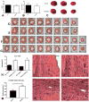
Figure 1. Fibroblast-specific Smad3 disruption accentuates adverse remodeling following reperfused myocardial infarction
A-B: Smad3fl/flPostn-Cre mice (FS3KO) exhibited reduced Smad3 expression in fibroblasts (A), but not in CD11b+ myeloid cells (B) harvested from the infarcted (I), or remodeling myocardium (R) after 1h of ischemia and 7 days of reperfusion. (*p<0.05, **p<0.01 vs. corresponding Smad3fl/fl n=5/group). C-F: Fibroblast-specific Smad3 loss increased adverse remodeling and worsened systolic dysfunction after 1h of ischemia and 28 days of reperfusion. In the model of reperfused myocardial infarction, FS3KO mice exhibited increased LVEDV (C) and LVESV (D), reduced LV fractional shortening (E) and increased LV mass (F) in comparison to Smad3fl/fl controls, after 28 days of reperfusion (*p<0.05) (ˆp<0.05, ˆˆp<0.01 vs. corresponding baseline values, n=11/group).
Figure 2
Figure 2. FS3KO mice exhibit increased late rupture-related mortality following non-reperfused myocardial infarction
A. In contrast to mice undergoing reperfused infarction protocols (that exhibit very low mortality rates), non-reperfused infarction is associated with a ∼50% mortality in WT mice, predominantly due to cardiac rupture. Control Smad3fl/fl mice had a 37.0% survival rate, compared with 18.7% for FS3KO animals (p=0.16, n=42-46 mice). In Smad3 fl/fl animals all deaths occurred during the first 5 days after coronary occlusion. Comparison of survival curves for late deaths (after the 6th day) suggested that FS3KO mice had increased late mortality (p=0.007). B. Systematic histological analysis of the heart of an FS3KO mouse that died 11 days after coronary occlusion shows H&E staining performed at 9 different levels, sectioned at 250μm partitions (1-9). The site of rupture is shown (arrows), filled with clot (scalebar=0.5 mm). Higher magnification images (Bi-iv) identify the site of rupture (arrows). Please note the presence of a dilated vascular structure within the healing scar (arrowhead) Scalebar=200μm. C-F: Echocardiographic analysis showed that surviving FS3KO mice had comparable LVEDV (C), and trends towards higher LVESV (D), lower fractional shortening (E) and increased LV mass (F) (Smad3fl/fl: 7days n=16, 28d n=9; FS3KO 7 days n=10, 28 days n=4; ˆˆp<0.01 vs corresponding baseline values (B)).
Figure 3
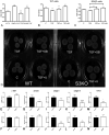
Figure 3. Fibroblast-specific loss of Smad3 does not affect acute infarct size, but is associated with larger scars and increased myofibroblast density
A-C: FS3KO and Smad3 fl/fl mice had comparable area at risk (AAR) (A) and infarct size:area at risk (IS:AAR) (B) (p=NS, n=10-11/group). D-H. However, despite comparable acute cardiomyocyte injury, FS3KO animals had significantly larger scars after 28 days of reperfusion. Scar size was assessed by sectioning the entire heart from base to apex and by staining for sirius red the first section from each 300μm partition. D-G: Representative sirius red-stained sections from Smad3fl/fl mouse and an FS3KO animal after 7 days of reperfusion (D and E respectively) and after 28 days of reperfusion (F and G respectively) (scalebar=1 mm). H: Despite similar segmental distribution of the infarct, the size of the collagenous scar was larger in FS3KO animals after 7 and 28 days of reperfusion (**p<0.01 vs. Smad3fl/fl, n=7-10/group). Fibroblast-specific Smad3 loss impaired contraction and remodeling of the scar. I-L. α-SMA immunohistochemistry was used to identify myofibroblasts in the infarct border zone after 7 days of reperfusion, as α-SMA+ cells located outside the vascular media (arrows). Scalebar=60μm. M. Quantitative analysis showed that FS3KO mice had higher myofibroblast density (*p<0.05, n=9/group).
Figure 4
Figure 4. Smad-dependent and Smad-independent pathways mediate fibroblast-induced collagen pad contraction
A. Smad3 KO fibroblasts exhibited impaired capacity to contract collagen pads (ˆˆp<0.01 vs. WT, n=6). TGF-β1 increases contraction in WT cells (**p<0.01, n=6), but has no significant effects on Smad3 KO cells. B. Both Erk inhibition (with U0126) and p38 MAPK inhibition (with SB203580) attenuated TGF-β-induced pad contraction (ˆˆp<0.01 vs. TGF, n=6), C. Incubation with U0126, or SB203580 reduced pad contraction in TGF-β-stimulated Smad3 KO cells, suggesting that Erk and p38 MAPK actions are independent of Smad3 (*p<0.05, **p<0.01 vs. TGF, n=6). D. Representative experiments illustrate the findings. E-N. Comparison of gene expression in infarct fibroblasts harvested from Smad3 fl/fl and FS3KO mice after 3 days (E, G, I, K, M), and 7 days (F, H, J, L, N) of reperfusion. Fibroblast-specific Smad3 loss did not affect α-SMA mRNA expression (E, F). FS3KO fibroblasts exhibited lower periostin (G) and TGF-β1 (M) expression after 3 days of reperfusion and lower levels of collagen I and III mRNA (J, L) after 7 days of reperfusion (*p<0.05, **p<0.01 vs. corresponding Smad3 fl/fl, n=5-6).
Figure 5
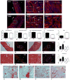
Figure 5. Fibroblast-specific Smad3 loss perturbs scar organization and attenuates TGF-β mediated changes in cell geometry
A-C. α-SMA staining of healing myocardial infarcts (7 days) shows that, in Smad3fl/fl mice, α-SMA+ myofibroblasts are spindle-shaped cells, localized in highly organized arrays (arrows, B-C). D-F. In contrast, distribution of α-SMA+ myofibroblasts in FS3KO mouse infarcts is disorganized and chaotic, as the cells have a rounded morphology and are malaligned (arrows, E-F). Scalebar=30μm. Although mean area of the myofibroblasts was comparable between groups (G), cell perimeter (H), perimeter:area ratio (I) and the long axis:short axis ratio (J) were lower in FS3KO animals (*p<0.05, n=5), reflecting the altered geometry of the cells. K. In order to quantitatively assess cell alignment in the healing infarct, we measured the angle between the long axis of the cell and the tangent of the ventricular wall (indicated by an arrow in panels A and D). The alignment angle was markedly higher in FS3KO mice, reflecting cellular malalignment in the absence of Smad3 (**p<0.01 vs fl/fl, n=5). Perturbed cellular alignment would be expected to reduce the tractional forces exerted by the cells, resulting in impaired scar contraction. L-S: Both light microscopy (L, N, P, R) and polarized light microscopy (M, O, Q, S) in sirius red-stained sections showed that while Smad3 fl/fl animals had aligned collagen fibers (P-Q, arrows), FS3KO animals exhibited areas of matrix disorganization (R-S, arrows). Scalebar=40μm. T-Y. Smad3 loss abrogated the effects of TGF-β1 on fibroblast shape. Fibroblasts from WT (V-W) and Smad3 KO mice (S3KO, X-Y) were cultured in collagen pads in the presence (W, Y) or absence (V, X) of TGF-β1. Sections of fibroblast-populated collagen pads were stained with sirius red/hematoxylin to identify fibroblasts (arrows) and the extracellular matrix. Scalebar=25μm. TGF-β1 stimulation increased cell area and perimeter in WT, but not in S3KO fibroblasts (T-U). (ˆp<0.05 vs. WT, **p<0.01 vs. corresponding WT, n=6-7).
Figure 6
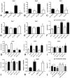
Figure 6. Smad3 loss markedly reduces expression of α2, α5 and β3 integrins in cardiac fibroblasts
In comparison to WT cells, Smad3 KO cardiac fibroblasts had significantly lower baseline expression of α2 (A, Itga2), α5 (B, Itga5) and β3 (C, Itgb3) mRNA. TGF-β1 stimulation markedly induced α2, α5 and β3 expression in cardiac fibroblasts in WT cells (**p<0.01 vs WT control, n=6), but not in Smad3 KO cells. α2, α5 and β3 integrin expression levels were significantly lower in Smad3 KO cells, in the presence or absence of TGF-β1 (ˆp<0.05, ˆˆp<0.01, n=6). D-E. In contrast, Smad3 loss did not affect α1 (D, Itga1) and β1 (E, Itgb1) integrin expression (expression normalized to C WT=1). F-J. Integrins mediate fibroblast activation inducing expression of NOX2. F. α2 and α5 integrin mediate pad contraction in both WT and Smad3 KO cells. α2, or α5 integrin blockade, reduces contraction of collagen pads populated with WT or Smad3 KO fibroblasts (ˆˆp<0.01 vs. corresponding control, n=6). KO cells exhibit impaired capacity to contract gels, in comparison to WT cells (**p<0.01). G. α5 blockade attenuates NOX2 mRNA synthesis in WT fibroblasts (ˆˆp<0.01 vs. WT control, n=3); in contrast α2 blockade does not affect NOX2 expression. Smad3 KO cells exhibited markedly lower expression of NOX2 (**p<0.01 vs. WT). In contrast to its effects on WT cells, α5 blockade did not affect NOX2 expression in Smad3 KO cells, suggesting that NOX2 transcription in cardiac fibroblasts is dependent on Smad3-mediated α5 integrin activation. H. Smad3 KO cells also had higher expression of SOD1 (**p<0.01 vs. corresponding WT); however, SOD1 levels were not affected by integrin inhibition. I-J: SOD2 and GSR expression was not affected by Smad3 loss or integrin blockade. K-L. α5 integrin overexpression does not reverse the contraction defect in Smad3 KO cells. K. α5 integrin overexpression in WT and Smad3 KO cells resulted in markedly increased α5 integrin expression (**p<0.01 vs. corresponding controls, ˆˆp<0.01 vs. WT C, n=6). L. Although α5 integrin overexpression (OE) increased contraction in WT cells, no effects on pad contraction were noted in Smad3 KO cells, indicating that α5 integrin is not sufficient to correct the contraction defect. Please note that untransfected cells exhibit increased contraction, suggesting that transfection modestly, but significantly reduces contraction in fibroblast-populated pads (##p<0.01 vs. corresponding cells transfected with control plasmid).
Figure 7
Figure 7. Cardiomyocyte-specific Smad3 loss attenuates post-infarction systolic dysfunction and reduces hypertrophic remodeling
Although LVEDV (A) and LVESV (B) were comparable between CMS3KO and Smad3fl/fl animals, cardiomyocyte-specific Smad3 loss attenuated systolic dysfunction (28 days) (C). D: LV mass was lower in CMS3KO mice after 7 days of reperfusion (*p<0.05, n=10-18/group; ˆp<0.05, ˆˆp<0.01 vs. corresponding baseline values). E-F. CMS3KO mice exhibited significantly lower ΔLVEDV and ΔLVESV after 7 days and trends towards reduced ΔLVEDV and ΔLVESV after 28 days of reperfusion, reflecting attenuated dilative remodeling. G. ΔLVFS was significantly lower in CMS3KO animals after 28 days, reflecting decreased systolic dysfunction (**p<0.01). H. ΔLVmass was markedly attenuated at both timepoints (*p<0.05).
Figure 8
Figure 8. Cardiomyocyte-specific Smad3 loss attenuated cardiomyocyte apoptosis in the viable remodeling myocardium, reduced NOX2 expression, and decreased nitrosative stress and MMP2 expression
A-B: TUNEL/WGA dual staining was used to identify apoptotic cardiomyocytes in the infarcted myocardium after 48h of reperfusion and in the viable remodeling myocardium after 7 days of reperfusion (scalebar=50μm). No statistically significant difference in the density of apoptotic cardiomyocytes was noted after 48h of reperfusion (p=0.21, n=6-7/group). After 7 days of reperfusion, cardiomyocyte-specific Smad3 loss attenuated cardiomyocyte apoptosis in the viable remodeling myocardium (**p<0.01 vs S3 fl/fl, n=6). C. CMS3KO mice had reduced NOX2 mRNA expression in the non-infarcted remodeling myocardium (NI) after 7 days of reperfusion (*p<0.05, n=5-9/group). D-E: NOX4 (D) and SOD1 (E) levels were comparable between CMS3KO and Smad3 fl/fl. NOX2, NOX4 and SOD1 expression in infarcted segments (I) was comparable between groups (C-E). F-G: CMS3KO mice exhibited reduced 3-nitrotyrosine levels in infarcted (I) and in non-infarcted (NI) areas, reflecting attenuated nitrosative stress (*p<0.05, **p<0.01, n=5/group). H-J: Moreover, CMS3KO animals had decreased levels of total and active MMP2 (aMMP2- cleaved) (*p<0.05, **p<0.01; n=8/group).
Figure 9
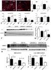
Figure 9. Schematic illustration of the novel findings of the study
Generation of active TGF-β in the infarcted myocardium triggers Smad3 activation in fibroblasts and cardiomyocytes. Our study investigates for the first time the role of fibroblast and cardiomyocyte-specific Smad3 signaling in the infarcted myocardium. Smad3 activation in cardiac fibroblasts restrains cell proliferation and controls alignment of fibroblasts in the infarct and formation of an organized collagen-based scar, preventing late cardiac rupture and adverse dilative remodeling. Our findings suggest that Smad3-dependent activation of a novel integrin (ITG)-NOX2 axis may stimulate extracellular matrix (ECM) protein deposition and organization in the infarcted heart. In contrast, activation of Smad3 signaling in cardiomyocytes has deleterious effects, promoting cardiomyocyte apoptosis and enhancing adverse remodeling and dysfunction. The effects of Smad3 on cardiomyocytes may also involve activation of a NOX2-mediated ROS-dependent axis. However, in cardiomyocytes, oxidative stress may promote cell death and accentuate MMP2 expression. Overactive MMP2 may cause adverse remodeling through its effects on the ECM, and may exacerbate systolic dysfunction by targeting proteins involved in sarcomere function, such as titin.

References

    1. Deten A, Holzl A, Leicht M, Barth W, Zimmer HG. Changes in extracellular matrix and in transforming growth factor beta isoforms after coronary artery ligation in rats. J Mol Cell Cardiol. 2001;33:1191–1207. - PubMed
    1. Dewald O, Ren G, Duerr GD, Zoerlein M, Klemm C, Gersch C, Tincey S, Michael LH, Entman ML, Frangogiannis NG. Of mice and dogs: species-specific differences in the inflammatory response following myocardial infarction. Am J Pathol. 2004;164:665–677. - PMC - PubMed
    1. Hao J, Ju H, Zhao S, Junaid A, Scammell-La Fleur T, Dixon IM. Elevation of expression of Smads 2, 3, and 4, decorin and TGF-beta in the chronic phase of myocardial infarct scar healing. J Mol Cell Cardiol. 1999;31:667–678. - PubMed
    1. Bujak M, Ren G, Kweon HJ, Dobaczewski M, Reddy A, Taffet G, Wang XF, Frangogiannis NG. Essential Role of Smad3 in Infarct Healing and in the Pathogenesis of Cardiac Remodeling. Circulation. 2007;116:2127–2138. - PubMed
    1. Dobaczewski M, Bujak M, Li N, Gonzalez-Quesada C, Mendoza LH, Wang XF, Frangogiannis NG. Smad3 signaling critically regulates fibroblast phenotype and function in healing myocardial infarction. Circ Res. 2010;107:418–428. - PMC - PubMed

Publication types

MeSH terms