. 2017 Dec 26;114(52):E11313-E11322.
doi: 10.1073/pnas.1711926115. Epub 2017 Dec 11.

FKBP12 contributes to α-synuclein toxicity by regulating the calcineurin-dependent phosphoproteome

Affiliations

FKBP12 contributes to α-synuclein toxicity by regulating the calcineurin-dependent phosphoproteome

Gabriela Caraveo et al. Proc Natl Acad Sci U S A. .

Abstract

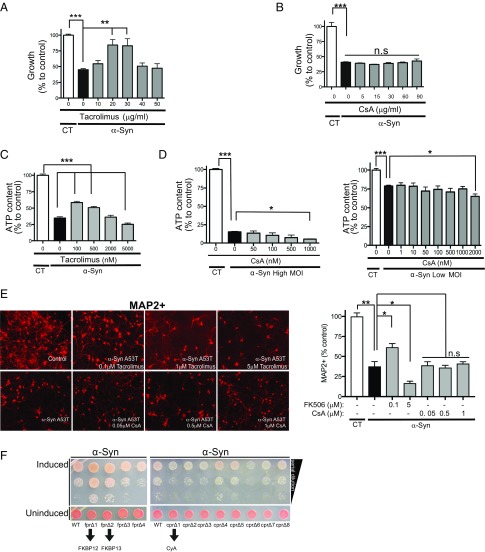
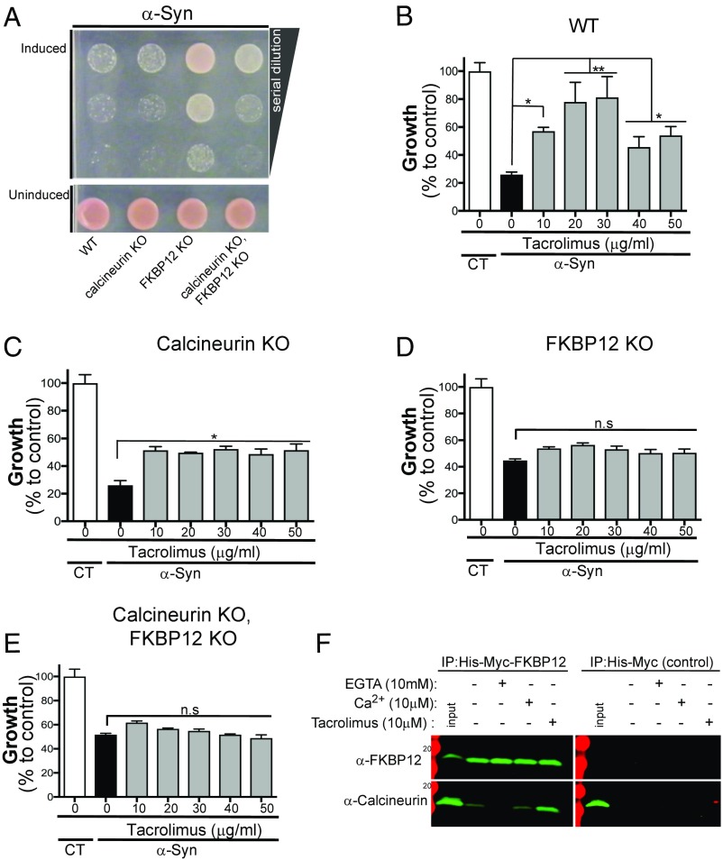
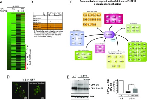
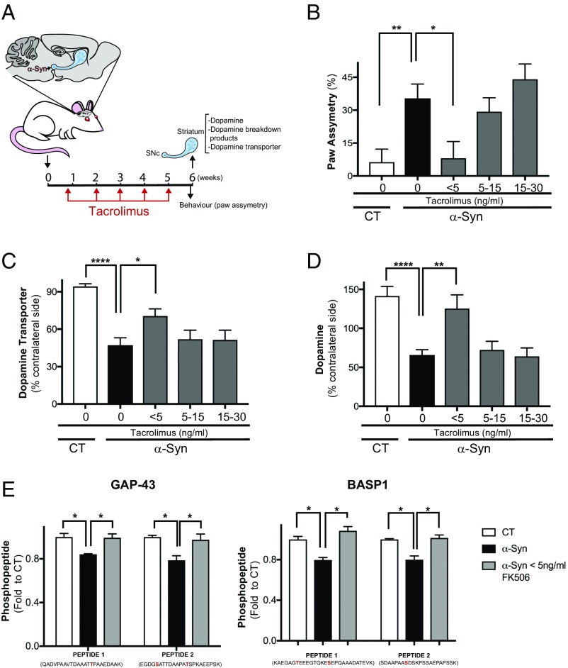
Calcineurin is an essential Ca2+-dependent phosphatase. Increased calcineurin activity is associated with α-synuclein (α-syn) toxicity, a protein implicated in Parkinson's Disease (PD) and other neurodegenerative diseases. Calcineurin can be inhibited with Tacrolimus through the recruitment and inhibition of the 12-kDa cis-trans proline isomerase FK506-binding protein (FKBP12). Whether calcineurin/FKBP12 represents a native physiologically relevant assembly that occurs in the absence of pharmacological perturbation has remained elusive. We leveraged α-syn as a model to interrogate whether FKBP12 plays a role in regulating calcineurin activity in the absence of Tacrolimus. We show that FKBP12 profoundly affects the calcineurin-dependent phosphoproteome, promoting the dephosphorylation of a subset of proteins that contributes to α-syn toxicity. Using a rat model of PD, partial elimination of the functional interaction between FKBP12 and calcineurin, with low doses of the Food and Drug Administration (FDA)-approved compound Tacrolimus, blocks calcineurin's activity toward those proteins and protects against the toxic hallmarks of α-syn pathology. Thus, FKBP12 can endogenously regulate calcineurin activity with therapeutic implications for the treatment of PD.

Keywords: FKBP12; Parkinson’s Disease; Tacrolimus; calcineurin; α-synuclein.


Conflict of interest statement

The authors declare no conflict of interest.

Figures

Fig. 1.
Fig. 1.
Inhibition of FKBP12 protects against α-syn toxicity. (A) Growth [described as percentage of control (CT)] of α-syn−expressing yeast cells grown for 48 h over a range of Tacrolimus concentrations. **P < 0.005 (one-way ANOVA, Fisher's test); ***P < 0.0005 (one-way ANOVA, Fisher's test). (B) Growth [described as percentage of control (CT)] of α-syn–expressing yeast cells grown for 48 h at the indicated CsA concentrations. ***P < 0.0005 (one-way ANOVA, Fisher's test). (C) Rat cortical neurons infected with high-titer (high MOI) α-syn A53T and/or LacZ as control (CT) treated with vehicle and/or increasing concentrations of Tacrolimus for 14 d and assayed for ATP content as a surrogate for viability. ***P < 0.005 (one-way ANOVA, Fisher’s test). (D) Same as in C, but neurons were infected with low titer (low MOI) and high titer (high MOI) of α-syn A53T and treated with various concentrations of CsA. Neuronal experiments performed in C and D represent data from six replicates in three independent experiments. The SE is present; it is very low. *P < 0.05 (one-way ANOVA, Dunnett’s multiple comparison test); ***P < 0.0005 (one-way ANOVA, Dunnett’s multiple comparison test). (E) Representative images of neuronal microtubule 2 (MAP2) red staining of rat primary neuronal cultures infected with either control lentivirus LacZ (control) and high-MOI α-syn A53T treated with various doses of FK506 or CsA for 14 d. Percentages of MAP2-positive neurons relative to control (LacZ infected) in the conditions described in C and D. *P < 0.05 (one-way ANOVA, Dunnett’s multiple comparison test); **P < 0.005 (one-way ANOVA, Fisher's test). (F) α-Syn–expressing yeast cells lacking individual FKBPs (fpr1-4Δ) and cyclophilins (cpr1-8Δ) were spotted onto plates containing uninducing media (SD−His,Trp–α-syn selective; Lower) and replica plated in threefold serial dilutions on α-syn–inducing plates containing selective media SGal (Upper). All yeast experiments were performed in triplicate with at least three biological replicates each time. n.s, nonsignificant; MOI, multiplicity of infection; SGal, synthetic galactose.
Fig. 2.
Fig. 2.
FKBP12 protects against α-syn toxicity in calcineurin-dependent and -independent manners. (A) α-Syn–expressing yeast cells either WT or lacking yeast FKBP12, fpr1 (FKBP12 KO); yeast calcineurin, cnb1 (calcineurin KO); or both yeast FKBP12 and calcineurin (calcineurin KO, FKBP12 KO) were spotted onto plates containing uninducing media (SD−His,Trp–α-syn selective; Lower) and replica plated in threefold serial dilutions on α-syn–inducing plates containing selective media synthetic galactose (SGal) (Upper). (BE) Growth (described as percentage over control) of α-syn–expressing yeast cells: WT (B), lacking yeast calcineurin (calcineurin KO; C), lacking yeast FKBP12 (FKBP12 KO; D), or lacking both FKBP12 and calcineurin (FKBP12 KO and calcineurin KO; E). Cells were grown for 48 h over a range of Tacrolimus concentrations. All yeast experiments were performed in triplicates with at least three biological replicate each time. All statistical comparisons were performed relative to no drug within each of the genetic conditions. *P < 0.05 (one-way ANOVA, Dunnett’s multiple comparison test); **P < 0.005 (one-way ANOVA, Dunnett’s multiple comparison test). (F) HEK 293 cells were stably transfected with His-Myc-FKBP12 and/or His-Myc, lysed, and treated with EGTA (10 mM), CaCl (10 μM), and/or Tacrolimus (10 μM). Lystes were subsequently immunoprecipitated (IP) with nickel beads and probed for calcineurin (α-CNB) and/or FKBP12 (α-Myc) by Western blot.
Fig. 3.
Fig. 3.
FKBP12 modulates calcineurin (CN) activity by altering its phospho-dependent proteome. (A) Heat map representing 527 hypophosphorylated phosphosites with abundances based on an MS shotgun approach. Cutoff was a |log2 FC| > 2 and q value < 0.05 between control and α-syn–expressing yeast cells across the different conditions. All of the Excel sheets associated with the data are available as tables in Dataset S1. (B) Diagram exemplifying how the CN- and FKBP12-dependent phosphosites were chosen. (C) Diagram of the proteins that contain all of the reverted CN/FKBP12-dependent phosphosites. Functions are annotated according to the SGD. (D) α-Syn–GFP localization in yeast cells in the presence (D, Right) and absence (D, Left) of protective dose of Tacrolimus (30 μg/mL). (E, Left) Western blot for CPY in α-syn−expressing cells in the presence and absence of a protective dose of Tacrolimus (30 μg/mL). Higher-molecular weight band represents the glycosylated form of CPY, which is typically found in the ER. The lower-molecular weight band (post-ER) is deglycosylated CPY, which has moved beyond the ER through the secretory pathway. Phosphoglycerate kinase (PGK) is used as a loading control. (E, Right) Quantitation of the ER and post-ER CPY bands from the Western blot; bands were quantitated using the Odyssey software. *P < 0.05 (one-way ANOVA, Dunnett's multiple comparison test).
Fig. 4.
Fig. 4.
Inhibition of calcineurin and FKBP12 by low doses of Tacrolimus protects against α-syn toxicity in vivo. (A) Diagram representing the rat in vivo experiment; briefly, n = 25–30 rats were injected unilaterally in the SNc with an AAV1/2 carrying either A53T α-syn or empty vector (CT). Four days after injection, rats were treated with different s.c. amounts of Tacrolimus on a weekly basis for 6 wk. Before the animals were killed, (B) the behavioral test for paw asymmetry was performed. Subsequently, striatal neurochemistry was performed. *P < 0.02 (one-way ANOVA, Dunnett’s multiple comparison test); **P < 0.007 (one-way ANOVA, Dunnett’s multiple comparison test). (C) DAT assayed by autoradiography at the striatum and normalized to its non–α-syn–injected contralateral side. *P < 0.03 (one-way ANOVA, Dunnett’s multiple comparison test); ****P < 0.0001 (one-way ANOVA, Dunnett’s multiple comparison test). (D) DA measured by HPLC. **P < 0.0016 (one-way ANOVA, Dunnett’s multiple comparison test); ****P < 0.0001 (one-way ANOVA, Dunnett’s multiple comparison test). (E) Striatal samples from CT, α-syn, and α-syn with <5 ng/mL of Tacrolimus were subjected to TMT MS (Materials and Methods), and phosphopeptides that were significantly rescued by Tacrolimus are shown. These phosphosites belong to two proteins: GAP-43 and BASP1. The phosphorylation site identified is highlighted in red. n = 3 rats. *P < 0.05 (two-tailed t test).

References

    1. Rusnak F, Mertz P. Calcineurin: Form and function. Physiol Rev. 2000;80:1483–1521. - PubMed
    1. Caraveo G, et al. Calcineurin determines toxic versus beneficial responses to α-synuclein. Proc Natl Acad Sci USA. 2014;111:E3544–E3552. - PMC - PubMed
    1. Martí MJ, Tolosa E, Campdelacreu J. Clinical overview of the synucleinopathies. Mov Disord. 2003;18(Suppl 6):S21–S27. - PubMed
    1. Hurwitz MY, Putkey JA, Klee CB, Means AR. Domain II of calmodulin is involved in activation of calcineurin. FEBS Lett. 1988;238:82–86. - PubMed
    1. Klee CB, Crouch TH, Krinks MH. Calcineurin: A calcium- and calmodulin-binding protein of the nervous system. Proc Natl Acad Sci USA. 1979;76:6270–6273. - PMC - PubMed

Publication types

MeSH terms