. 2017 Dec 26;114(52):13673-13678.
doi: 10.1073/pnas.1717168115. Epub 2017 Dec 11.

Kinetics of drug-ribosome interactions defines the cidality of macrolide antibiotics

Affiliations

Kinetics of drug-ribosome interactions defines the cidality of macrolide antibiotics

Maxim S Svetlov et al. Proc Natl Acad Sci U S A. .

Abstract

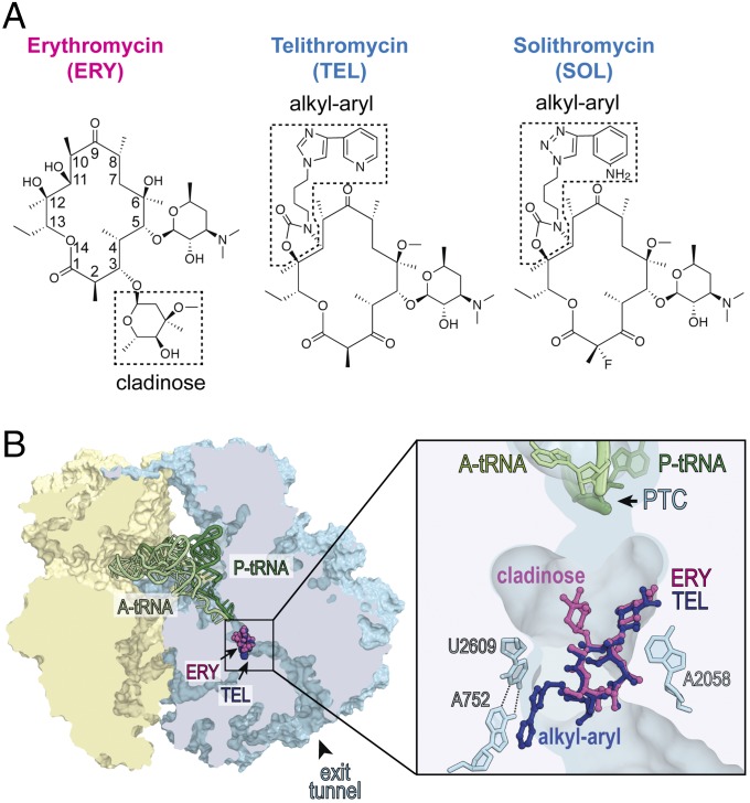
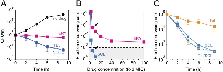
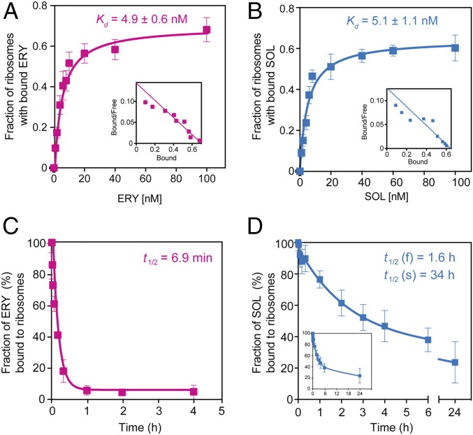
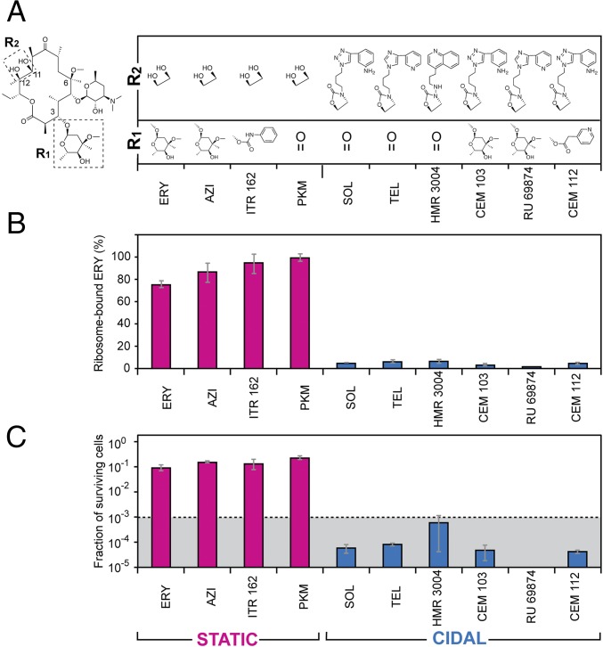
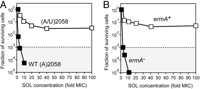
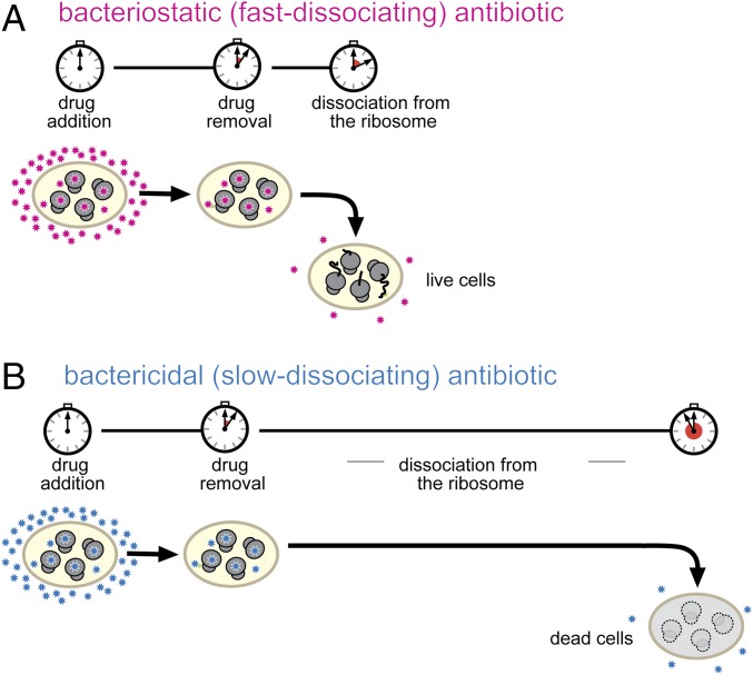
Antibiotics can cause dormancy (bacteriostasis) or induce death (cidality) of the targeted bacteria. The bactericidal capacity is one of the most important properties of antibacterial agents. However, the understanding of the fundamental differences in the mode of action of bacteriostatic or bactericidal antibiotics, especially those belonging to the same chemical class, is very rudimentary. Here, by examining the activity and binding properties of chemically distinct macrolide inhibitors of translation, we have identified a key difference in their interaction with the ribosome, which correlates with their ability to cause cell death. While bacteriostatic and bactericidal macrolides bind in the nascent peptide exit tunnel of the large ribosomal subunit with comparable affinities, the bactericidal antibiotics dissociate from the ribosome with significantly slower rates. The sluggish dissociation of bactericidal macrolides correlates with the presence in their structure of an extended alkyl-aryl side chain, which establishes idiosyncratic interactions with the ribosomal RNA. Mutations or chemical alterations of the rRNA nucleotides in the drug binding site can protect cells from macrolide-induced killing, even with inhibitor concentrations that significantly exceed those required for cell growth arrest. We propose that the increased translation downtime due to slow dissociation of the antibiotic may damage cells beyond the point where growth can be reinitiated upon the removal of the drug due to depletion of critical components of the gene-expression pathway.

Keywords: erythromycin; inhibitors; ketolides; solithromycin; translation.


Conflict of interest statement

Conflict of interest statement: In the previous years, A.S.M. had grants from Cempra, Inc. and Melinta Therapeutics, which were involved in the development of macrolide antibiotics.

Figures

Fig. 1.
Fig. 1.
Bacteriostatic and bactericidal macrolide and ketolide antibiotics bind to the same site in the bacterial ribosome. (A) Chemical structures of bacteriostatic macrolide ERY and bactericidal ketolides TEL and SOL. The C3-cladinose sugar of ERY and the alkyl-aryl side chains in TEL and SOL are boxed. (B) Binding site of macrolides and ketolides in the ribosome. A cross section of the bacterial (Thermus thermophilus) 70S ribosome with ERY (purple) and TEL (blue) bound in the nascent peptide exit tunnel (PDB ID codes: 4V7X and 4V7Z, respectively, from ref. 11). Small subunit is yellow, large subunit is light blue, A-site tRNA is light green, and P-site tRNA is dark green. The zoomed-in image shows interactions of the C5 desosamine of both antibiotics with A2058 and of the alkyl-aryl side chain of TEL with the A752-U2609 base pair in the nascent peptide exit tunnel. The peptidyl-transferase center (PTC) is indicated.
Fig. 2.
Fig. 2.
Macrolide antibiotics differ significantly in their bactericidal activity. (A) Killing effect of fourfold MIC concentrations of ERY (magenta), TEL (dark blue), or SOL (light blue) against S. pneumoniae cells. The viable cells counts are indicated as colony forming units (CFU). (B) Survival of S. pneumoniae cells exposed for 18 h to various concentrations of ERY (magenta) or SOL (light blue). The experimental points determined at the fourfold MIC concentration of ERY or SOL are indicated by arrows. The dotted line marks the three-orders reduction in CFU, representing the operational definition of the bactericidal activity (1). (C) Survival of cells exposed for the indicated time periods to fourfold MIC of SOL (light blue), 40-fold MIC of Tet (orange), or fourfold MIC of SOL and 40-fold MIC of Tet (light blue/orange). In the latter case, cells were preincubated with Tet for 30 min before the addition of SOL. The error bars in A and C show SD in three independent experiments.
Fig. 3.
Fig. 3.
Thermodynamic and kinetic parameters of interaction of bacteriostatic and bactericidal macrolides with the bacterial ribosome. (A and B) Equilibrium binding of [14C]-ERY (A) or [14C]-SOL (B) to S. pneumoniae ribosomes. Ribosomes were mixed with varying concentrations of the radiolabeled drugs and incubated for 2 h at 37 °C before determining the amount of bound antibiotic (see Materials and Methods for details). Insets show the Scatchard plots for ERY and SOL equilibrium binding. (C and D) Kinetics of dissociation of [14C]-ERY (C) or [14C]-SOL (D) from the ribosome. The Inset in D shows the complete curve. Ribosomes were preequilibrated with the [14C]-labeled antibiotics and after addition of an excess of the respective nonlabeled drug the amount of ribosome-associated radioactivity was monitored over time. The data were fitted to a one-phase (ERY) or two-phase (SOL) exponential functions that yielded dissociation rate constants of (10 ± 1.4) × 10−2 min−1 for ERY, (0.72 ± 0.25) × 10−2 min−1 for the faster (f) phase of SOL, and (0.034 ± 0.02) × 10−2 min−1 for the slower (s) phase of SOL. The values of dissociation rate constants were used for calculating the half-life (t1/2) of the complexes (shown in the figure). All experiments were performed in triplicates. Error bars represent standard deviation (SD).
Fig. 4.
Fig. 4.
Cidality of macrolides correlates with slow dissociation and the presence of an extended alkyl-aryl side chain. (A) Chemical structures of the C3 or C11, C12 side chains (boxed in the depicted ERY structure) of the macrolide and ketolide antibiotics. Note that ERY, AZI, and PKM carry a C6 hydroxyl instead of the shown metoxy group present in the rest of the compounds. (B) Displacement of nonlabeled macrolides bound to S. pneumoniae ribosomes by [14C]-ERY. After pre-equilibration with the corresponding antibiotic, ribosomes were incubated for 30 min at 37 °C with a 20-fold molar excess of [14C]-ERY and the amount of labeled ERY replacing the prebound inhibitor was measured. Binding of [14C]-ERY to the vacant drug-free ribosomes was taken as 100%. The blue bars represent slow-dissociating antibiotics that after 30-min incubation remain bound to more than 90% of the ribosomes; the fast-dissociating inhibitors that vacate more than 90% of the ribosomes over a 30-min incubation time are indicated by the magenta bars. (C) Surviving of S. pneumoniae cells after 18-h exposure to the corresponding fourfold MIC concentration of the indicated antibiotics. The dotted line indicates the three-orders reduction in the viable cell count representing the operational definition of the bactericidal activity. Error bars indicate SD in three independent experiments. The graph bars representing bactericidal antibiotics are blue and those corresponding to bacteriostatic inhibitors are magenta.
Fig. 5.
Fig. 5.
Alterations of the ribosomal drug-binding site protect bacteria from killing by bactericidal antibiotics. (A) Survival of the WT S. pneumoniae strain Cp2000 and the killing-resistant mutant upon exposure for 18 h to varying concentrations of SOL. All four 23S rRNA gene alleles in the WT cells carry A2058, whereas in the mutant cells the same 23S rRNA position in two of the alleles has been mutated to U. (B) Survival of the parental (ermA) S. pneumoniae strain CPM1 and its ermA+ variant (Cp1290) upon exposure for 18 h to varying concentrations of SOL. In both panels the dotted line indicates the three-orders reduction in the viable cell count. The concentrations of SOL for the treatment of WT and altered strains were adjusted according to the respective MIC values (Table S2).
Fig. 6.
Fig. 6.
Prolonged inhibition of protein synthesis leads to cell death. (A) With fast-dissociating inhibitors (magenta stars), the duration of protein synthesis shut-down is determined primarily by the timing of exposure to the antibiotic. Translation readily resumes upon the removal of the drug and cells can restart their growth. (B) With slowly dissociating antibiotics (blue stars), the translation downtime is prolonged due to the slow off-rate of the drug. During the extended translation down time depletion of critical cellular components prevents the resumption of translation and of cell growth.

References

    1. Clinical and Laboratory Standards Institute . Methods for Determining Bactericidal Activity of Antimicrobial Agents; Approved Guideline. NCCLS Document M26-A. NCCLS; Wayne, PA: 1999.
    1. Goldstein FW, Emirian MF, Coutrot A, Acar JF. Bacteriostatic and bactericidal activity of azithromycin against Haemophilus influenzae. J Antimicrob Chemother. 1990;25(Suppl A):25–28. - PubMed
    1. Woosley LN, Castanheira M, Jones RN. CEM-101 activity against Gram-positive organisms. Antimicrob Agents Chemother. 2010;54:2182–2187. - PMC - PubMed
    1. Kosowska K, et al. Activities of two novel macrolides, GW 773546 and GW 708408, compared with those of telithromycin, erythromycin, azithromycin, and clarithromycin against Haemophilus influenzae. Antimicrob Agents Chemother. 2004;48:4113–4119. - PMC - PubMed
    1. Gaynor M, Mankin AS. Macrolide antibiotics: Binding site, mechanism of action, resistance. Curr Top Med Chem. 2003;3:949–961. - PubMed

Publication types