. 2017 Dec 26;114(52):E11141-E11150.
doi: 10.1073/pnas.1717891115. Epub 2017 Dec 11.

Rapid, direct activity assays for Smoothened reveal Hedgehog pathway regulation by membrane cholesterol and extracellular sodium

Affiliations

Rapid, direct activity assays for Smoothened reveal Hedgehog pathway regulation by membrane cholesterol and extracellular sodium

Benjamin R Myers et al. Proc Natl Acad Sci U S A. .

Abstract

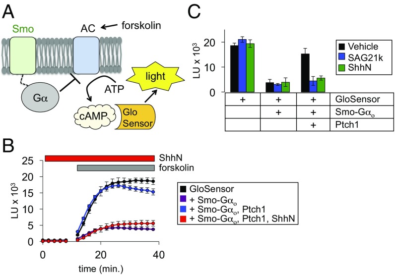
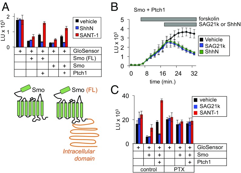
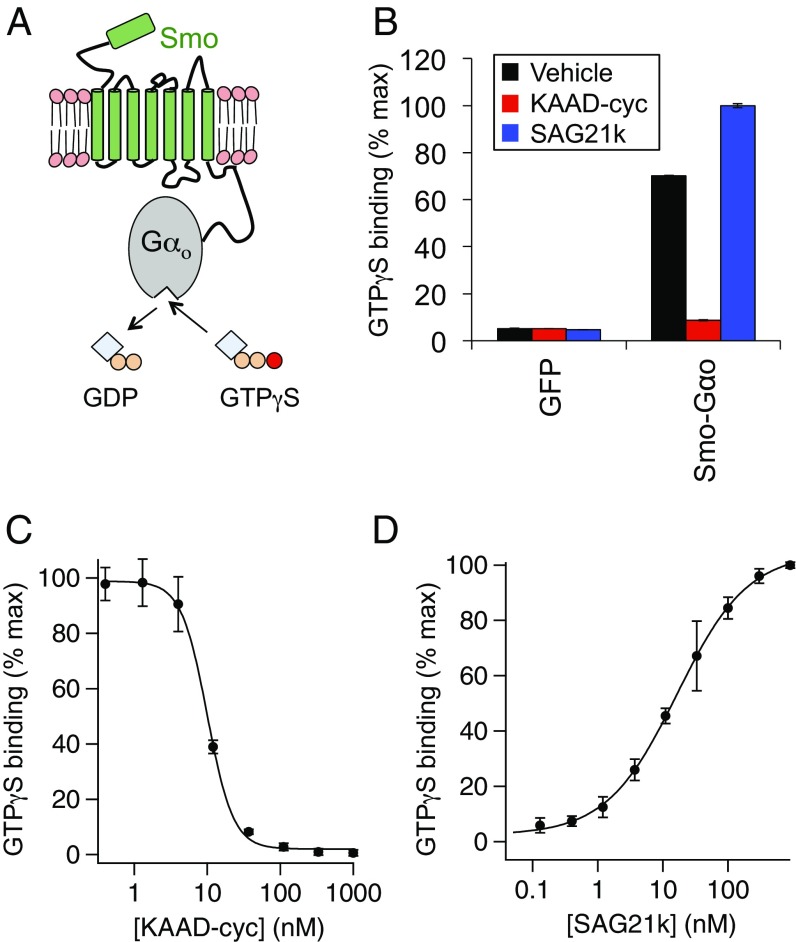
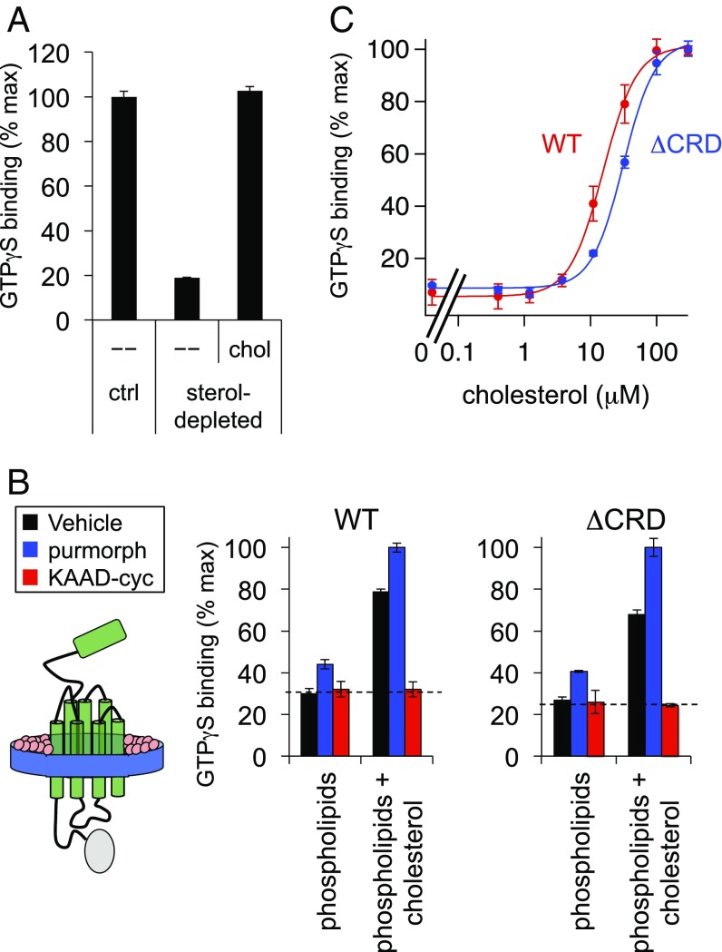
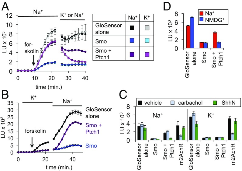
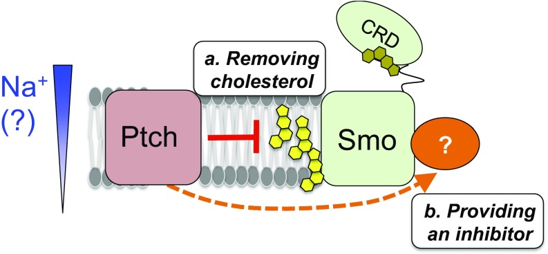
Hedgehog signaling specifies tissue patterning and renewal, and pathway components are commonly mutated in certain malignancies. Although central to ensuring appropriate pathway activity in all Hedgehog-responsive cells, how the transporter-like receptor Patched1 regulates the seven-transmembrane protein Smoothened remains mysterious, partially due to limitations in existing tools and experimental systems. Here we employ direct, real-time, biochemical and physiology-based approaches to monitor Smoothened activity in cellular and in vitro contexts. Patched1-Smoothened coupling is rapid, dynamic, and can be recapitulated without cilium-specific proteins or lipids. By reconstituting purified Smoothened in vitro, we show that cholesterol within the bilayer is sufficient for constitutive Smoothened activation. Cholesterol effects occur independently of the lipid-binding Smoothened extracellular domain, a region that is dispensable for Patched1-Smoothened coupling. Finally, we show that Patched1 specifically requires extracellular Na+ to regulate Smoothened in our assays, raising the possibility that a Na+ gradient provides the energy source for Patched1 catalytic activity. Our work suggests a hypothesis wherein Patched1, chemiosmotically driven by the transmembrane Na+ gradient common to metazoans, regulates Smoothened by shielding its heptahelical domain from cholesterol, or by providing an inhibitor that overrides this cholesterol activation.

Keywords: Hedgehog; Na+; Patched; Smoothened; cholesterol.


Conflict of interest statement

The authors declare no conflict of interest.

Figures

Fig. 1.
Fig. 1.
A live-cell assay shows rapid, cilium-independent Ptch1 regulation of Smo. (A) Schematic diagram of the GloSensor cAMP assay. Smo couples to endogenous (or fused) inhibitory Gα proteins, which block forskolin-induced AC activity, thereby reducing cAMP. Increased Smo activity is therefore reflected as a decrease in luminescence. (B) Live-cell luminescence traces from HEK293 cells transfected with the indicated plasmids. Baseline luminescence was recorded for 10 min at 2-min intervals, followed by forskolin treatment and continued monitoring. For ShhN treatment, ShhN was added 10 min before measurement of baseline and forskolin-induced luminescence; ShhN remained present at 200 nM for the entire experiment. (C) Bar graph shows steady state luminescence for HEK293 cells transfected with GloSensor, Smo, and Ptch1 expression plasmids. Cells were preincubated for 10 min with ShhN or SAG21k as indicated in B.
Fig. 2.
Fig. 2.
Ptch1-mediated regulation of Smo conformation is fast, nonrate-limiting, and occurs independently of Smo ciliary trafficking. (A, Top) Ptch1 blocked the activity of full-length Smo [Smo (FL)] and the cytotail-deleted form (Smo) to similar degrees in the GloSensor assay. (Bottom) A topology diagram of the two Smo expression constructs; the cytoplasmic tail required for transcriptional coupling and ciliary trafficking is indicated in orange. (B) To demonstrate the rapid onset of ShhN or SAG21k effects, cells expressing GloSensor, Smo, and Ptch1 were stimulated with forskolin, and ShhN or SAG21k (vs. a vehicle control) were then added as indicated by the gray bar. (C) Overnight pretreatment with PTX (100 ng/mL) prevents Smo from inhibiting GloSensor luminescence.
Fig. 3.
Fig. 3.
An in vitro assay for Smo conformational state reflects modulation by exogenous small molecules and membrane cholesterol. (A) Diagram of the in vitro Smo–Gαo assay, in which Smo activity promotes GDP dissociation and nucleotide exchange, measured as binding of nonhydrolyzable 35S-GTPγS. (B) Membranes were prepared from HEK293 cells transfected with plasmids encoding GFP or Smo–Gαo, and incubated with the Smo inverse agonist KAAD-cyclopamine (KAAD-cyc, 300 nM), the Smo agonist SAG21k (50 nM), or a DMSO control, followed by 35S-GTPγS binding and scintillation counting. Data were normalized to maximum SAG21k-stimulated binding. (C and D) Concentration-response relationships for KAAD-cyclopamine and SAG21k in the 35S-GTPγS binding assay, revealing a KAAD-cyclopamine IC50 of 9.99 ± 0.36 nM and a SAG21k EC50 of 16.53 ± 1.45 nM, in reasonable agreement with previously published values. Note that D was conducted at high [GDP] (300 µM) to expand the window between basal and SAG21k-induced activity (Materials and Methods).
Fig. 4.
Fig. 4.
Smo reconstitution reveals that cholesterol and phospholipids are sufficient for constitutive activity. (A) Membranes from cells expressing Smo–Gαo were depleted of endogenous sterols using methyl-β-cyclodextrin (MβCD) or incubated with buffer alone as a control (ctrl). Sterol-depleted membranes were subsequently incubated with a cholesterol-MβCD complex (chol) or a buffer control (–), then processed for GTPγS binding. Data were normalized to the amount of Smo constitutive GTPγS binding under control (nonsterol-depleted) conditions. (B) Diagram of the Smo–Gαo fusion (green) reconstituted into nanodiscs with a membrane scaffold protein (MSP1D1, blue) and defined lipids (pink). Nanodiscs were formed from purified Smo–Gαo or SmoΔCRD–Gαo (a form lacking the CRD) in combination with synthetic phospholipids alone, or a mixture of phospholipids and 8 mol% cholesterol. Each nanodisc preparation was stimulated with purmorphamine, KAAD-cyc, or a vehicle control, followed by measurement of GTPγS binding. The dashed line represents the baseline level of G protein coupling in this assay, as defined by treatment with KAAD-cyc (a full inverse antagonist). (C) Membranes derived from HEK293-cells transfected with Smo–Gαo or SmoΔCRD–Gαo were stripped of endogenous cholesterol with MβCD and replenished with a cholesterol–MβCD complex as in A. For each data point, the cholesterol concentration in the assay buffer was derived by assuming a 10:1 ratio of cyclodextrin to cholesterol in our saturated cholesterol–MβCD complexes, as defined by previous measurements (67). Data are normalized to GTPγS binding stimulated by the maximal dose of cholesterol–MβCD for each construct. Curve-fitting with the Hill equation revealed EC50s of 15.55 ± 0.83 μM and 31.39 ± 1.73 μM for wild-type and CRD-deleted Smo, respectively.
Fig. 5.
Fig. 5.
Cholesterol derivatives show a similar structure–activity relationship on wild-type and CRD-deleted Smo in G protein coupling assays. Membranes from HEK293 cells transfected with (A) wild-type or (B) CRD-deleted Smo–Gαo fusions were depleted of endogenous sterols, and then treated with the indicated sterols (in complex with cyclodextrin) as in Fig. 4. Values were normalized to the level of rescue produced by the sterol biosynthesis intermediate desmosterol (which consistently gave slightly higher values of GTPγS binding than cholesterol).
Fig. 6.
Fig. 6.
Ptch1 can inhibit Smo in the absence of CRD-cholesterol binding. (A) Smo−/− mouse embryonic fibroblasts (MEFs) were transfected with the indicated Smo and Ptch1 expression constructs along with an 8×Gli-luciferase reporter, and stimulated with control (black) or ShhN (green) conditioned medium. Relative luciferase units (RLU) are plotted as a fold-increase over the baseline value, defined as reporter activity from the negative control transfection (no Smo). (B) A similar experiment, in which cells transfected with wild-type Smo (red) or SmoA1 D99A Y134F (SmoA1 DAYF) + Ptch1 (blue) were stimulated with increasing concentrations of ShhN conditioned medium. (C) SmoΔCRD is constitutively active and suppressible by Ptch1. Suppression is evident when Ptch1 is coexpressed according to standard conditions (Materials and Methods), and becomes even more obvious when increasing amounts of Ptch1 cDNA are transfected. The standard 10-min ShhN pretreatment fully reverses the effects of Ptch1 coexpression on both wild-type and CRD-deleted Smo, even at the highest amounts of Ptch1 cDNA transfected. The observed suppression of SmoΔCRD is therefore truly dependent on Ptch1 activity and not simply an artifact of protein overexpression in this assay. (D) Simulation of the functional behavior of wild-type and various CRD-mutated forms of Smo, taking into account observations from Gli transcriptional assays in this and several other studies (22, 27, 68). Smo activity is represented on the y axis, while Ptch1 activity (blocked by binding of Hh) is on the x axis. The possible ranges of Ptch1 activity encompassed by endogenous vs. transfected Ptch are indicated below the x axis.
Fig. 7.
Fig. 7.
Ptch1 activity depends on conserved residues that mediate intramembrane ion flux in bacterial RNDs. (A) Top-down views of the prototypical bacterial RND AcrB (green, PDB ID code 2DHH) and NPC1 (blue, PDB ID code 5U73), with conserved charged residues in TM4 and TM10 highlighted in red. (B) Sequence alignment of transmembrane helices 4 and 10 of several RND family proteins. ecAcrB, Escherichia coli AcrB; mmNpc1, Mus musculus (mouse) NPC1; mmPtch1, Mus musculus (mouse) Ptch1; ttSecDF, Thermus thermophiles secDF; vaSecD1, Vibrio alginolyticus secD-1; vaSecF1, Vibrio alginolyticus secF-1. SecD/F in certain organisms, including E. coli and V. alginolyticus, is a heterodimer composed of D + F, with each subunit corresponding to either the first or last six-transmembrane helices of the RND family. Asterisks indicate key residues for ion flux, with those neutralized by the Ptch1 “NNQ” mutation colored green. The activity of different Ptch1 constructs was tested in a Gli-luciferase transcriptional reporter assay in Ptch1−/− cells (C) or the GloSensor assay in HEK293 cells (D). In both assays, wild-type Ptch1 suppressed Smo activity and the suppression was reversed by addition of ShhN or SAG21k, while the NNQ mutation diminished Ptch1 activity.
Fig. 8.
Fig. 8.
A role for extracellular Na+ in Ptch1–Smo regulation. (A) HEK293 cells were transfected with GloSensor + the indicated plasmids and stimulated with forskolin in physiological saline (Na+). The bath was replaced at the indicated time with a low Na+/high K+ buffer (K+), in which all Na+ was replaced by K+, and vice versa. A control buffer replacement maintained physiological Na+-based saline for the entire experiment. (B) Cells were transfected with GloSensor alone (black), or in the additional presence of Smo (blue), or Smo and Ptch1 (purple). After loading with luciferin in K+ buffer, cells were switched at the indicated time to physiological Na+ buffer. While Ptch1 failed to suppress Smo in the K+ buffer (note the overlapping blue and purple traces), Ptch1 activity increased almost immediately after the buffer change and plateaued within 20 min. See Fig. S10A for quantification. (C) Steady-state luminescence in HEK293 cells expressing GloSensor + the indicated plasmids (m2AchR, m2 muscarinic acetylcholine receptor, stimulated by its ligand carbachol). Cells were bathed in Na+ (Left) vs. K+ (Right) for all steps following substrate loading, and forskolin-induced luminescence was measured as in Fig. 1C. (D) Steady-state forskolin-induced luminescence in cells bathed in saline solution based on Na+ or the organic cation NMDG+, revealing that NMDG+ cannot sustain Ptch1 activity.
Fig. 9.
Fig. 9.
Model for the roles of membrane cholesterol in the Hh pathway. When the pathway is on, following Hh binding to and inactivation of Ptch1, membrane cholesterol activates Smo by binding to an as yet unidentified site within the Smo heptahelical bundle (or by modulating the membrane’s properties). In the “off” state of the pathway, Ptch1 could repress Smo by either: (a) altering cholesterol availability to Smo (yellow); or (b) providing a negative regulator (orange) that overrides the cholesterol activating effect on Smo. In either case, the modulatory influences of Ptch1 activity and membrane cholesterol on Smo function are distinct from the CRD-cholesterol interactions highlighted in previous structural studies (see main text). Based on the observed Ptch1 requirement for extracellular Na+, we hypothesize that Ptch1 depends on the transmembrane Na+ gradient to power its activity. See Discussion for more details.

Comment in

Similar articles

Cited by

References

    1. Briscoe J, Thérond PP. The mechanisms of Hedgehog signalling and its roles in development and disease. Nat Rev Mol Cell Biol. 2013;14:416–429. - PubMed
    1. Pak E, Segal RA. Hedgehog signal transduction: Key players, oncogenic drivers, and cancer therapy. Dev Cell. 2016;38:333–344. - PMC - PubMed
    1. Wu F, Zhang Y, Sun B, McMahon AP, Wang Y. Hedgehog signaling: From basic biology to cancer therapy. Cell Chem Biol. 2017;24:252–280. - PMC - PubMed
    1. Shin K, et al. Hedgehog/Wnt feedback supports regenerative proliferation of epithelial stem cells in bladder. Nature. 2011;472:110–114. - PMC - PubMed
    1. Zhao C, et al. Stromal Gli2 activity coordinates a niche signaling program for mammary epithelial stem cells. Science. 2017;356:eaal3485. - PubMed

Publication types

MeSH terms

LinkOut - more resources