. 2017 Dec 26;114(52):13720-13725.
doi: 10.1073/pnas.1718471115. Epub 2017 Dec 11.

Architecture of the human PI4KIIIα lipid kinase complex

Affiliations

Architecture of the human PI4KIIIα lipid kinase complex

Joshua A Lees et al. Proc Natl Acad Sci U S A. .

Abstract

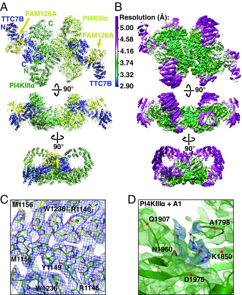
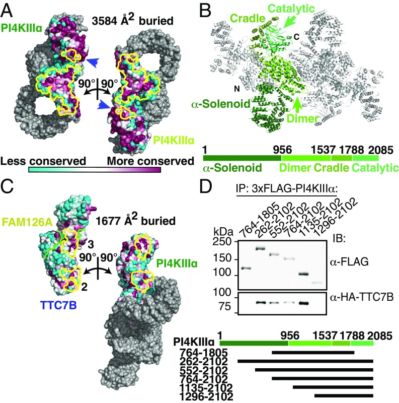
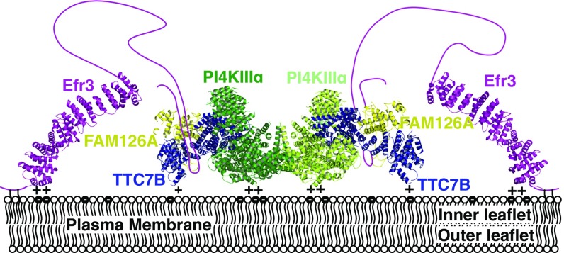
Plasma membrane (PM) phosphoinositides play essential roles in cell physiology, serving as both markers of membrane identity and signaling molecules central to the cell's interaction with its environment. The first step in PM phosphoinositide synthesis is the conversion of phosphatidylinositol (PI) to PI4P, the precursor of PI(4,5)P2 and PI(3,4,5)P3 This conversion is catalyzed by the PI4KIIIα complex, comprising a lipid kinase, PI4KIIIα, and two regulatory subunits, TTC7 and FAM126. We here report the structure of this complex at 3.6-Å resolution, determined by cryo-electron microscopy. The proteins form an obligate ∼700-kDa superassembly with a broad surface suitable for membrane interaction, toward which the kinase active sites are oriented. The structural complexity of the assembly highlights PI4P synthesis as a major regulatory junction in PM phosphoinositide homeostasis. Our studies provide a framework for further exploring the mechanisms underlying PM phosphoinositide regulation.

Keywords: lipid kinase; phosphoinositides; signaling.


Conflict of interest statement

The authors declare no conflict of interest.

Figures

Fig. 1.
Fig. 1.
The PI4KIIIα/TTC7/FAM126 complex forms a ∼700-kDa homodimeric superassembly. (A) Ribbon model of PI4KIIIα/TTC7B/FAM126A complex, colored and labeled by subunit, and superimposed with map density. (B) Single-particle cryo-EM model of PI4KIIIα/TTC7B/FAM126A complex at 3.6-Å resolution, contoured at 8.5 σ. Each view is colored by local resolution according to the scale at Top Left. (C) Model and typical density from the core of the PI4KIIIα map, contoured at 8.5 σ, with resolution approaching 3.0 Å. Prominent residues are labeled for orientation purposes. (D) Ribbon diagram (green) and density (green mesh) from the active site of PI4KIIIa, with the A1 inhibitor in stick representation. Map density used to place the A1 ligand is in blue. The resolution of this region is ∼4–5 Å, with the density contoured at 8.5 σ. Alpha carbons of indicated residues are denoted by red dots for orientation purposes.
Fig. 2.
Fig. 2.
PI4KIIIα homodimerizes and interacts stably with TTC7 via large conserved surfaces. (A) Solvent-accessible surface representation of PI4KIIIα/TTC7B/FAM126A complex, with the PI4KIIIα surface (excluding the α-solenoid) colored by conservation. Each heterotrimer is rotated by ±90° as indicated to reveal the homodimerization interface. PI4KIIIα surface residues participating in the dimerization are outlined in yellow. Blue arrows indicate regions of the kinase catalytic domains that contact the opposite copy of PI4KIIIα. (B) Ribbon representation of the PI4KIIIα complex, with one copy of PI4KIIIα colored by domain. Boundary residue numbers for each domain are indicated on the schematic below. (C) Solvent-accessible surface representation of PI4KIIIα/TTC7B/FAM126A complex, with the TTC7/FAM126 complex and PI4KIIIα surfaces colored by conservation, as in A. PI4KIIIα and the TTC7B/FAM126A complex are each rotated by ±90° as indicated to reveal the PI4KIIIα/TTC7B dimerization interface. Interacting residues are outlined in yellow, while the TTC7B/FAM126A boundary is indicated by a green line. TTC7 surfaces 2 and 3 interact with the dimerization and cradle domains of PI4KIIIα, respectively. The additional contact between TTC7B and the tip of the PI4KIIIα α-solenoid, which was not sufficiently resolved to model by a Cα trace, is neither shown nor included in the surface area calculation. (D) Truncation constructs of 3xFLAG-tagged PI4KIIIα were overexpressed alongside HA-TTC7B and EGFP-FAM126A(2–289) in Expi293 cells. α-FLAG-immunoprecipitated samples were resolved by SDS/PAGE and immunoblotted with anti-FLAG and anti-HA antibodies to probe coprecipitation of TTC7B with the different PI4KIIIα constructs. Boundaries of PI4KIIIα truncation constructs are indicated above the blot and in the schematic at bottom.
Fig. 3.
Fig. 3.
The interaction between TTC7 and PI4KIIIα is required for PI4KIIIα stability in vivo. (A) Patient fibroblasts or B cells were isolated and immunoblotted with antibodies against components of the PI4KIIIα complex. The levels of other complex subunits are reduced in the absence of intact TTC7A. (B) TTC7B mRNA is less abundant than TTC7A mRNA in human fibroblasts and nearly absent in B cells. cDNA from control human fibroblasts and B cells used in C was analyzed by qRT-PCR with TTC7A- and TTC7B-specific primer pairs. The ratio of TTC7B expression to that of TTC7A is shown for each cell type. Error bars represent SD (n = 3). (C) Spleens isolated from “flaky skin” mice (17, 18) were immunoblotted for PI4KA complex subunits. Loss of TTC7A caused reduction in the levels of other complex components. (D) CID-MIA patient 4 carries a mutation that truncates TTC7A at residue 823 (corresponding to residue 809 of TTC7B). The corresponding truncated residues are indicated in light blue on the structure of TTC7B in the context of the kinase complex. This mutation likely abolishes the interaction between TTC7 and the tip of the PI4KIIIα α-solenoid, causing the pathology of CID-MIA. (E) CID-MIA patient 4 carries an intact, though mildly truncated version of TTC7A. Immunoblots for other PI4KA complex components indicate little change in protein levels. Immunoblots against B cells from patient 3 are shown for comparison. Ctrl, Control; IB, immunoblot; Pat., Patient.
Fig. 4.
Fig. 4.
PI4KIIIα complex interacts with the plasma membrane via a large conserved surface that orients the catalytic domains toward substrate. (A, Top) Surface representation of PI4KIIIα complex, colored by subunit, as in Fig. 1A. Surfaces designated side 1 and side 2 are indicated by labels. (A, Lower Left) Side 1 and side 2 surfaces of PI4KIIIα complex, colored by conservation. Regions built as polyalanine helices are indicated in gray. (A, Lower Right) Side 1 and side 2 surfaces of PI4KIIIα complex, colored by surface charge. Regions built as polyalanine helices are indicated in gray. (B) Active site structures of PI4KIIIα + A1 inhibitor and PI3Kγ + ATP (PDB ID code 1E8X) are shown after structural alignment for comparison and oriented such that the putative membrane-binding surface of PI4KIIIα lies directly below the indicated view. In this orientation, the PI-binding site of each molecule lies directly below the bound ligand in each molecule.
Fig. 5.
Fig. 5.
A model for PI4KIIIα complex recruitment to the plasma membrane. EFR3, for which the structure from Saccharomyces cerevisiae (PDB ID code 4N5A) is shown, localizes to the membrane via a basic patch at its N terminus as well as N-terminal palmitoylation (33). The PI4KIIIα complex is recruited to the plasma membrane by an interaction between TTC7/FAM126 and the EFR3 unstructured C terminus (5, 6). The PI4KIIIα/TTC7/FAM126 complex interacts with the acidic inner leaflet of the plasma membrane via a flat, basic surface, orienting its active sites optimally for reaction with phosphatidylinositol in the membrane.

References

    1. Di Paolo G, De Camilli P. Phosphoinositides in cell regulation and membrane dynamics. Nature. 2006;443:651–657. - PubMed
    1. Balla T. Phosphoinositides: Tiny lipids with giant impact on cell regulation. Physiol Rev. 2013;93:1019–1137. - PMC - PubMed
    1. Tan J, Brill JA. Cinderella story: PI4P goes from precursor to key signaling molecule. Crit Rev Biochem Mol Biol. 2014;49:33–58. - PubMed
    1. Baird D, Stefan C, Audhya A, Weys S, Emr SD. Assembly of the PtdIns 4-kinase Stt4 complex at the plasma membrane requires Ypp1 and Efr3. J Cell Biol. 2008;183:1061–1074. - PMC - PubMed
    1. Wu X, et al. Structural insights into assembly and regulation of the plasma membrane phosphatidylinositol 4-kinase complex. Dev Cell. 2014;28:19–29. - PMC - PubMed

Publication types

MeSH terms

Substances