

Interferon-beta represses cancer stem cell properties in triple-negative breast cancer
- PMID: 29229854
- PMCID: PMC5748193
- DOI: 10.1073/pnas.1713728114
Interferon-beta represses cancer stem cell properties in triple-negative breast cancer
Abstract
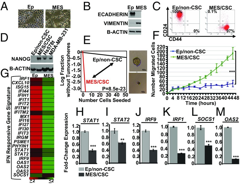
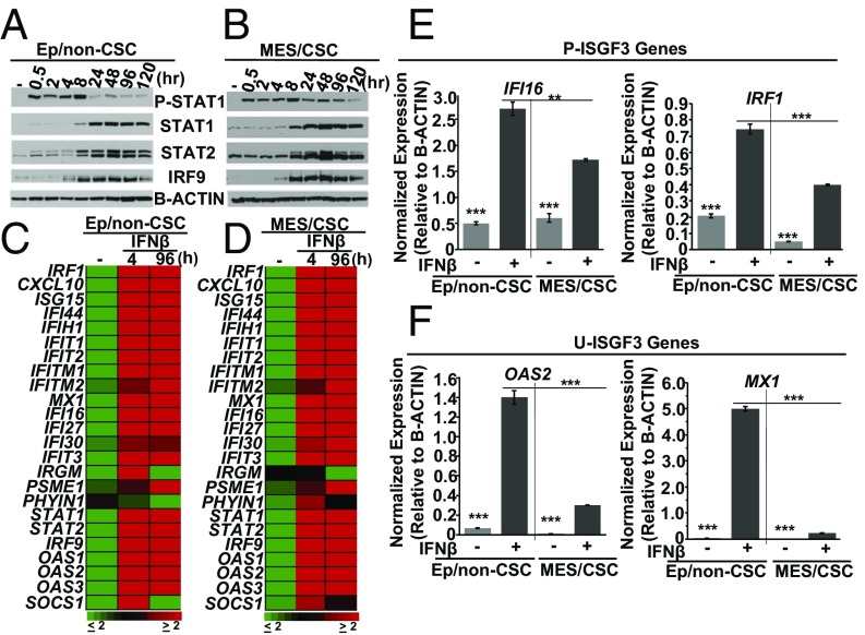
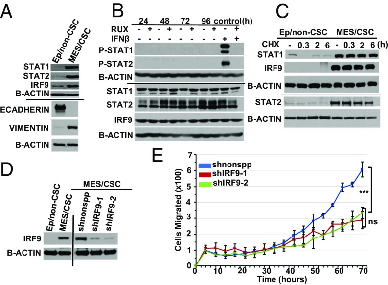
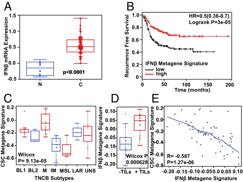
Triple-negative breast cancer (TNBC), the deadliest form of this disease, lacks a targeted therapy. TNBC tumors that fail to respond to chemotherapy are characterized by a repressed IFN/signal transducer and activator of transcription (IFN/STAT) gene signature and are often enriched for cancer stem cells (CSCs). We have found that human mammary epithelial cells that undergo an epithelial-to-mesenchymal transition (EMT) following transformation acquire CSC properties. These mesenchymal/CSCs have a significantly repressed IFN/STAT gene expression signature and an enhanced ability to migrate and form tumor spheres. Treatment with IFN-beta (IFN-β) led to a less aggressive epithelial/non-CSC-like state, with repressed expression of mesenchymal proteins (VIMENTIN, SLUG), reduced migration and tumor sphere formation, and reexpression of CD24 (a surface marker for non-CSCs), concomitant with an epithelium-like morphology. The CSC-like properties were correlated with high levels of unphosphorylated IFN-stimulated gene factor 3 (U-ISGF3), which was previously linked to resistance to DNA damage. Inhibiting the expression of IRF9 (the DNA-binding component of U-ISGF3) reduced the migration of mesenchymal/CSCs. Here we report a positive translational role for IFN-β, as gene expression profiling of patient-derived TNBC tumors demonstrates that an IFN-β metagene signature correlates with improved patient survival, an immune response linked with tumor-infiltrating lymphocytes (TILs), and a repressed CSC metagene signature. Taken together, our findings indicate that repressed IFN signaling in TNBCs with CSC-like properties is due to high levels of U-ISGF3 and that treatment with IFN-β reduces CSC properties, suggesting a therapeutic strategy to treat drug-resistant, highly aggressive TNBC tumors.
Keywords: cancer stem cells; interferon-beta; triple-negative breast cancer; tumor microenvironment.
Copyright © 2017 the Author(s). Published by PNAS.
Conflict of interest statement
The authors declare no conflict of interest.
Figures

Similar articles
-
The Critical, Clinical Role of Interferon-Beta in Regulating Cancer Stem Cell Properties in Triple-Negative Breast Cancer.DNA Cell Biol. 2018 Jun;37(6):513-516. doi: 10.1089/dna.2018.4247. Epub 2018 May 11. DNA Cell Biol. 2018. PMID: 29750542 Free PMC article.
-
The opposing effects of interferon-beta and oncostatin-M as regulators of cancer stem cell plasticity in triple-negative breast cancer.Breast Cancer Res. 2019 Apr 29;21(1):54. doi: 10.1186/s13058-019-1136-x. Breast Cancer Res. 2019. PMID: 31036052 Free PMC article.
-
GSK3β regulates epithelial-mesenchymal transition and cancer stem cell properties in triple-negative breast cancer.Breast Cancer Res. 2019 Mar 7;21(1):37. doi: 10.1186/s13058-019-1125-0. Breast Cancer Res. 2019. PMID: 30845991 Free PMC article.
-
DNMT1: A key drug target in triple-negative breast cancer.Semin Cancer Biol. 2021 Jul;72:198-213. doi: 10.1016/j.semcancer.2020.05.010. Epub 2020 May 24. Semin Cancer Biol. 2021. PMID: 32461152 Review.
-
The hedgehog pathway in triple-negative breast cancer.Cancer Med. 2016 Oct;5(10):2989-3006. doi: 10.1002/cam4.833. Epub 2016 Aug 18. Cancer Med. 2016. PMID: 27539549 Free PMC article. Review.
Cited by
-
The growth inhibitory effect of human gingiva-derived mesenchymal stromal cells expressing interferon-β on tongue squamous cell carcinoma cells and xenograft model.Stem Cell Res Ther. 2019 Jul 29;10(1):224. doi: 10.1186/s13287-019-1320-z. Stem Cell Res Ther. 2019. PMID: 31358054 Free PMC article.
-
DNA Damage-Induced Inflammatory Microenvironment and Adult Stem Cell Response.Front Cell Dev Biol. 2021 Oct 8;9:729136. doi: 10.3389/fcell.2021.729136. eCollection 2021. Front Cell Dev Biol. 2021. PMID: 34692684 Free PMC article. Review.
-
Intrinsic Interferon Signaling Regulates the Cell Death and Mesenchymal Phenotype of Glioblastoma Stem Cells.Cancers (Basel). 2021 Oct 21;13(21):5284. doi: 10.3390/cancers13215284. Cancers (Basel). 2021. PMID: 34771447 Free PMC article.
-
Transcriptional factor six2 promotes the competitive endogenous RNA network between CYP4Z1 and pseudogene CYP4Z2P responsible for maintaining the stemness of breast cancer cells.J Hematol Oncol. 2019 Mar 4;12(1):23. doi: 10.1186/s13045-019-0697-6. J Hematol Oncol. 2019. PMID: 30832689 Free PMC article.
-
Elevated Type I Interferon Signaling Defines the Proliferative Advantage of ARF and p53 Mutant Tumor Cells.Mol Cell Biol. 2025;45(6):246-261. doi: 10.1080/10985549.2025.2497817. Epub 2025 May 12. Mol Cell Biol. 2025. PMID: 40351125
References
-
- Nguyen D-X, Bos P-D, Massagué J. Metastasis: From dissemination to organ-specific colonization. Nat Rev Cancer. 2009;9:274–284. - PubMed
Publication types
MeSH terms
Substances
Grants and funding
LinkOut - more resources
Full Text Sources
Other Literature Sources
Molecular Biology Databases
Research Materials
