. 2017 Dec 11;7(1):17354.
doi: 10.1038/s41598-017-17638-x.

Myosin-X knockout is semi-lethal and demonstrates that myosin-X functions in neural tube closure, pigmentation, hyaloid vasculature regression, and filopodia formation

Affiliations

Myosin-X knockout is semi-lethal and demonstrates that myosin-X functions in neural tube closure, pigmentation, hyaloid vasculature regression, and filopodia formation

Ernest G Heimsath Jr et al. Sci Rep. .

Abstract

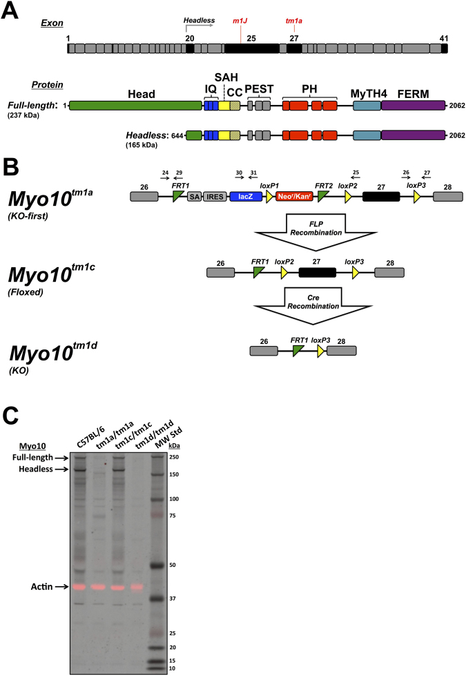
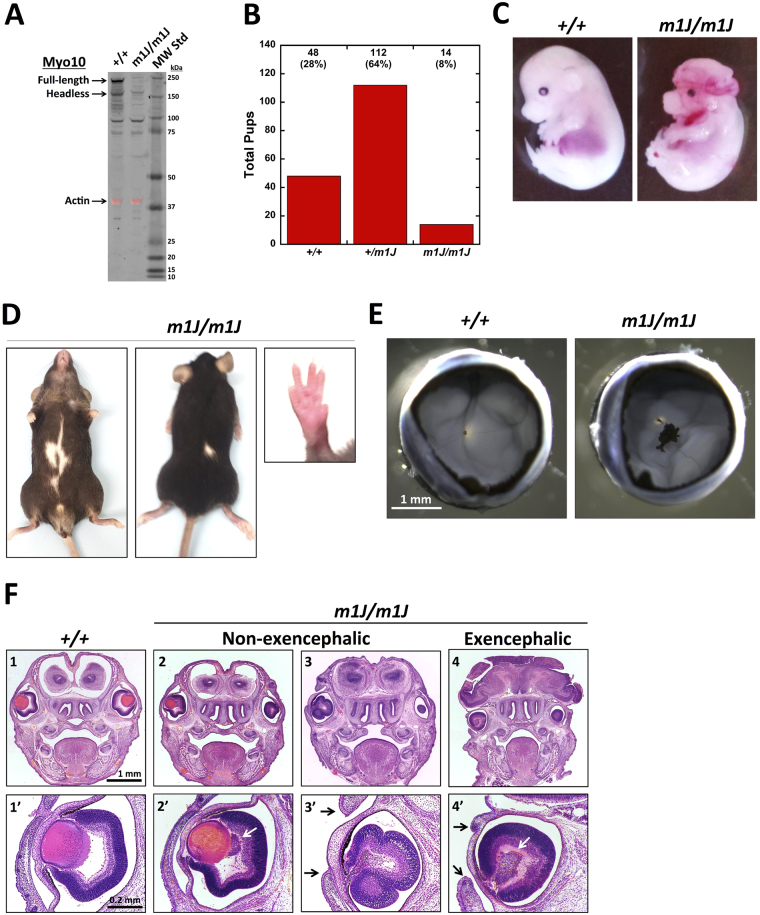
Myosin-X (Myo10) is an unconventional myosin best known for its striking localization to the tips of filopodia. Despite the broad expression of Myo10 in vertebrate tissues, its functions at the organismal level remain largely unknown. We report here the generation of KO-first (Myo10 tm1a/tm1a ), floxed (Myo10 tm1c/tm1c ), and KO mice (Myo10 tm1d/tm1d ). Complete knockout of Myo10 is semi-lethal, with over half of homozygous KO embryos exhibiting exencephaly, a severe defect in neural tube closure. All Myo10 KO mice that survive birth exhibit a white belly spot, all have persistent fetal vasculature in the eye, and ~50% have webbed digits. Myo10 KO mice that survive birth can breed and produce litters of KO embryos, demonstrating that Myo10 is not absolutely essential for mitosis, meiosis, adult survival, or fertility. KO-first mice and an independent spontaneous deletion (Myo10 m1J/m1J ) exhibit the same core phenotypes. During retinal angiogenesis, KO mice exhibit a ~50% decrease in endothelial filopodia, demonstrating that Myo10 is required to form normal numbers of filopodia in vivo. The Myo10 mice generated here demonstrate that Myo10 has important functions in mammalian development and provide key tools for defining the functions of Myo10 in vivo.


Conflict of interest statement

The authors declare that they have no competing interests.

Figures

Figure 1
Figure 1
Generation of Myo10 KO mice. (A) Aligned bar diagrams showing exon boundaries relative to the protein domain structure of full-length and headless Myo10. The gene for full-length Myo10 in Mus musculus is located on chromosome 15 and contains 41 exons (NCBI Gene ID: 17909). Transcripts for headless Myo10 are produced by use of three alternative transcription start sites located in intron 19 followed by splicing to exons 20–41. The Myo10 tm1a mutation targets exon 27 while the spontaneous Myo10 m1J mutation is due to an 8 bp deletion in exon 25. Exons of particular interest are shaded black. The protein domain structure includes a myosin motor domain (Head), IQ motifs (IQ), stable α-helix (SAH), coiled coil (CC), PEST region, Pleckstrin homology (PH) domains, and a MyTH4-FERM domain. (B) Design of the Myo10 tm1a , Myo10 tm1c, and Myo10 tm1d alleles. Myo10 expression in the KO-first allele (Myo10 tm1a) is disrupted due to insertion of a KO first cassette that includes a strong splice acceptor (SA), a stop codon, an internal ribosome entry site (IRES), and LacZ upstream of exon 27. Because the KO-first cassette is flanked by FRT sites, it can be excised by FLP recombinase, restoring Myo10 expression and generating the conditional Myo10 tm1c allele. Because exon 27 is flanked by loxP sites, exposure to Cre recombinase deletes exon 27 and generates the Myo10 tm1d KO allele. Black arrows indicate genotyping primers as described in the Methods. (C) Immunoblot of whole brain lysates from P5 mice. As expected, both full-length and headless Myo10 are detected in the wild-type C57BL/6 and neither is detected in the KO-first Myo10 tm1a/tm1a. Expression of full-length and headless is restored in Myo10 tm1c/tm1c, and neither is detected in the Myo10 tm1d/tm1d KO. Note that at these exposure settings numerous faintly stained bands below the molecular weights of full-length and headless Myo10 are visible in the C57BL/6 sample and that most of these putative breakdown products are absent in the KO-first Myo10 tm1a/tm1a and Myo10 tm1d/tm1d KO samples. Some non-specifically stained bands are also visible, such as the faint band just below 37 kDa. The signal from the same blot stained with anti-actin (43 kDa) is shown in the red channel as a loading control and has been overlaid onto the black and white Myo10 signal.
Figure 2
Figure 2
Myo10 tm1d/tm1d KO mice have a white belly spot and persistent hyaloid vasculature plus a high frequency of exencephaly, webbed digits, and other abnormalities. (A) Total number of pups obtained for each genotype from heterozygous matings showing that homozygous nulls are obtained at less than half the expected number (n = 42 litters, 260 pups total, mean litter size 6.1). (B) E14.5 embryos showing a normal Myo10 +/+ control and an exencephalic Myo10 tm1d/tm1d littermate. Note that the Myo10 tm1d/tm1d embryo also exhibits microphthalmia. (C) Adult mice showing an example of the white belly spot observed in all Myo10 tm1d/tm1d mice (white arrow). (D) Left forepaws from adult mice showing an example of the webbed digits observed in ~50% of Myo10 tm1d/tm1d mice. (E) Eyecups from 6-week-old mice showing a control eye from a wild-type mouse and an example of the pigmented mass present in Myo10 tm1d/tm1d mice (black arrow). For each eye, the cornea, iris and lens were dissected away and then four incisions were made in each eyecup (dotted lines) to allow the lens to be carefully removed and the eyecup to be partially flattened. (F) Tails from adult mice showing an example of a kinked tail in a Myo10 tm1d/tm1d mouse. Kinked tails were present in most Myo10 tm1d/tm1d mice, but were often much more subtle.
Figure 3
Figure 3
Eyes from Myo10 tm1d/tm1d KO mice have persistent hyaloid vasculature. (A) Eyecups from P5 mice showing a control eye from a wild-type mouse and an example of the pigmented persistent hyaloid vasculature present in a Myo10 tm1d/tm1d mouse. The white spot in the center of each eye is the optic disk. An irregular black mass is located above and to the right of the optic disc in the Myo10 tm1d/tm1d eye. (B) The same eyecups in (A) were fluorescently stained with a PECAM-1 antibody (green) to label endothelial cells. In the control at left, several long and relatively straight branches (dotted white arrow) of the hyaloid vasculature can be seen extending from their origin near the optic disk towards the rim of the eyecup. Consistent with their identification as hyaloid vasculature, they are located within the vitreous and slightly above the developing retinal vasculature, which forms reticulum on the surface of the retina (solid white arrow). In the Myo10 tm1d/tm1d eye, a relatively thick hyaloid artery extends from the optic disk to the pigmented mass, while long and relatively straight branches of the hylaloid vasculature extend outward from arms of the pigmented mass. Because the eyecups curve upwards at their outer edges, the view there is foreshortened. The hyaloid and retinal vasculatures can be best distinguished by enlarging the images on a digital display. (C) Schematic cross-section of a normal P5 eye. At this stage the hyaloid vasculature has begun to regress, but still perfuses the vitreous and forms a plexus on the posterior of the lens, while the retinal vasculature is undergoing angiogenesis and extending on the surface of the retina. Illustration created by EGH. (D) Close-up view of the pigmented mass in (A) and (B) showing that it includes a dense plexus of hyaloid vasculature intermixed with pigment cells. The plexus can be seen most clearly after enlargement on a digital display.
Figure 4
Figure 4
Loss of Myo10 decreases the number of filopodia during retinal angiogenesis. (A) Fluorescence image of flat mounted retinas showing retinal vasculature at P5. Dissected retinas were stained with a PECAM-1 antibody (green) and counterstained with phalloidin (red). The retinal vasculature extended to similar positions in control and Myo10 tm1d/tm1d eyes. The image represents a stitch of micrographs taken at 20X and is best visualized if the images are enlarged on a digital display. (B) High resolution images of the angiogenic expansion front from the P5 retinas in (A) showing filopodia radiating from endothelial tips cells. Loss of Myo10 results in a decreased number of filopodia and leads to a less dense vascular network. Images were captured as Z-stacks at 60X and displayed as maximum projections with the PECAM-1 channel displayed in inverted grayscale to highlight endothelial filopodia. (C) Quantification showing that loss of Myo10 decreases the branch point density/mm2 by 32%. (D) Quantification showing that loss of Myo10 decreased the number of filopodia per mm of vascular front by ~50%. N = 3 mice for both (C) and (D) and error bars denote standard deviation.
Figure 5
Figure 5
Myo10 m1J /m1J mice have a white belly spot and persistent hyaloid vasculature plus a high frequency of exencephaly and other abnormalities. (A) Immunoblot of whole brain lysates from 1 month old mice showing deletion of full-length and headless Myo10. Although bands corresponding to full-length and headless Myo10 are absent in the Myo10 m1J /m1J lysate, it should be noted that the Myo10 antibody shows some non-specific staining, including one band that is slightly larger than headless and another that is near the 100 kDa standard. The signal from the same blot stained with anti-actin (43 kDa) is shown in the red channel as a loading control and has been overlaid onto the black and white Myo10 signal. (B) Total number of pups obtained for each genotype from heterozygous matings showing that Myo10 m1J /m1J homozygotes were obtained at less than half the expected number (n = 28 litters, 174 pups total; mean litter size 6.2). (C) E15.5 embryos showing a Myo10 +/+ control and an exencephalic Myo10 m1J /m1J littermate. (D) Adult Myo10 m1J/m1J mouse showing examples of a white belly spot, a white spot on the back, and syndactyly. (E) Eyecups from 6-week old mice showing a normal eye from a Myo10 +/+ mouse and an example of the pigmented mass of persistent hyaloid vasculature present in the eyes of Myo10 m1J/m1J mice. (F) Rostral coronal sections from E16.5 embryos showing a wild-type, two nulls that did not have exencephaly, and one null that did have exencephaly. Note the relatively normal appearance of the brain and cranium in the null embryos without exencephaly and the lack of a cranium and gross defects in brain morphology in the embryo with exencephaly. The null embryo on the left was selected for sectioning in part because it had normal sized eyes while the two embryos on right had microphthalmia. Note that slight differences in the plane of section between the left and right side of the skull make some of the eyes on the right side appear smaller than they actually are. Higher magnification images of sections through the middle of the eyes showing the abnormal accumulations of cells (white arrows) behind the lens and defects in lens development in the two eyes on the right (at this stage the lens should be visible as red stained sphere). Note also that the null eyes in 3′ and 4′ failed to undergo eyelid closure (black arrows), a process that normally occurs between E15.5 and E16.5.

References

    1. Berg JS, Derfler BH, Pennisi CM, Corey DP, Cheney RE. Myosin-X, a novel myosin with pleckstrin homology domains, associates with regions of dynamic actin. J Cell Sci. 2000;113:3439–3451. - PubMed
    1. Ropars V, et al. The myosin X motor is optimized for movement on actin bundles. Nature communications. 2016;7:12456. doi: 10.1038/ncomms12456. - DOI - PMC - PubMed
    1. Kerber ML, Cheney RE. Myosin-X: a MyTH-FERM myosin at the tips of filopodia. J Cell Sci. 2011;124:3733–3741. doi: 10.1242/jcs.023549. - DOI - PMC - PubMed
    1. Mercer JA, Seperack PK, Strobel MC, Copeland NG, Jenkins NA. Novel myosin heavy chain encoded by murine dilute coat colour locus [publishederratum appears in Nature 1991 Aug 8; 352(6335):547] Nature. 1991;349:709–713. doi: 10.1038/349709a0. - DOI - PubMed
    1. Avraham KB, et al. The mouse Snell’s waltzer deafness gene encodes an unconventional myosin required for structural integrity of inner ear hair cells. Nat Genet. 1995;11:369–375. doi: 10.1038/ng1295-369. - DOI - PubMed

Publication types

LinkOut - more resources