Circulating CCL20 as a New Biomarker of Abdominal Aortic Aneurysm
- PMID: 29229985
- PMCID: PMC5725593
- DOI: 10.1038/s41598-017-17594-6
Circulating CCL20 as a New Biomarker of Abdominal Aortic Aneurysm
Abstract
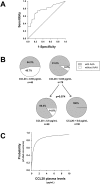
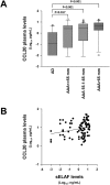
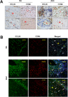
Autoimmunity appears to play a role in abdominal aortic aneurysm (AAA) pathology. Although the chemokine CCL20 has been involved in autoimmune diseases, its relationship with the pathogenesis of AAA is unclear. We investigated CCL20 expression in AAA and evaluated it as a potential biomarker for AAA. CCL20 was measured in plasma of AAA patients (n = 96), atherosclerotic disease (AD) patients (n = 28) and controls (n = 45). AAA presence was associated with higher plasma levels of CCL20 after adjustments for confounders in the linear regression analysis. Diagnostic performance of plasma CCL20 was assessed by ROC curve analysis, AUC 0.768 (CI:0.678-0.858; p<0.001). Classification and regression tree analysis classified patients into two CCL20 plasma level groups. The high-CCL20 group had a higher number of AAA than the low-CCL20 group (91% vs 54.3%, p< 0.001). mRNA of CCL20 and its receptor CCR6 were higher in AAA (n = 89) than in control aortas (n = 17, p<0.001). A positive correlation was found between both mRNA in controls (R = 0674; p = 0.003), but not in AAA. Immunohistochemistry showed that CCR6 and CCL20 colocalized in the media and endothelial cells. Infiltrating leukocytes immunostained for both proteins but only colocalized in some of them. Our data shows that CCL20 is increased in AAA and circulating CCL20 is a high sensitive biomarker of AAA.
Conflict of interest statement
The authors declare that they have no competing interests.
Figures


Similar articles
-
Plasma profiling by a protein array approach identifies IGFBP-1 as a novel biomarker of abdominal aortic aneurysm.Atherosclerosis. 2012 Apr;221(2):544-50. doi: 10.1016/j.atherosclerosis.2012.01.009. Epub 2012 Jan 25. Atherosclerosis. 2012. PMID: 22325929
-
Serum matrix metalloproteinase-9 is a valuable biomarker for identification of abdominal and thoracic aortic aneurysm: a case-control study.BMC Cardiovasc Disord. 2018 Oct 29;18(1):202. doi: 10.1186/s12872-018-0931-0. BMC Cardiovasc Disord. 2018. PMID: 30373522 Free PMC article.
-
Plasma levels of chemokine ligand 20 and chemokine receptor 6 in patients with sepsis: A case control study.Eur J Anaesthesiol. 2016 May;33(5):348-55. doi: 10.1097/EJA.0000000000000388. Eur J Anaesthesiol. 2016. PMID: 26771764
-
Lipoprotein(a) Levels in Patients With Abdominal Aortic Aneurysm.Angiology. 2017 Feb;68(2):99-108. doi: 10.1177/0003319716637792. Epub 2016 Sep 29. Angiology. 2017. PMID: 26980774
-
Surrogate Markers of Abdominal Aortic Aneurysm Progression.Arterioscler Thromb Vasc Biol. 2016 Feb;36(2):236-44. doi: 10.1161/ATVBAHA.115.306538. Epub 2015 Dec 29. Arterioscler Thromb Vasc Biol. 2016. PMID: 26715680 Review.
Cited by
-
Novel plasma protein biomarkers from critically ill sepsis patients.Clin Proteomics. 2022 Dec 27;19(1):50. doi: 10.1186/s12014-022-09389-3. Clin Proteomics. 2022. PMID: 36572854 Free PMC article.
-
Identifying novel mechanisms of abdominal aortic aneurysm via unbiased proteomics and systems biology.Front Cardiovasc Med. 2022 Aug 3;9:889994. doi: 10.3389/fcvm.2022.889994. eCollection 2022. Front Cardiovasc Med. 2022. PMID: 35990960 Free PMC article.
-
An iron oxide nanoworm hybrid on an interdigitated microelectrode silica surface to detect abdominal aortic aneurysms.Mikrochim Acta. 2021 May 11;188(6):185. doi: 10.1007/s00604-021-04836-8. Mikrochim Acta. 2021. PMID: 33977395
-
Discovery of novel biomarkers for atherosclerotic aortic aneurysm through proteomics-based assessment of disease progression.Sci Rep. 2020 Apr 14;10(1):6429. doi: 10.1038/s41598-020-63229-8. Sci Rep. 2020. PMID: 32286426 Free PMC article.
-
Current-Volt Biosensing "Cystatin C" on Carbon Nanowired Interdigitated Electrode Surface: A Clinical Marker Analysis for Bulged Aorta.J Anal Methods Chem. 2022 May 23;2022:8160502. doi: 10.1155/2022/8160502. eCollection 2022. J Anal Methods Chem. 2022. PMID: 35655788 Free PMC article.
References
-
- Johnston KW, et al. Suggested standards for reporting on arterial aneurysms. Subcommittee on Reporting Standards for Arterial Aneurysms, Ad Hoc Committee on Reporting Standards, Society for Vascular Surgery and North American Chapter, International Society for Cardiovascular Surgery. J Vasc Surg. 1991;13:452. doi: 10.1067/mva.1991.26737. - DOI - PubMed
Publication types
MeSH terms
Substances
LinkOut - more resources
Full Text Sources
Other Literature Sources
Medical

