. 2018 Jan;13(1):82-89.
doi: 10.1038/s41565-017-0012-z. Epub 2017 Dec 11.

Nanoparticle orientation to control RNA loading and ligand display on extracellular vesicles for cancer regression

Affiliations

Nanoparticle orientation to control RNA loading and ligand display on extracellular vesicles for cancer regression

Fengmei Pi et al. Nat Nanotechnol. 2018 Jan.

Abstract

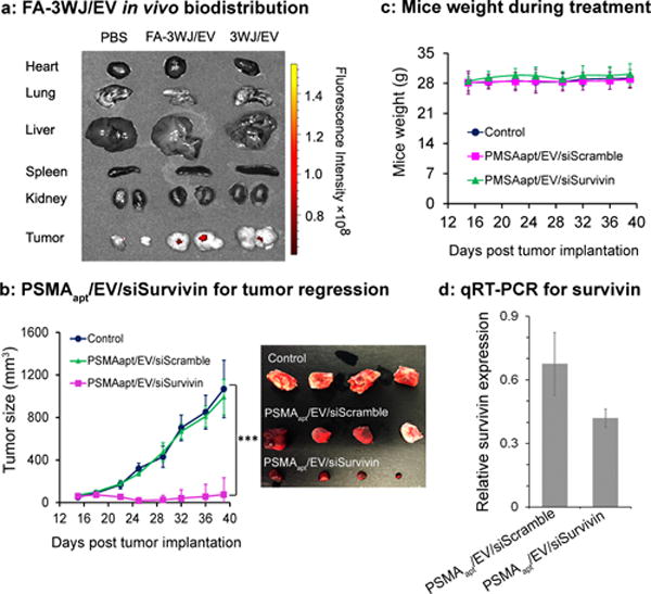
Nanotechnology offers many benefits, and here we report an advantage of applying RNA nanotechnology for directional control. The orientation of arrow-shaped RNA was altered to control ligand display on extracellular vesicle membranes for specific cell targeting, or to regulate intracellular trafficking of small interfering RNA (siRNA) or microRNA (miRNA). Placing membrane-anchoring cholesterol at the tail of the arrow results in display of RNA aptamer or folate on the outer surface of the extracellular vesicle. In contrast, placing the cholesterol at the arrowhead results in partial loading of RNA nanoparticles into the extracellular vesicles. Taking advantage of the RNA ligand for specific targeting and extracellular vesicles for efficient membrane fusion, the resulting ligand-displaying extracellular vesicles were capable of specific delivery of siRNA to cells, and efficiently blocked tumour growth in three cancer models. Extracellular vesicles displaying an aptamer that binds to prostate-specific membrane antigen, and loaded with survivin siRNA, inhibited prostate cancer xenograft. The same extracellular vesicle instead displaying epidermal growth-factor receptor aptamer inhibited orthotopic breast cancer models. Likewise, survivin siRNA-loaded and folate-displaying extracellular vesicles inhibited patient-derived colorectal cancer xenograft.


Conflict of interest statement

CONFLICT OF INTEREST

P. Guo’s Sylvan G. Frank Endowed Chair position in Pharmaceutics and Drug Delivery is funded by the CM Chen Foundation, and he is a consultant of Oxford Nanopore, Nanobio Delivery Pharmaceutical Co., Ltd, and the cofounder of P&Z Biological Technology LLC. Pi now works for Nanobio Delivery Pharmaceutical Co., Ltd, S. Wang and F. Haque now work for P&Z Biological Technology LLC.

Figures

Figure 1
Figure 1. RNA nanotechnology for decorating native EVs
(a) AFM image of extended 3WJ of the motor pRNA of bacteriophage phi29. (b) Illustration of the location for cholesterol labeling of the arrow-head or arrow-tail of 3WJ. (c) Negative-stained EM image of EVs from HEK293T cells purified with differential ultracentrifugation method and cushion modified ultracentrifugation method. (d–g) NTA for size analysis and DLS for Zeta potential measurements. (h) 2D structure (left panel) and native PAGE for testing 3WJ assembly from three component strands, as indicated. (i). EVs loading and RNA aptamer display.
Figure 2
Figure 2. Comparison of the role between arrow-head and arrow-tail 3WJ
(a–b) Illustration showing the difference between arrow-head and arrow-tail display. (c) Syner gel to test arrow-head and arrow-tail Alexa647-3WJ/EV degradation by RNase in FBS. The gel was imaged at Alexa647 channel (d) and the bands were quantified by Image J. (e–i) Assay to compare cell binding of folate-3WJ arrow-tail (e–g) and arrow-head (h–i) on folate receptor positive and negative cells.
Figure 3
Figure 3. Specific binding and siRNA delivery to cells in vitro using PSMA aptamer-displaying EVs
(a) Flow cytometry (left) and confocal images (right) showing the binding of PSMA RNA aptamer-displaying EVs to PSMA-receptor positive and negative cells. Nucleus (Blue), cytoskeleton (Green), and RNA (Red) in confocal images. (b) RT-PCR assay for PSMA aptamer-mediated delivery of survivin siRNA by EVs to PSMA(+) prostate cancer cells. Statistics: n=4; experiment was run in four biological replicates and two to four technical repeats with an ANOVA analysis; holm adjusted p = 0.0120, 0.0067 comparing PSMAapt/EV/siSurvivin to PSMAapt/EV/siScramble and 3WJ/EV/siSurvivin, respectively. (c) MTT assay showing reduced cellular proliferation. n=3, p = 0.003, 0.031 comparing PSMAapt/EV/siSurvivin to PSMAapt/EV/siScramble and 3WJ/EV/siSurvivin respectively. *p<0.05, **p<0.01.
Figure 4
Figure 4. Animal trials using ligands displaying EV for tumor inhibition
(a) Organ images showing specific tumor targeting 8 hrs after systemic injection of folate displaying EVs to mice with subcutaneous KB cell xenografts. n = 2, two independent experiments. (b) Intravenous treatment of nude mice bearing LNCaP-LN3 subcutaneous xenografts with PSMAapt/EV/siSurvivin or PSMAapt/EV/siScramble (both with 0.6 mg/kg, siRNA/mice body weight), and PBS, injected twice per week for three weeks. n=10 biological replicates, 2 independent experiments, and statistics were calculated using a two-sided t-test expressed as averages and with standard deviation. p = 0.347, 0.6–2, 1.5e–6, 8.2e–8, 2.1e–7, 1.0e–7, 1.9e–7, 1.8e–6 for days 15, 18, 22, 25, 29, 32, 36, and 39 respectively for PSMAapt/EV/siSurvivin compared to control. (c) Body weight of mice during the time course of EVs treatment. (d) RT-PCR showing the trend of knockdown survivin mRNA expression in prostate tumors after EV treatment.
Figure 5
Figure 5. EGFR aptamer displaying EVs can deliver survivin siRNA to breast cancer orthotopic xenograft mouse model
(a) EGFR aptamer displaying EVs showed enhanced targeting effect to breast tumor in orthotopic xenograft mice models. (b) Intravenous treatment of nude mice bearing breast cancer orthotopic xenografts with EGFRapt/EV/siSurvivin and controls (n=5). After 6 weeks, EGFRapt/EV/siSurvivin treated group had significantly smaller tumor size than other controls. p = 0.008 comparing EGFRapt/EV/siSurvivin to EGFRapt/EV/siScramble. (c) Analysis of the protein expression in tumor extracts showed that EGFRapt/EV/siSurvivin treatment significantly reduced the expression of Survivin. p=0.0004 comparing EGFRapt/EV/siSurvivin to EGFRapt/EV/siScramble. (d) Quantitative real-time PCR on extracted RNA from tumors showed the reduction of Survivin mRNA in the EGFRapt/EV/siSurvivin treated mice compared to controls. p=0.024 comparing EGFRapt/EV/siSurvivin to EGFRapt/EV/siScramble. Error bars indicate s.e.m. * p < 0.05, ** p < 0.01, *** p<0.001.
Figure 6
Figure 6. Folate displaying EVs can deliver survivin siRNA to patient derived colorectal cancer xenograft (PDX-CRC) mouse model
(a) Intravenous treatment of nude mice bearing PDX-CRC xenografts with FA/EV/siSurvivin and controls (n=4). After 6 weeks, FA/EV/siSurvivin treated group had significantly smaller tumor size, p = 0.0098 and 0.0387 comparing FA/EV/siSurvivin to FA/EV/siScramble at week 4 and week 5 respectively. (b) Lower tumor weight than controls. p = 0.0024 comparing FA/EV/siSurvivin to FA/EV/siScramble. Error bars indicate s.e.m. * p < 0.05, ** p < 0.01.

References

    1. Shu D, Shu Y, Haque F, Abdelmawla S, Guo P. Thermodynamically stable RNA three-way junctions for constructing multifuntional nanoparticles for delivery of therapeutics. Nature Nanotechnology. 2011;6:658–667. - PMC - PubMed
    1. Zhang H, et al. Crystal Structure of 3WJ Core Revealing Divalent Ion-promoted Thermostability and Assembly of the Phi29 Hexameric Motor pRNA. RNA. 2013;19:1226–1237. - PMC - PubMed
    1. Guo P, Erickson S, Anderson D. A small viral RNA is required for in vitro packaging of bacteriophage phi29 DNA. Science. 1987;236:690–694. - PubMed
    1. Guo P, Zhang C, Chen C, Trottier M, Garver K. Inter-RNA interaction of phage phi29 pRNA to form a hexameric complex for viral DNA transportation. Mol Cell. 1998;2:149–155. - PubMed
    1. Lamichhane TN, Raiker RS, Jay SM. Exogenous DNA Loading into Extracellular Vesicles via Electroporation is Size-Dependent and Enables Limited Gene Delivery. Mol Pharm. 2015;12:3650–3657. - PMC - PubMed

Publication types

MeSH terms