. 2017 Dec 11;7(1):17288.
doi: 10.1038/s41598-017-17304-2.

Three-dimensional (3D) culture of adult murine colon as an in vitro model of cryptosporidiosis: Proof of concept

Affiliations

Three-dimensional (3D) culture of adult murine colon as an in vitro model of cryptosporidiosis: Proof of concept

Martha Baydoun et al. Sci Rep. .

Abstract

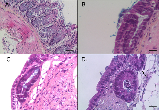
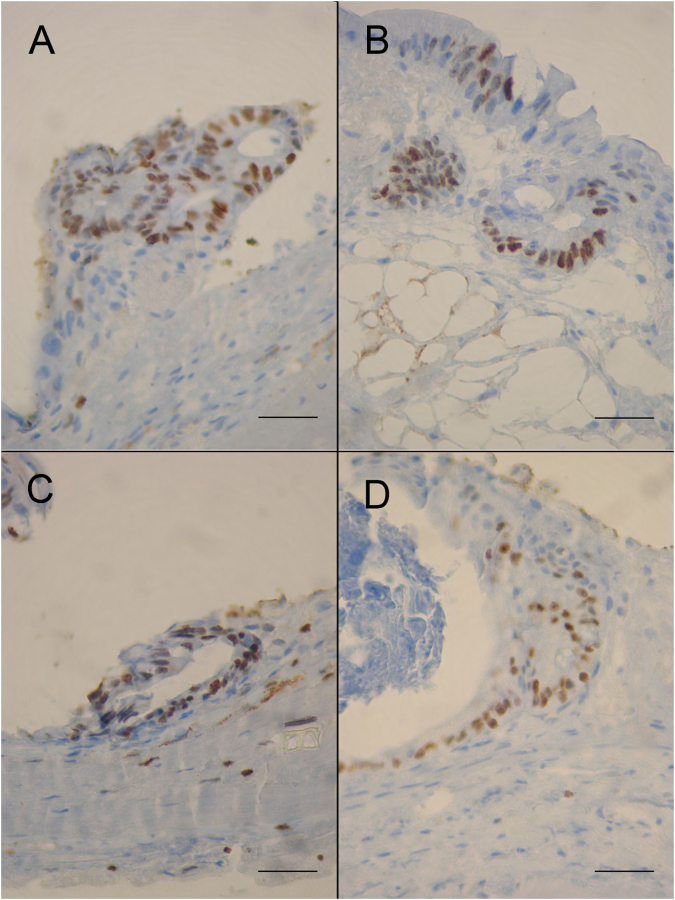
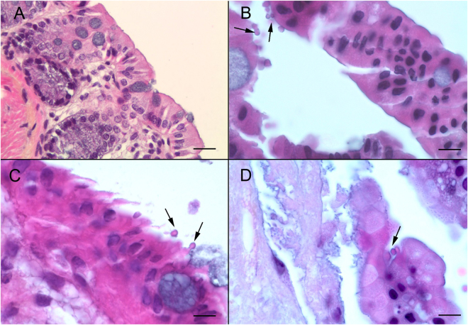
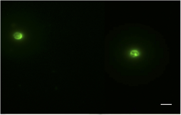
Cryptosporidium parvum is a major cause of diarrheal illness and was recently potentially associated with digestive carcinogenesis. Despite its impact on human health, Cryptosporidium pathogenesis remains poorly known, mainly due to the lack of a long-term culture method for this parasite. Thus, the aim of the present study was to develop a three-dimensional (3D) culture model from adult murine colon allowing biological investigations of the host-parasite interactions in an in vivo-like environment and, in particular, the development of parasite-induced neoplasia. Colonic explants were cultured and preserved ex vivo for 35 days and co-culturing was performed with C. parvum. Strikingly, the resulting system allowed the reproduction of neoplastic lesions in vitro at 27 days post-infection (PI), providing new evidence of the role of the parasite in the induction of carcinogenesis. This promising model could facilitate the study of host-pathogen interactions and the investigation of the process involved in Cryptosporidium-induced cell transformation.


Conflict of interest statement

The authors declare that they have no competing interests.

Figures

Figure 1
Figure 1
Histological validation of the murine colonic explant culture system (A) Hematoxylin eosin safranin (HES) staining of a normal colon section of an uninfected SCID mouse. Scale bar, 40 µm. (B) HES staining of a colonic explant section after 14 days of culture showing good tissue preservation with a characteristic collagen fiber and crypt-like structures. Scale bar, 20 µm. (C,D) HES staining of a colonic explant section after 21 and 30 days of culture, respectively. Tissue preservation was confirmed with a presence of high prismatic epithelium, basal position of nucleus, a well-organized basal lamina and the presence of crypt-like structures (black arrow (D)). Presence of large cyst-like formations (white arrow (C)). Scale bars, 20 µm.
Figure 2
Figure 2
Expression of the Ki-67 marker of proliferation in the epithelium of murine colonic explants (A–D) Ki-67 expression is maintained within the epithelial cell layer throughout the culture period in explant sections of mouse colon after 8 (A), 12 (B), 14 (C) and 30 (D) days of culture respectively. Ki67 labeling seems to gradually diminish with increasing culture time. Scale bars, 25 µm.
Figure 3
Figure 3
C. parvum infection of the murine colonic explant system (A) Hematoxylin eosin safranin (HES) staining of an uninfected SCID mouse colon section. Scale bar, 15 µm (B) HES staining of a colonic explant section after 12 days of culture followed of 4 days after infection with C. parvum. Developmental stages of C. parvum are observed in the apical position (arrows) within the intestinal epithelial cells. Scale bar, 10 µm (C) HES staining of cultured colonic explant sections after 14 days post-infection with C. parvum showing extracellular and intracellular stages of the parasite (arrows). (D) HES staining of cultured colonic explant sections at 22 days post-infection with C. parvum showing parasites within the intestinal epithelial cells. Scale bar, 25 µm.
Figure 4
Figure 4
Presence of Cryptosporidium parvum oocysts in the medium culture pooled after 27 days PI and stained by immunofluorescence (fluorescein isothiocyanate (FITC)-conjugated anti-Cryptosporidium). Scale bar, 5 µm.
Figure 5
Figure 5
Quantification of Cryptosporidium 18s rDNA gene copies by qPCR in the supernatant of explants. (A) Standard curves corresponding to the amplification plots and (B) Comparison between explants infected with 25 oocysts (1) and 250 oocysts (2), respectively. Sample t-test was performed to compare values according to parasite concentrations at day 0 (D0) and 22 days PI (22D). Data is presented as mean number of copies ± SEM of three individual samples. *Indicates significant differences between the mean number of copies on the day of infection and the mean number of copies 22 days PI for tissues infected with 100 sporozoites (25 oocysts) and between explants infected with 100 sporozoites (25 oocysts) and 1000 sporozoites (250 oocysts).
Figure 6
Figure 6
Development of neoplasia in the murine colonic explant model. (A) Hematoxylin eosin safranin (HES) staining of an uninfected colonic explant section showing a normal epithelial structure. Scale bar, 65 µm. (B) HES staining showing a low-grade intraepithelial lesion in a colonic explant after 27 days of infection with C. parvum (grey arrow) characterized by: (i) loss of cell-to-cell contact (ii) reduction of the interglandular space (white arrow) (iii) loss of nuclear polarity with slight pseudostratification (black arrow). Scale bar, 25 µm.

References

    1. Ryan U, Fayer R, Xiao L. Cryptosporidium species in humans and animals: current understanding and research needs. Parasitology. 2014;141:1667–1685. doi: 10.1017/S0031182014001085. - DOI - PubMed
    1. Squire SA, Ryan U. Cryptosporidium and Giardia in Africa: current and future challenges. Parasit. Vectors. 2017;10:195. doi: 10.1186/s13071-017-2111-y. - DOI - PMC - PubMed
    1. Ryan U, Hijjawi N. New developments in Cryptosporidium research. Int. J. Parasitol. 2015;45:367–373. doi: 10.1016/j.ijpara.2015.01.009. - DOI - PubMed
    1. Checkley W, et al. A review of the global burden, novel diagnostics, therapeutics, and vaccine targets for cryptosporidium. Lancet Infect. Dis. 2015;15:85–94. doi: 10.1016/S1473-3099(14)70772-8. - DOI - PMC - PubMed
    1. Morada M, et al. Continuous culture of Cryptosporidium parvum using hollow fiber technology. Int. J. Parasitol. 2016;46:21–29. doi: 10.1016/j.ijpara.2015.07.006. - DOI - PubMed

Publication types