. 2017 Dec 11;7(1):17355.
doi: 10.1038/s41598-017-17729-9.

The Toll-Like Receptor 2/6 Agonist, FSL-1 Lipopeptide, Therapeutically Mitigates Acute Radiation Syndrome

Affiliations

The Toll-Like Receptor 2/6 Agonist, FSL-1 Lipopeptide, Therapeutically Mitigates Acute Radiation Syndrome

Cathryn J Kurkjian et al. Sci Rep. .

Abstract

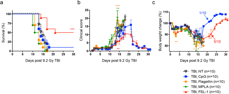
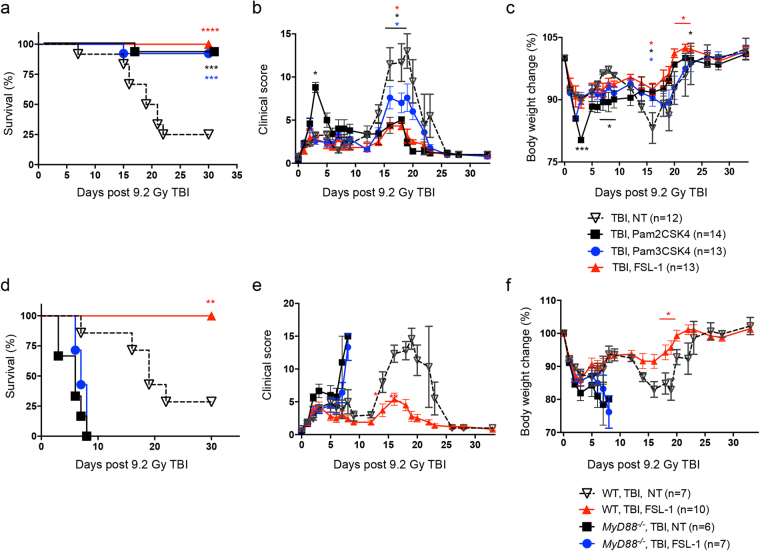
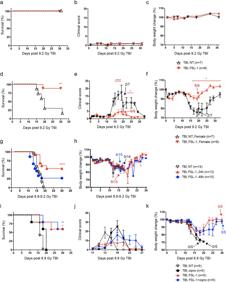
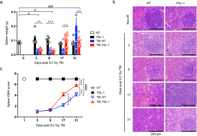
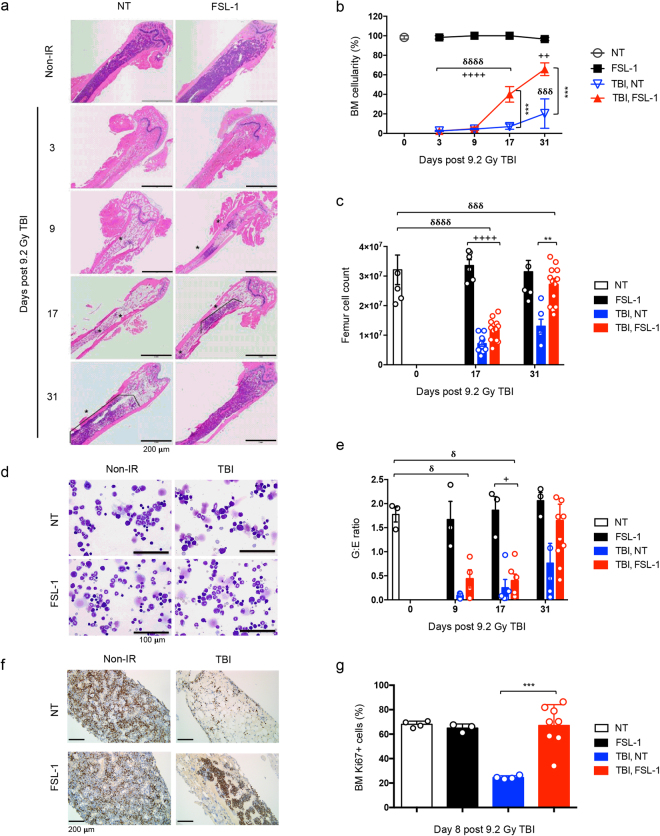
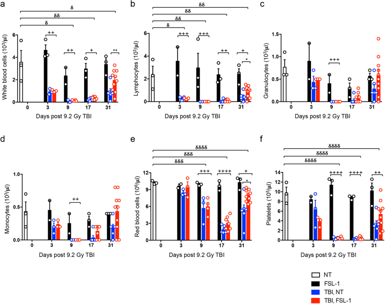
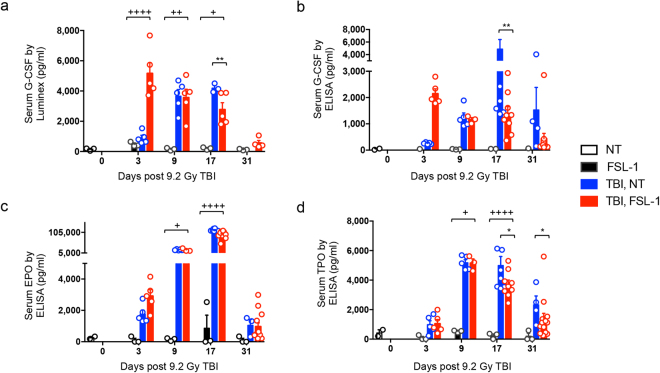
Risks of radiation exposure from nuclear incidents and cancer radiotherapy are undeniable realities. These dangers urgently compel the development of agents for ameliorating radiation-induced injuries. Biologic pathways mediated by myeloid differentiation primary response gene 88 (MyD88), the common adaptor for toll-like receptor (TLR) and Interleukin-1 receptor signaling, are critical for radioprotection. Treating with agonists prior to radiation enhances survival by activating TLR signaling, whereas radiomitigating TLR-activating therapeutics given after exposure are less defined. We examine the radiomitigation capability of TLR agonists and identify one that is superior for its efficacy and reduced toxic consequences compared to other tested agonists. We demonstrate that the synthetic TLR2/6 ligand Fibroblast-stimulating lipopeptide (FSL-1) substantially prolongs survival in both male and female mice when administered 24 hours after radiation and shows MyD88-dependent function. FSL-1 treatment results in accelerated hematopoiesis in bone marrow, spleen and periphery, and augments systemic levels of hematopoiesis-stimulating factors. The ability of FSL-1 to stimulate hematopoiesis is critical, as hematopoietic dysfunction results from a range of ionizing radiation doses. The efficacy of a single FSL-1 dose for alleviating radiation injury while protecting against adverse effects reveals a viable radiation countermeasures agent.


Conflict of interest statement

The authors declare that they have no competing interests.

Figures

Figure 1
Figure 1
Treatment with TLR ligands impacts survival of irradiated mice. Male C57BL/6 mice were irradiated (9.2 Gy TBI) and 24 hours later, received a single i.p. injection of physiological water (no treatment, NT), 2.5 mg/kg CpG–ODN2395 (CpG), 0.2 mg/kg Flagellin (Fla), 0.25 mg/kg MPLA, or 0.25 mg/kg FSL–1 (n = 10 per group). (a) Survival over 30 days post TBI, (b) clinical score and (c) percent body weight change of each treatment group was compared to irradiated, non–treated mice (TBI, NT). Data are representative of 2 independent experiments and are depicted as mean ± s.e.m. *P < 0.05, **P < 0.01 or ***P < 0.001 between treated and TBI, NT mice, with colors correlating to treatment group. Arrows indicate time post TBI in which remaining mice in group began to recover.
Figure 2
Figure 2
Radiomitigation is TLR2 and MyD88 specific. Male C57BL/6 mice were administered physiological water (NT), 0.25 mg/kg FSL–1, 0.25 mg/kg Pam2CSK4 or 0.25 mg/kg Pam3CSK4 at 24 hours after 9.2 Gy TBI exposure. (a) Survival was recorded through 30 days post TBI. Data is cumulative of 2 independent experiments with n = 4–6 mice per group per experiment. (b) Clinical score and (c) percent body weight change were assessed in comparison to irradiated, non–treated controls (TBI, NT). C57BL/6 or MyD88 −/− male and female mice were administered physiological water (NT) or 0.25 mg/kg FSL–1 at 24 hours post 9.2 Gy TBI. (d) Survival, (e) clinical score, and (f) percent body weight change were assessed for 30 days post TBI. Data is representative of 2 experiments with n = 6 to 10 as indicated in legend. Data are shown as mean ± s.e.m. *P < 0.05, **P < 0.01, ***P < 0.001 or ****P < 0.0001 between treated and TBI, NT mice, with colors correlating to treatment group.
Figure 3
Figure 3
FSL–1, a TLR2/6 ligand, demonstrates radiomitigation properties. Toxicity was tested in male C57BL/6 mice, comparing physiologic water (NT) to 0.25 mg/kg FSL–1 treatment. (a) Survival, (b) clinical score, and (c) percent body weight change of mice were monitored over 30 days. Sex independency was tested by treating female C57BL/6 mice exposed to 9.2 Gy TBI with physiological water (NT) or 0.25 mg/kg FSL–1 given 24 hours post TBI. (d) Survival, (e) clinical score and (f) percent body weight change of female mice were assessed for 30 days. The effectiveness of FSL–1 treatment was compared by delivering physiological water (NT) or 0.25 mg/kg FSL–1 at 24 or 48 hours after 8.8 or 9.2 Gy TBI. (g) Survival and (h) percent body weight change were assessed for 30 days. Efficacy of FSL–1 combined with antibiotics treatment was evaluated by delivering physiological water (NT) or 0.25 mg/kg FSL–1 at 24 hours after 8.8 Gy TBI. On days 4 to 30 post TBI, ciprofloxacin was provided in autoclaved acidified water ad libitum from sipper tubes and also in wetted feed. Controls received no ciprofloxacin support, but did receive acidified water and moistened regular feed. (i) Survival, (j) clinical score and (k) percent body weight change were assessed for 30 days. Data (mean ± s.e.m.) are representative of 2 independent experiments with n = 5 to 15 as indicated in panel legends. *P < 0.05, **P < 0.01 or ****P < 0.0001 between treated and TBI, NT mice, with color representing treatment group. Arrows indicate time post TBI in which remaining mice in group began to recover.
Figure 4
Figure 4
FSL–1 drives splenic EMH. Male C57BL/6 mice were administered physiological water (NT) or 0.25 mg/kg FSL–1 at 24 hours post 9.2 Gy TBI (n = 6–15 mice per group). (a) Spleens were harvested at 3, 9, 17 and 31 days after TBI, with weights shown. Each symbol represents one mouse. (b) Representative images of H&E stained spleen sections are shown. (c) Spleen EMH is quantified based on a scale described in Methods and represented as mean ± s.e.m. All data are representative of 3 independent experiments. ****P < 0.0001 between TBI, NT and TBI, FSL–1–treated mice. ++ P < 0.01 or +++ P < 0.001 between TBI, FSL–1 and FSL–1–treated mice. δ P < 0.05, δδδ P < 0.001 or δδδδ P < 0.0001 between TBI, NT and NT control mice.
Figure 5
Figure 5
FSL–1 drives medullary hematopoiesis. Male C57BL/6 mice were given physiological water (NT) or FSL–1 at 24 hours post TBI. Femurs were collected at days 3, 9, 17 and 31 post TBI (n = 6–15 per group). (a) Representative images of H&E stained femur sections are shown. (b) Quantitative analysis of bone marrow (BM) percent cellularity and (c) BM cell counts per femur are presented. NT, non–radiated and not treated with FSL–1; FSL–1, non–radiated, but treated with FSL–1; TBI, NT, radiated and not treated with FSL–1; TBI, FSL–1, radiated and treated with FSL–1. (d) Representative modified Wright stained cytology images are shown. (e) BM cellular differentiation is represented as ratio of Granulocytic to Erythroid cells (G:E ratio). (f) Representative images of Ki67 stained femur sections are shown. (g) Quantitative analysis of Ki67 positive cells (% of total cells) is summarized from 3 random fields of view. The data includes 713 cells counted for NT samples, 1370 cells counted for FSL–1–treated samples, 858 cells counted for TBI, NT samples and 1979 cells counted for TBI, FSL–1 samples. Data are representative of 3 independent experiments. Graphed data are mean ± s.e.m. **P < 0.01 or ***P < 0.001 between TBI, NT and TBI, FSL–1–treated mice. + P < 0.05, ++ P < 0.01 or ++++ P < 0.0001 between TBI, FSL–1–treated and FSL–1 only treated mice. δ P < 0.05, δδδ P < 0.001 or δδδδ P < 0.0001 between TBI, NT and NT control mice. In c, e and g, each symbol represents one mouse.
Figure 6
Figure 6
FSL–1 enhances peripheral blood repopulation. Male C57BL/6 mice were administered physiological water (NT) or FSL–1 at 24 hours post 9.2 Gy TBI. Peripheral blood was collected with (a) white blood cell, (b) lymphocyte, (c) granulocyte, (d) monocyte, (e) red blood cell and (f) platelet populations counted. The number of mice examined are: n = 3 for NT and FSL–1 only groups; n = 6 for TBI, NT mice; n = 6–9 (days 3 and 9); n = 7 (day 17) and n = 10 (day 31) for TBI, FSL–1–treated mice. Data are represented by mean ± s.e.m., and are tallied from 2–3 independent experiments. Each symbol represents one mouse. *P < 0.05 or **P < 0.01 between TBI, NT and TBI, FSL–1–treated mice. + P < 0.05, ++ P < 0.01, +++ P < 0.001 or ++++ P < 0.0001 between TBI, FSL–1 and FSL–1 only treated mice. δ P < 0.05, δδ P < 0.01, δδδ P < 0.001 or δδδδ P < 0.0001 between TBI, NT and NT control mice.
Figure 7
Figure 7
G–CSF biomarker is elevated early in FSL–1–treated mice. Male C57BL/6 mice were administered physiological water (NT) or FSL–1 at 24 hours post 9.2 Gy TBI. Peripheral blood was collected on days 3, 9, 17 and 31 post TBI. G–CSF levels were assessed by (a) luminex or (b) ELISA. (c) EPO and (d) TPO levels were assessed by ELISA. Luminex data represents one experiment, while ELISA data are cumulative of 2 independent experiments. Each symbol represents one mouse. *P < 0.05 or **P < 0.01 between TBI, NT and TBI, FSL–1–treated mice. + P < 0.05, ++ P < 0.01 or ++++ P < 0.0001 between FSL–1 and TBI, FSL–1–treated mice.

References

    1. Singh VK, Romaine PLP, Newman VL. Biologics as countermeasures for acute radiation syndrome: where are we now? Expert Opin Biol Ther. 2015;15:465–471. doi: 10.1517/14712598.2015.986453. - DOI - PMC - PubMed
    1. Singh VK, Romaine PLP, Seed TM. Medical Countermeasures for Radiation Exposure and Related Injuries: Characterization of Medicines, FDA-Approval Status and Inclusion into the Strategic National Stockpile. Health Phys. 2015;108:607–630. doi: 10.1097/HP.0000000000000279. - DOI - PMC - PubMed
    1. US Department of Homeland Security. https://www.dhs.gov/topic/nuclear-security (2017).
    1. NIAID-National Institutes of Health. https://www.niaid.nih.gov/topics/radnuc/program/Pages/FocusedResearchDev... (2017).
    1. Hafer N, et al. NIAID/NIH radiation/nuclear medical countermeasures product research and development program. Health Phys. 2010;98:903–905. doi: 10.1097/HP.0b013e3181bbc4df. - DOI - PubMed

Publication types

MeSH terms