

Embryonal tumor with multilayered rosettes, C19MC-altered: Report of an extremely rare malignant pediatric central nervous system neoplasm
- PMID: 29230288
- PMCID: PMC5718304
- DOI: 10.1177/2050313X17745208
Embryonal tumor with multilayered rosettes, C19MC-altered: Report of an extremely rare malignant pediatric central nervous system neoplasm
Abstract
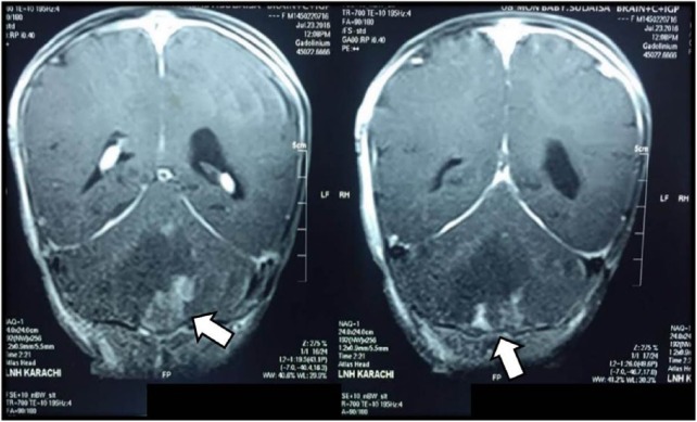
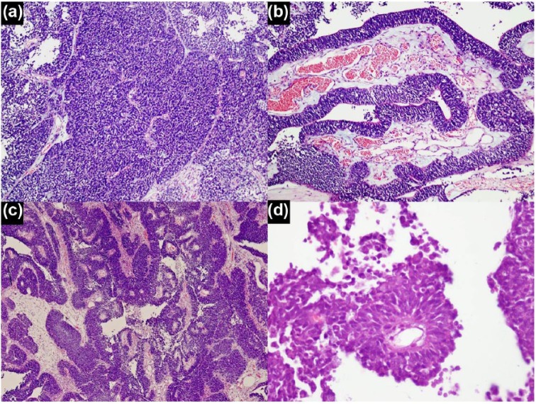
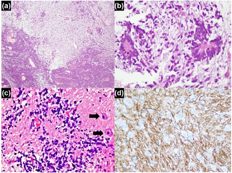
The 2016 update of the WHO Classification of Tumours of the Central Nervous System has redefined a number of tumors. Embryonal tumor with multilayered rosettes, C19MC-altered is one such tumor entity which has been newly defined on the basis of a characteristic molecular alteration. We report, to our knowledge, the first case of this rare pediatric brain neoplasm in the Pakistani population. An 8-month-old girl was presented with vomiting and left-sided ptosis, and magnetic resonance imaging scan showed a cerebellar tumor. Histologically, a highly cellular population of primitive cells was seen alternating with hypocellular neuropil-rich regions containing multilayered true rosettes and cells with glial and neuronal differentiation. Amplification of 19q13. 42 chromosome region on fluorescence in situ hybridization analysis confirmed the diagnosis. Post-operative radiological examination revealed widespread central nervous system involvement. Adjuvant treatment was not offered due to complications. Patient expired a week after diagnosis.
Keywords: C19MC; Central nervous system–primitive neuroectodermal tumor; LIN28A; embryonal tumor with multilayered rosettes; multilayered rosettes.
Conflict of interest statement
Declaration of conflicting interests: The author(s) declared no potential conflicts of interest with respect to the research, authorship and/or publication of this article.
Figures
References
-
- Korshunov A, McLendon RE, Judkins AR, et al. Embryonal tumour with multilayered rosettes, C19MC-altered. In: Louis DN, Ohgaki H, Wiestler OD, et al. (eds) WHO classification of tumours of the central nervous system. 4th ed Lyon: IARC Press, 2016, pp. 201–205.
-
- McLendon RE, Judkins AR, Eberhart CG, et al. CNS primitive neuroectodermal tumours (PNETs). In: Louis DN, Ohgaki H, Wiestler OD, et al. (eds) WHO classification of tumours of the central nervous system. 4th ed Lyon: IARC Press, 2007, pp. 141–146.
-
- Korshunov A, Remke M, Gessi M, et al. Focal genomic amplification at 19q13.42 comprises a powerful diagnostic marker for embryonal tumors with ependymoblastic rosettes. Acta Neuropathol 2010; 120(2): 253–260. - PubMed
-
- Eberhart CG, Brat DJ, Cohen KJ, et al. Pediatric neuroblastic brain tumors containing abundant neuropil and true rosettes. Pediatr Dev Pathol 2000; 3(4): 346–352. - PubMed
Publication types
LinkOut - more resources
Full Text Sources
Other Literature Sources
Research Materials
