Review
. 2017 Nov 20:4:200.
doi: 10.3389/fmed.2017.00200. eCollection 2017.

Non-Invasive Monitoring of Cardiac Output in Critical Care Medicine

Affiliations
Review

Non-Invasive Monitoring of Cardiac Output in Critical Care Medicine

Lee S Nguyen et al. Front Med (Lausanne). .

Abstract

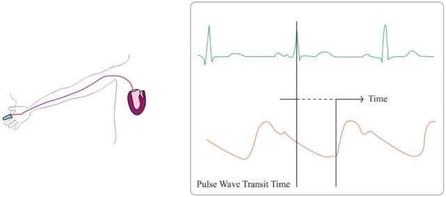
Critically ill patients require close hemodynamic monitoring to titrate treatment on a regular basis. It allows administering fluid with parsimony and adjusting inotropes and vasoactive drugs when necessary. Although invasive monitoring is considered as the reference method, non-invasive monitoring presents the obvious advantage of being associated with fewer complications, at the expanse of accuracy, precision, and step-response change. A great many methods and devices are now used over the world, and this article focuses on several of them, providing with a brief review of related underlying physical principles and validation articles analysis. Reviewed methods include electrical bioimpedance and bioreactance, respiratory-derived cardiac output (CO) monitoring technique, pulse wave transit time, ultrasound CO monitoring, multimodal algorithmic estimation, and inductance thoracocardiography. Quality criteria with which devices were reviewed included: accuracy (closeness of agreement between a measurement value and a true value of the measured), precision (closeness of agreement between replicate measurements on the same or similar objects under specified conditions), and step response change (delay between physiological change and its indication). Our conclusion is that the offer of non-invasive monitoring has improved in the past few years, even though further developments are needed to provide clinicians with sufficiently accurate devices for routine use, as alternative to invasive monitoring devices.

Keywords: bioreactance; cardiac output; critical care medicine; hemodynamics; non-invasive monitoring.


Figures

Figure 1
Figure 1
Bioimpedance and bioreactance signal. Upper part, in orange the input constant alternating current: Io = 5 mA, frequency 75 kHz (ω = 150,000 radians/s). In blue the output voltage. V(t) = 200 ± 2 mV, frequency F(t) = 75 kHz ± 5 Hz. The instantaneous changes in phase are figured in green. In the middle, the Vo envelope (AM component) is extracted from the envelope of V = 4 mV, corresponding to the bioimpedance signal (Z = 4/5 = 0.8 Ω). The lower part shows the corresponding changes in frequency as obtained by the sum of instant phase shift (FM signal) figuring the bioreactance signal: F = 10 Hz (ω = 20 radians/s). Using appropriate scaling the shape of the AM and FM signals is the same.
Figure 2
Figure 2
Partial rebreathing principles. Left panel represents baseline state, where the rebreathing valve is off and every parameter is at baseline levels. Middle panel represents early rebreathing time, when the valve is put; there is a decrease of VCO2 with simultaneous rise in PaCO2 and PETCO2. Right panel represents late rebreathing time when valve is off again and all parameters return to baseline levels, while mixed venous PCO2 has varied.
Figure 3
Figure 3
Pulse wave transit time principles. Pulse wave transit time is based on the delay of the generation of a stroke volume (orange line) after the generation of the R-wave on the electrocardiogram (green line).
Figure 4
Figure 4
Echocardiographic monitoring. Aortic flow velocity time integral (VTI) multiplied by the cross-sectional area (CSA) allows to compute stroke volume (SV) ejected by the left ventricle (LV). Heart rate (HR) then allows to compute cardiac output (CO) = VTI × CSA × HR.

References

    1. Cruz K, Franklin C. The pulmonary artery catheter: uses and controversies. Crit Care Clin (2001) 17:271–91.10.1016/S0749-0704(05)70167-6 - DOI - PubMed
    1. Huttemann E, Schelenz C, Kara F, Chatzinikolaou K, Reinhart K. The use and safety of transoesophageal echocardiography in the general ICU – a minireview. Acta Anaesthesiol Scand (2004) 48:827–36.10.1111/j.0001-5172.2004.00423.x - DOI - PubMed
    1. Frezza EE, Mezghebe H. Indications and complications of arterial catheter use in surgical or medical intensive care units: analysis of 4932 patients. Am Surg (1998) 64:127–31. - PubMed
    1. Marik PE, Baram M. Noninvasive hemodynamic monitoring in the intensive care unit. Crit Care Clin (2007) 23:383–400.10.1016/j.ccc.2007.05.002 - DOI - PubMed
    1. Squara P, Imhoff M, Cecconi M. Metrology in medicine: from measurements to decision, with specific reference to anesthesia and intensive care. Anesth Analg (2015) 120:66–75.10.1213/ANE.0000000000000477 - DOI - PMC - PubMed