Review
. 2017 Dec 11;6(1):44.
doi: 10.1186/s40169-017-0175-0.

Cancer nanomedicine: a review of recent success in drug delivery

Affiliations
Review

Cancer nanomedicine: a review of recent success in drug delivery

Stephanie Tran et al. Clin Transl Med. .

Abstract

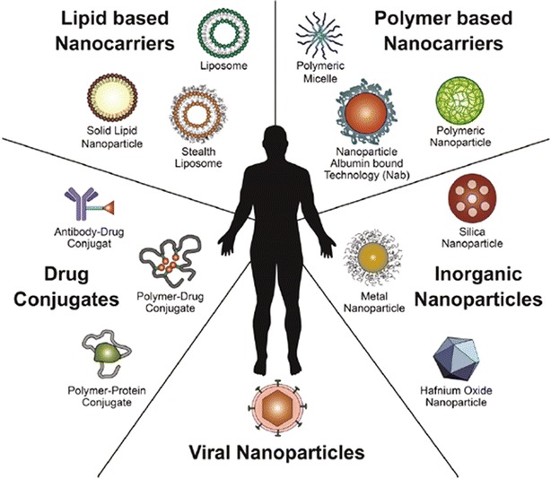
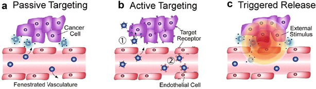
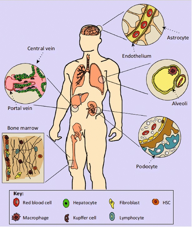
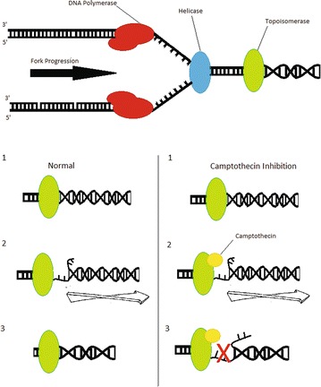
Cancer continues to be one of the most difficult global healthcare problems. Although there is a large library of drugs that can be used in cancer treatment, the problem is selectively killing all the cancer cells while reducing collateral toxicity to healthy cells. There are several biological barriers to effective drug delivery in cancer such as renal, hepatic, or immune clearance. Nanoparticles loaded with drugs can be designed to overcome these biological barriers to improve efficacy while reducing morbidity. Nanomedicine has ushered in a new era for drug delivery by improving the therapeutic indices of the active pharmaceutical ingredients engineered within nanoparticles. First generation nanomedicines have received widespread clinical approval over the past two decades, from Doxil® (liposomal doxorubicin) in 1995 to Onivyde® (liposomal irinotecan) in 2015. This review highlights the biological barriers to effective drug delivery in cancer, emphasizing the need for nanoparticles for improving therapeutic outcomes. A summary of different nanoparticles used for drug delivery applications in cancer are presented. The review summarizes recent successes in cancer nanomedicine in the clinic. The clinical trials of Onivyde leading to its approval in 2015 by the Food and Drug Adminstration are highlighted as a case study in the recent clinical success of nanomedicine against cancer. Next generation nanomedicines need to be better targeted to specifically destroy cancerous tissue, but face several obstacles in their clinical development, including identification of appropriate biomarkers to target, scale-up of synthesis, and reproducible characterization. These hurdles need to be overcome through multidisciplinary collaborations across academia, pharmaceutical industry, and regulatory agencies in order to achieve the goal of eradicating cancer. This review discusses the current use of clinically approved nanomedicines, the investigation of nanomedicines in clinical trials, and the challenges that may hinder development of the nanomedicines for cancer treatment.

Keywords: Clinical trials; Combination treatment; MM-398; Nanoparticles; Oncology; Theranostics; Therapeutics.


Figures

Fig. 1
Fig. 1
Overview of established nanomedicines in the clinic. This diagram shows an overview of the nanomedicines currently being investigated in the clinic for cancer treatment. Lipid-based, polymer-based, inorganic, viral, and drug-conjugated nanoparticles are examples of platforms that have been established in clinical research (Reproduced with permission from [3])
Fig. 2
Fig. 2
Types of targeting for nanoparticle delivery to tumor tissue. a Passive targeting relies on the leaky vasculature that is exhibited by tumors, allowing nanoparticles to travel through the fenestrations and reach tumors. b Active targeting can be used when nanoparticles have ligands on their surface that can recognize and bind receptors that are overexpressed on tumor cells. c Triggered release allows nanoparticles to congregate if exposed to an external stimulus such as a magnetic field or light (Reproduced with permission from [3])
Fig. 3
Fig. 3
Organ systems that affect nanoparticle delivery. The method of entry affects circulation time, organ processing, and overall efficacy. Intravenously injected nanoparticles can extravasate from the bloodstream and enter organs such as the liver, spleen, bone marrow, and central nervous system. Nanomedicines that are administered orally can enter the gut and pass through the liver via the hepatic portal system. Inhaled nanomedicines may contact macrophages in pulmonary alveoli. Following circulation in organs, nanoparticles may encounter renal clearance in the kidneys (Reproduced with permission from [11])
Fig. 4
Fig. 4
A schematic illustrating replication fork arrest by a drug-aborted topoisomerase I-DNA cleavable complex. In this model, the camptothecin trapped topoisomerase I-DNA cleavable complex is viewed as a bulky DNA lesion which arrests the replication fork by blocking the movement of replication machinery. This blockage also alters the physical state of the cleavable complex and possibly leads to fork breakage at the complex site. At low levels of cleavable complexes, when only one replication fork is arrested, continued replication by the other unimpeded fork on the same plasmid DNA leads to the formation of linearized replication products. The irreversible replication arrest and fork breakage may be the cause of camptothecin’s S-phase-specific cytotoxicity [62, 63]
Fig. 5
Fig. 5
Metabolic pathway of irinotecan activation into SN-38 (Reproduced with permission from [81])
Fig. 6
Fig. 6
Depiction of the exchange of triethylamine for irinotecan, which forms a stable complex with sucrose octasulfate inside the liposome (Reproduced with permission from [66])
Fig. 7
Fig. 7
Kaplan–Meier curves of overall and progression-free survival. Abbreviations used are m for months, OS for overall survival, and PFS for progression-free survival (Reproduced with permission from [76])
Fig. 8
Fig. 8
The Kaplan–Meier estimates of overall and progression-free survival (a, b respectively) in the intent-to-treat population for liposomal irinotecan (PEP02), irinotecan, and docetaxal (Reproduced with permission from [69])
Fig. 9
Fig. 9
Kaplan–Meier survival analyses for phase III trial. HR hazard ratio. a Overall survival with nanoliposomal irinotecan plus fluorouracil and folinic acid versus fluorouracil and folinic acid. b Overall survival with nanoliposomal irinotecan monotherapy versus fluorouracil and folinic acid. c Progression-free survival with nanoliposomal irinotecan plus fluorouracil and folinic acid versus fluorouracil and folinic acid. d Progression-free survival with nanoliposomal irinotecan monotherapy versus fluorouracil and folinic acid (Reproduced with permission from [77])

References

    1. Siegel RL, Miller KD, Fedewa SA, Ahnen DJ, Meester RG, Barzi A, Jemal A. Colorectal cancer statistics, 2017. CA Cancer J Clin. 2017;67(3):177–193. doi: 10.3322/caac.21395. - DOI - PubMed
    1. Hanahan D, Weinberg RA. Hallmarks of cancer: the next generation. Cell. 2011;144(5):646–674. doi: 10.1016/j.cell.2011.02.013. - DOI - PubMed
    1. Wicki A, Witzigmann D, Balasubramanian V, Huwyler J. Nanomedicine in cancer therapy: challenges, opportunities, and clinical applications. J Control Release. 2015;200:138–157. doi: 10.1016/j.jconrel.2014.12.030. - DOI - PubMed
    1. Sinha R, Kim GJ, Nie S, Shin DM. Nanotechnology in cancer therapeutics: bioconjugated nanoparticles for drug delivery. Mol Cancer Ther. 2006;5(8):1909–1917. doi: 10.1158/1535-7163.MCT-06-0141. - DOI - PubMed
    1. Albanese A, Tang PS, Chan WC. The effect of nanoparticle size, shape, and surface chemistry on biological systems. Annu Rev Biomed Eng. 2012;14:1–16. doi: 10.1146/annurev-bioeng-071811-150124. - DOI - PubMed