

A case report of proliferative glomerulonephritis with monoclonal immunoglobulin M-kappa deposits without associated lymphoproliferative disorder or detectable paraproteinemia
- PMID: 29230710
- PMCID: PMC5886923
- DOI: 10.1007/s13730-017-0291-0
A case report of proliferative glomerulonephritis with monoclonal immunoglobulin M-kappa deposits without associated lymphoproliferative disorder or detectable paraproteinemia
Abstract
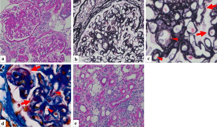
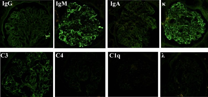
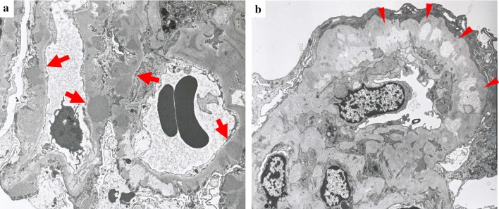
A 53-year-old man presented with proteinuria and hematuria. No significant abnormality was detected in his physical examination or laboratory tests, including evidence of paraprotein in serum and urine. Renal biopsy revealed mesangial proliferation, thickened glomerular basement membranes, and spike formation. Immunofluorescence revealed deposition of immunoglobulin (Ig) M heavy chain, kappa (κ) light chain, and complement component C3 along capillary walls in the glomeruli. Light chain staining indicated significant restriction, because only κ chain, not lambda chain, was present in glomeruli. Aggregated electron dense deposits were observed in the subepithelial area and within the lamina densa on electron-microscopic examination. Cryoglobulinemia and amyloidosis were ruled out. Clinically, steroid therapy was not initiated due to patient preference, and the only prescribed medication was an angiotensin II receptor blocker. At the approximately 3-year follow-up, estimated glomerular filtration rate had decreased very mildly. The present case demonstrates that deposition of monoclonal IgM-κ may be associated with membranoproliferative glomerulonephritis-like changes in the glomeruli. Although no underlying hematological abnormality or paraproteinemia was observed in this case within the range of limited clinical examination, the patient's condition is consistent with proliferative glomerulonephritis with monoclonal IgM deposits, similar to the recently established proliferative glomerulonephritis with monoclonal IgG deposits. Further elucidation of the pathophysiology and effective treatments of the disorder should be expected in the future through the accumulation of similar cases.
Keywords: IgM; MPGN; Monoclonal; PGNMID; PGNMIMD.
Conflict of interest statement
Conflict of interest
The authors have declared that no conflict of interest exists.
Human and animal rights
This article does not contain any studies with human participants or animals performed by any of the authors.
Informed Consent
Informed consent was obtained from the patient included in this article.
Figures

References
-
- Nasr SH, Markowitz GS, Stokes MB, Seshan SV, Valderrama E, Appel GB, Aucouturier P, D’Agati VD. Proliferative glomerulonephritis with monoclonal IgG deposits: a distinct entity mimicking immune-complex glomerulonephritis. Kidney Int. 2004;65:85–96. doi: 10.1111/j.1523-1755.2004.00365.x. - DOI - PubMed
-
- Oe Y, Joh K, Sato M, Taguma Y, Onishi Y, Nakayama K, Sato T. Proliferative glomerulonephritis with monoclonal IgM-κ deposits in chronic lymphocytic leukemia/small lymphocytic leukemia: case report and review of the literature. CEN Case Reports. 2013;2:222–227. doi: 10.1007/s13730-013-0068-z. - DOI - PMC - PubMed
-
- Audard V, Georges B, Vanhille P, Toly C, Deroure B, Fakhouri F, Cuvelier R, Belenfant X, Surin B, Aucouturier P, Mougenot B, Ronco P. Renal lesions associated with IgM-secreting monoclonal proliferations: revisiting the disease spectrum. Clin J Am Soc Nephrol. 2008;3:1339–1349. doi: 10.2215/CJN.01600408. - DOI - PMC - PubMed
LinkOut - more resources
Full Text Sources
Other Literature Sources
Miscellaneous
