. 2018 Aug;80(2):756-766.
doi: 10.1002/mrm.27036. Epub 2017 Dec 11.

Placenta microstructure and microcirculation imaging with diffusion MRI

Affiliations

Placenta microstructure and microcirculation imaging with diffusion MRI

Paddy J Slator et al. Magn Reson Med. 2018 Aug.

Abstract

Purpose: To assess which microstructural models best explain the diffusion-weighted MRI signal in the human placenta.

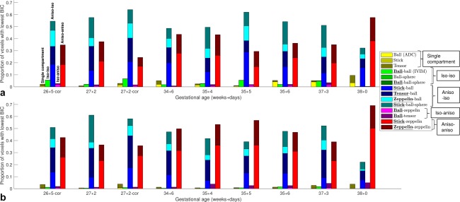
Methods: The placentas of nine healthy pregnant subjects were scanned with a multishell, multidirectional diffusion protocol at 3T. A range of multicompartment biophysical models were fit to the data, and ranked using the Bayesian information criterion.

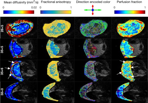
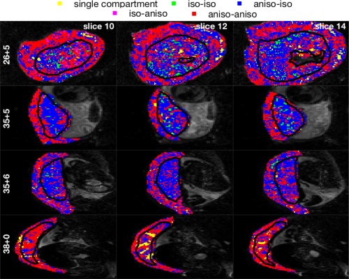
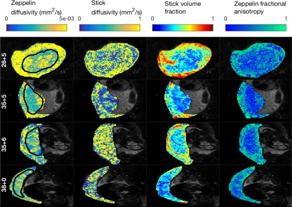
Results: Anisotropic extensions to the intravoxel incoherent motion model, which consider the effect of coherent orientation in both microvascular structure and tissue microstructure, consistently had the lowest Bayesian information criterion values. Model parameter maps and model selection results were consistent with the physiology of the placenta and surrounding tissue.

Conclusion: Anisotropic intravoxel incoherent motion models explain the placental diffusion signal better than apparent diffusion coefficient, intravoxel incoherent motion, and diffusion tensor models, in information theoretic terms, when using this protocol. Future work will aim to determine if model-derived parameters are sensitive to placental pathologies associated with disorders, such as fetal growth restriction and early-onset pre-eclampsia. Magn Reson Med 80:756-766, 2018. © 2017 The Authors Magnetic Resonance in Medicine published by Wiley Periodicals, Inc. on behalf of International Society for Magnetic Resonance in Medicine. This is an open access article under the terms of the Creative Commons Attribution License, which permits use, distribution and reproduction in any medium, provided the original work is properly cited.

Keywords: Bayesian information criterion; diffusion MRI; intravoxel incoherent motion; microstructure; model selection; placenta.


Figures

Figure 1
Figure 1
Schematic representation of blood flow through the placenta and surrounding tissue. Blue and red arrows show the flow directions of oxygenated (red) and deoxygenated (blue) fetal blood through the placental vasculature. For clarity, only the largest villi are included (for normal placentas terminal villi make up 40% of villous tree volume [21]). Dashed white arrows show idealized flow lines through intervillous space for maternal blood. Idealized oxygenation states are represented by the red to blue color gradient.
Figure 2
Figure 2
Mean diffusivity maps for three slices of a single placental diffusion‐weighted MRI scan. Gestational age: 35 + 6 (weeks + days). The columns b = 40 s mm−2 to b = 2000 s mm−2 show the mean diffusivities derived from a diffusion tensor fit only to the images at that nonzero b‐value and b = 0. Red and black outline the uterine wall and placenta ROIs respectively. Color represents mean diffusivity in mm2 s−1 (note that the scale is a factor of 10 higher for the b = 40 s mm−2 maps).
Figure 3
Figure 3
Parameter maps derived from diffusion tensor and ball‐ball model fits. Each row displays maps for a single slice from one subject, labeled by gestational age (weeks + days). Slices are displayed in the EPI acquisition plane, corresponding to the coronal plane (row 1) and axial plane (remaining rows). Arrows in row 3 highlight areas of high diffusivity and high perfusion at the boundary of the placenta. https://onlinelibrary.wiley.com/action/downloadSupplement?doi=10.1002%2Fmrm.27036&attachmentId=213714997 is the complete version of this figure, containing these maps for all subjects.
Figure 4
Figure 4
Model selection results across all subjects. Bar plots showing the proportion of voxels where each model had the lowest Bayesian information criterion for nine placental scans. Subjects are labeled by gestational age, with “‐cor” indicating that the placenta was scanned coronally. The perfusion model compartment is emphasized in the legend text. a: Placenta ROI. b: Uterine wall ROI.
Figure 5
Figure 5
Mapping the spatial pattern of model selection results. Each row displays three slices for a single subject, labeled by gestational age (weeks + days). Voxels are colored according to the category of the model with the lowest Bayesian information criterion in that voxel. Models are labeled according to the isotropy of the perfusion and diffusion compartments, respectively, for example “aniso‐iso” refers to models with anisotropic perfusion compartment and isotropic diffusion compartment. Slices are displayed in the EPI acquisition plane (coronal plane for row 1, axial plane for other rows). https://onlinelibrary.wiley.com/action/downloadSupplement?doi=10.1002%2Fmrm.27036&attachmentId=213714997 is the complete version of this figure, containing these maps for all subjects.
Figure 6
Figure 6
Parameter maps derived from stick‐zeppelin model. Each row displays maps for a single axial slice from one subject, labeled by gestational age (weeks + days). Slices are displayed in the EPI acquisition plane (coronal plane for row 1, axial plane for other rows). https://onlinelibrary.wiley.com/action/downloadSupplement?doi=10.1002%2Fmrm.27036&attachmentId=213714997 is the complete version of this figure, containing these maps for all subjects.

Similar articles

Cited by

References

    1. Nelson DM. How the placenta affects your life, from womb to tomb. Am J Obstet Gynecol 2015;213:S12–S13. - PubMed
    1. Vedmedovska N, Rezeberga D, Teibe U, Melderis I, Donders GGG. Placental pathology in fetal growth restriction. Eur J Obstet Gynecol Reprod Biol 2011;155:36–40. - PubMed
    1. Mifsud W, Sebire NJ. Placental pathology in early‐onset and late‐onset fetal growth restriction. Fetal Diagn Therapy 2014;36:117–128. - PubMed
    1. Veerbeek JHW, Nikkels PGJ, Torrance HL, Gravesteijn J, Post Uiterweer ED, Derks JB, Koenen SV, Visser GHA, Van Rijn BB, Franx A. Placental pathology in early intrauterine growth restriction associated with maternal hypertension. Placenta 2014;35:696–701. - PubMed
    1. Myatt L. Role of placenta in preeclampsia. Endocrine 2002;19:103–111. - PubMed

Publication types

LinkOut - more resources