. 2018 Aug;23(7):624-633.
doi: 10.1177/2472555217746324. Epub 2017 Dec 12.

High-Throughput Screening Approach for Identifying Compounds That Inhibit Nonhomologous End Joining

Affiliations

High-Throughput Screening Approach for Identifying Compounds That Inhibit Nonhomologous End Joining

Andrea L Bredemeyer et al. SLAS Discov. 2018 Aug.

Abstract

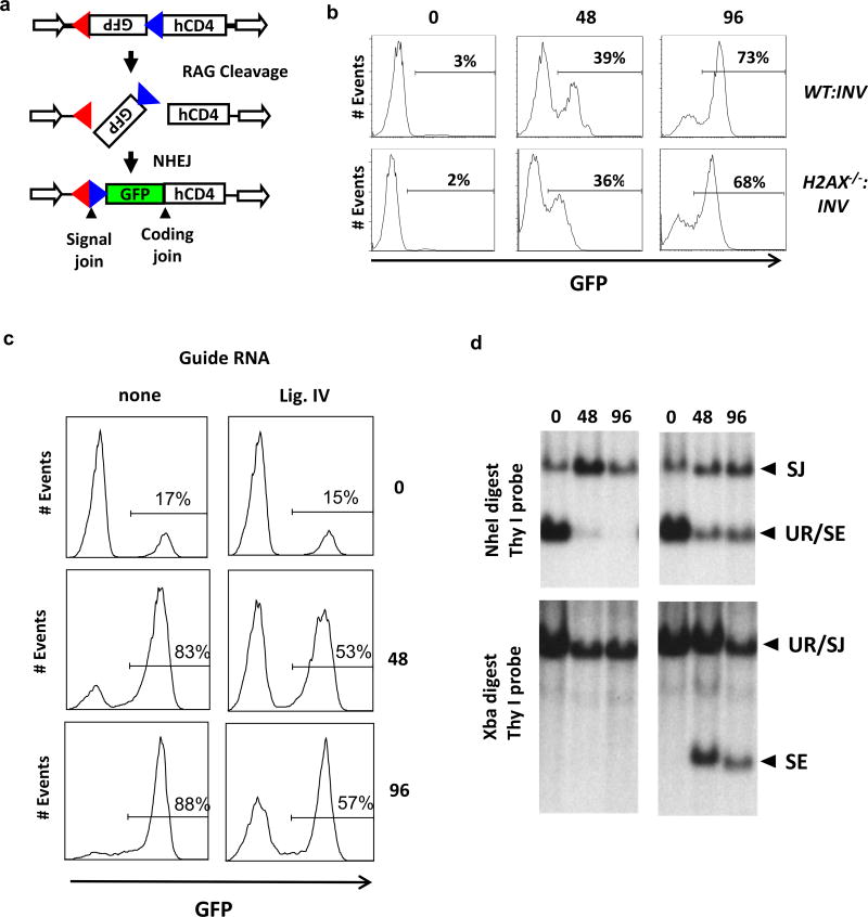
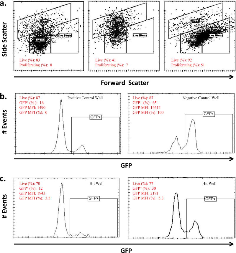
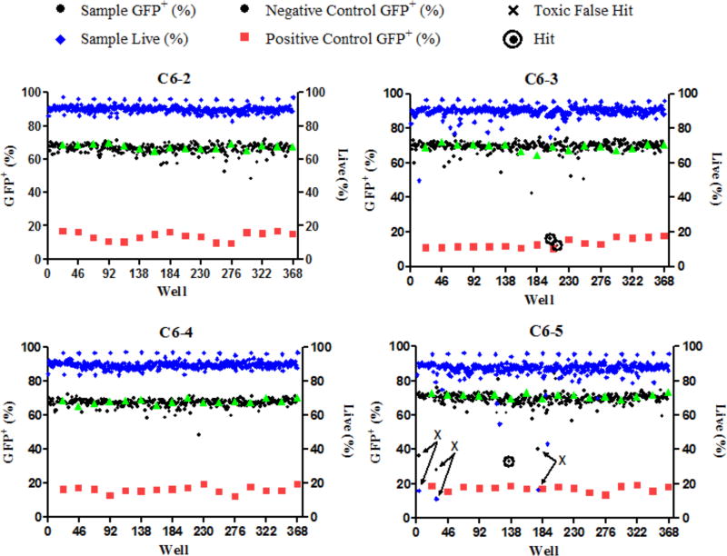
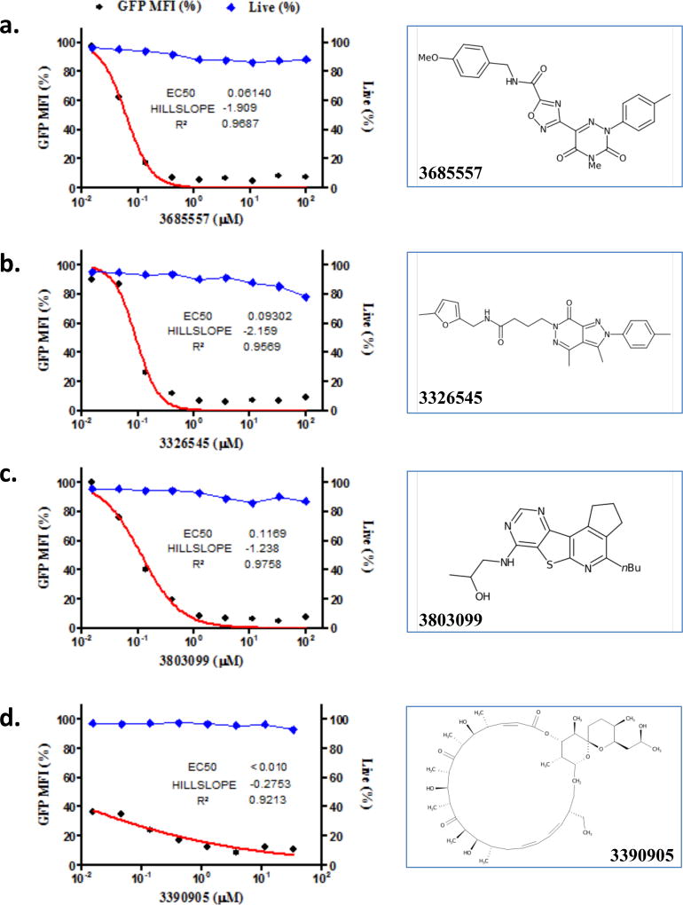
DNA double-strand breaks (DSBs) are repaired primarily by homologous recombination (HR) or nonhomologous end joining (NHEJ). Compounds that modulate HR have shown promise as cancer therapeutics. The V(D)J recombination reaction, which assembles antigen receptor genes in lymphocytes, is initiated by the introduction of DNA DSBs at two recombining gene segments by the RAG endonuclease, followed by the NHEJ-mediated repair of these DSBs. Here, using HyperCyt automated flow cytometry, we develop a robust high-throughput screening (HTS) assay for NHEJ that utilizes engineered pre-B-cell lines where the V(D)J recombination reaction can be induced and monitored at a single-cell level. This approach, novel in processing four 384-well plates at a time in parallel, was used to screen the National Cancer Institute NeXT library to identify compounds that inhibit V(D)J recombination and NHEJ. Assessment of cell light scattering characteristics at the primary HTS stage (83,536 compounds) enabled elimination of 60% of apparent hits as false positives. Although all the active compounds that we identified had an inhibitory effect on RAG cleavage, we have established this as an approach that could identify compounds that inhibit RAG cleavage or NHEJ using new chemical libraries.

Keywords: cancer and cancer drugs; cell-based assays; immune system diseases; oncology.


Figures

Figure 1
Figure 1. Retroviral V(D)J recombination substrate
a) Schematic of the pMX-INV retroviral recombination substrate after RAG DNA cleavage and normal NHEJ-mediated DSB repair placing GFP in the sense orientation. b) Flow cytometric analysis of GFP expression (an indicator of RAG DNA cleavage and normal NHEJ-mediated DSB repair) in wild type (WT:INV) and H2AX-deficient (H2AX−/−:INV) abl pre-B cells after 0, 48 or 96 hours of imatinib treatment. c) Flow cytometric analysis of GFP expression in WT:MGINV:Cas9 abl pre-B cells that were treated with doxycycline for 7 days followed by imatinib treatment for 0, 48 or 96 hours. Cells that had the no (none) or the DNA Ligase 4 guide RNA are indicated. d) Southern blot analysis of genomic DNA from cells in panel c. Bands due to unrearranged (UR) pMG-INV or a pMG-INV signal join (SJ) or unrepaired signal end (SE) indicated.
Figure 2
Figure 2. Gating configuration for flow cytometry analysis
A) Forward vs. side light scatter gates were used to determine cell viability and proliferation. Higher side scatter characteristics (middle panel) indicates cell death whereas higher forward scatter characteristics (right panel) indicates proliferation. Normal forward and side scatter parameters for imatininb treated abl pre-B cells that are viable and not proliferating is shown (left panel). B) GFP fluorescence intensity profiles (FL1-H) of cells from representative positive (left panel) and negative (right panel) control wells. C) GFP fluorescence intensity profiles of cells from representative hit wells containing active compounds that block V(D)J recombination.
Figure 3
Figure 3. Representative results from parallel HTS analysis
Each panel represents analysis of a separate 384 well plate by each of the four flow cytometers (C6-2, C6-3, C6-4, C6-5) in the Cluster Cytometry platform. Illustrated are GFP expression in positive control wells (red squares), negative control wells (green triangles) and wells containing test compounds (black circles). The percentage of live cells in test compound wells was also quantified on the basis of light scatter profiles (blue triangles). False positive results are indicated by significantly diminished GFP expression associated with low viability (Xs with arrows indicating decreases in both percent live and percent GFP+ for cells in the wells). Active "hit" compounds are indicated by diminished GFP expression without diminished viability (circled black circles).
Figure 4
Figure 4. Dose response profiles
Examples of compounds with good viability and dose response kinetics (a–d) and one that exhibits toxicity at the effective dose (D). The structures of the compounds are also indicated and a representative toxic, false positive compound (d). Structures of the active compounds are illustrated to the right of the respective response profile. Compound 3685557 (a) was not in the original library but was selected for testing on the basis of structural similarity to another active library compound. %Live cells, blue diamonds. % GFP Median Fluorescence Intensity (MFI), black circles.
Figure 5
Figure 5. Assessment of active compounds
a) Flow cytometric analysis of GFP expression H2AX−/−:INV abl pre-B cells treated with imatinib and 40uM of the test compounds for 4 days. b) Southern blot analysis of pMX-INV showing bands that are due to un-rearranged pMX-INV (NR, GFP−), pMX-INV with a normal coding join (CJ GFP+), or pMX-INV with an un-repaired coding end (CE). The Thy 1 probe used hybridizes to the endogenous Thy1 gene (asterisk).

Similar articles

Cited by

  • Suppression of fumarate hydratase activity increases the efficacy of cisplatin-mediated chemotherapy in gastric cancer.
    Yu HE, Wang F, Yu F, Zeng ZL, Wang Y, Lu YX, Jin Y, Wang DS, Qiu MZ, Pu HY, Kang TB, Xie D, Ju HQ, Xu RH, Luo HY. Yu HE, et al. Cell Death Dis. 2019 May 28;10(6):413. doi: 10.1038/s41419-019-1652-8. Cell Death Dis. 2019. Retraction in: Cell Death Dis. 2023 Jan 20;14(1):46. doi: 10.1038/s41419-023-05604-7. PMID: 31138787 Free PMC article. Retracted.
  • MYSM1 attenuates DNA damage signals triggered by physiologic and genotoxic DNA breaks.
    Mathias B, O'Leary D, Saucier N, Ahmad F, White LS, Russell L, Shinawi M, Smith MJ, Abraham RS, Cooper MA, Kitcharoensakkul M, Green AM, Bednarski JJ. Mathias B, et al. J Allergy Clin Immunol. 2024 Apr;153(4):1113-1124.e7. doi: 10.1016/j.jaci.2023.12.001. Epub 2023 Dec 6. J Allergy Clin Immunol. 2024. PMID: 38065233 Free PMC article.
  • Guidelines for the use of flow cytometry and cell sorting in immunological studies (second edition).
    Cossarizza A, Chang HD, Radbruch A, Acs A, Adam D, Adam-Klages S, Agace WW, Aghaeepour N, Akdis M, Allez M, Almeida LN, Alvisi G, Anderson G, Andrä I, Annunziato F, Anselmo A, Bacher P, Baldari CT, Bari S, Barnaba V, Barros-Martins J, Battistini L, Bauer W, Baumgart S, Baumgarth N, Baumjohann D, Baying B, Bebawy M, Becher B, Beisker W, Benes V, Beyaert R, Blanco A, Boardman DA, Bogdan C, Borger JG, Borsellino G, Boulais PE, Bradford JA, Brenner D, Brinkman RR, Brooks AES, Busch DH, Büscher M, Bushnell TP, Calzetti F, Cameron G, Cammarata I, Cao X, Cardell SL, Casola S, Cassatella MA, Cavani A, Celada A, Chatenoud L, Chattopadhyay PK, Chow S, Christakou E, Čičin-Šain L, Clerici M, Colombo FS, Cook L, Cooke A, Cooper AM, Corbett AJ, Cosma A, Cosmi L, Coulie PG, Cumano A, Cvetkovic L, Dang VD, Dang-Heine C, Davey MS, Davies D, De Biasi S, Del Zotto G, Dela Cruz GV, Delacher M, Della Bella S, Dellabona P, Deniz G, Dessing M, Di Santo JP, Diefenbach A, Dieli F, Dolf A, Dörner T, Dress RJ, Dudziak D, Dustin M, Dutertre CA, Ebner F, Eckle SBG, Edinger M, Eede P, Ehrhardt GRA, Eich M, Engel P, Engelhardt B, Erdei A, Esser C, Everts B, Evrard M, Falk CS, Fehniger TA, Felipo-Benavent M, Fer… See abstract for full author list ➔ Cossarizza A, et al. Eur J Immunol. 2019 Oct;49(10):1457-1973. doi: 10.1002/eji.201970107. Eur J Immunol. 2019. PMID: 31633216 Free PMC article.

References

    1. Swift LH, Golsteyn RM. Genotoxic anti-cancer agents and their relationship to DNA damage, mitosis, and checkpoint adaptation in proliferating cancer cells. Int. J. Mol. Sci. 2014;15:3403–3431. - PMC - PubMed
    1. Ciccia A, Elledge SJ. The DNA damage response: making it safe to play with knives. Mol. Cell. 2010;40:179–204. - PMC - PubMed
    1. Lieber MR. The mechanism of double-strand DNA break repair by the nonhomologous DNA end-joining pathway. Annu. Rev. Biochem. 2010;79:181–211. - PMC - PubMed
    1. Bryant HE, Schultz N, Thomas HD, et al. Specific killing of BRCA2-deficient tumours with inhibitors of poly(ADP-ribose) polymerase. Nature. 2005;434:913–917. - PubMed
    1. Farmer H, McCabe N, Lord CJ, et al. Targeting the DNA repair defect in BRCA mutant cells as a therapeutic strategy. Nature. 2005;434:917–921. - PubMed

Publication types

MeSH terms

LinkOut - more resources