. 2018 Feb;28(2):164-177.
doi: 10.1002/hipo.22819. Epub 2017 Dec 27.

Reconstructing relational information

Affiliations

Reconstructing relational information

Kevin M Horecka et al. Hippocampus. 2018 Feb.

Abstract

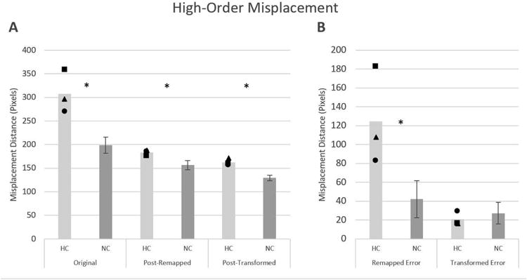
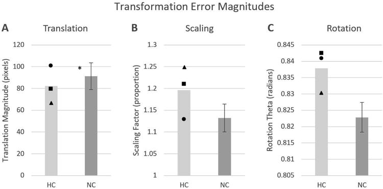
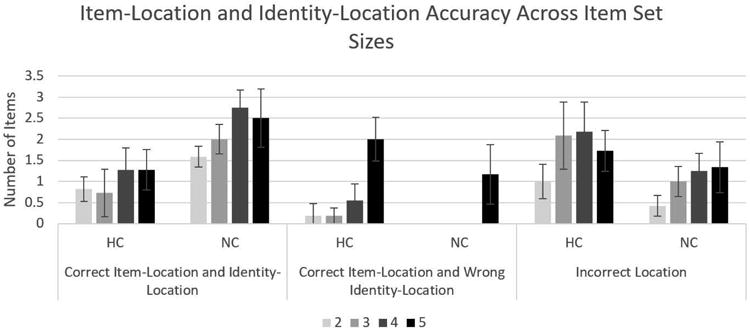
Hippocampal involvement in learning and remembering relational information has an extensive history, often focusing specifically on spatial information. In humans, spatial reconstruction (SR) paradigms are a powerful tool for evaluating an individuals' spatial-relational memory. In SR tasks, participants study locations of items in space and subsequently reconstruct the studied display after a short delay. Previous work has revealed that patients with hippocampal damage are impaired both in overall placement accuracy as well as on a specific measure of relational memory efficacy, "swaps" (i.e., when the relative location of two items is reversed). However, the necessity of the hippocampus for other types of spatial-relational information involved in reconstruction behaviors (e.g., where in the environment and relative to which other items an item was located) have not yet been investigated systematically. In this work, three patients with hippocampal damage and nine healthy matched comparison participants performed an SR task. An analysis framework was developed to independently assess three first-order types of relations: (1) memory for the binding of specific item identities to locations, (2) memory for arrangement of items in relation to each other or the environment bounds, regardless of memory for the item identity, and (3) higher-order, compound relational errors (i.e., errors involving multiple pieces of relational information). Reconstruction errors were evaluated to determine the degree to which patients and comparisons differed (or not) on each type of spatial-relational information. Data revealed that the primary group difference in performance was for identity-location information. However, when the locations of items were evaluated without regarding the identities, no group difference was found in the number of item placements to studied locations. The present work provides a principled approach to analysis of SR data and clarifies our understanding of the types of spatial relations impaired in hippocampal damaged.

Keywords: binding; declarative memory; online processing; relational memory; spatial reconstruction.


Figures

Figure 1
Figure 1
Magnetic resonance scans of hippocampal patients. Images are coronal slices through four points along the hippocampus from T1-weighed scans. Volume changes can be noted in the hippocampal region for patient 1846, and significant bilateral MTL damage including the hippocampus can be noted in patient 1951. A = anterior, P = posterior, NC = healthy comparison brain.
Figure 2
Figure 2
A sample trial from the spatial reconstruction task; A, B, and C show different interpretations of the same reconstruction where the objects are in the reconstructed locations and the dots are the target locations from the study phase. (A) shows item-item and item-environment relations (dotted lines), which might be used to determine the locations of the dots. (B) shows reconstructed item to target location relations where gray shaded circles indicate a failure in placement of items to those locations (note that identity is ignored as, for example, the bus is not in its target location). (C) shows compound relations where some items roughly swap locations (although not necessarily precisely, in this case being a “partial swap”), some show cycle relationships (i.e. Bus◊Bed◊Bicycle◊Bus), and some show single-item placement in the correct target location.
Figure 3
Figure 3
The analysis procedure as defined in the above sections. (A) shows the correct locations (dots with shadowed identities) and the reconstructed locations (items) along with shadowded dotted lines showing the original misplacement. Note that large amounts of misplacement error is accumulated via improper assignment of identities to locations. (B) shows the global remapping (dark dots are targets and light dots are reconstructed points) wherein identity is stripped away and a minimal one-to-one mapping is formed. (C) shows the global transformation (in this case a slight rotation and scaling error) where dark dots are targets, medium dots are reconstructed points, and light dots are post-transformation reconstructed points (i.e. points where the global error have been subtracted from the reconstructed points). (D) shows the binary accuracy evaluation on the post-transformed points (where the dark shaded circle signifies an inaccurate item). (E) shows the compound errors identified and labelled according to the previous steps (where shaded regions are considered “partial” groups where at least one item was inaccurately placed).
Figure 4
Figure 4
Comparisons of Patient (HC) vs Comparison Participant (NC) groups for A) Misplacement Across Analysis Steps, i.e. misplacement magnitudes at each step of analysis and B) Reduction in Misplacement by High-Order Error Type, i.e. differences in misplacement accounted for by each step (i.e. the subtraction of A). For Patients, individual symbols are shown such that: Square is 1951, Triangle is 1846, and Circle is 2563.
Figure 5
Figure 5
Comparisons of Patient (HC) vs Comparison Participant (NC) in the magnitude of the three types of global transformation error; A) the Translation magnitude, B) the Scaling magnitude (where 1 means no scaling, <1 means shrinking, and >1 means stretching), and C) the Rotation angle (in radians). For Patients, individual symbols are shown such that: Square is 1951, Triangle is 1846, and Circle is 2563.
Figure 6
Figure 6
Comparisons of Patient (HC) vs Comparison Participant (NC) in the number of items in which itemlocation and identity-location was correct and incorrect. Note that items which were not placed in any studied location are collapsed together as ‘Inaccurate Location’. For Patients, individual symbols are shown such that: Square is 1951, Triangle is 1846, and Circle is 2563.
Figure 7
Figure 7
Comparisons of Patient (HC) vs Comparison Participant (NC) Item-location and identity-location accuracy across set sizes for patients vs comparisons. Note that items which were not placed in any studied location are collapsed together as ‘Inaccurate Location’.

References

    1. Aggleton JP, Brown MW. Episodic memory, amnesia, and the hippocampal-anterior thalamic axis. The Behavioral and Brain Sciences. 1999;22(3):425–44-89. Retrieved from http://www.ncbi.nlm.nih.gov/pubmed/11301518. - PubMed
    1. Allen JS, Tranel D, Bruss J, Damasio H. Correlations between Regional Brain Volumes and Memory Performance in Anoxia. Journal of Clinical and Experimental Neuropsychology. 2006;28(4):457–476. http://doi.org/10.1080/13803390590949287. - DOI - PubMed
    1. Allen RJ, Vargha-Khadem F, Baddeley AD. Item-location binding in working memory: Is it hippocampus-dependent? Neuropsychologia. 2014;59(1):74–84. http://doi.org/10.1016/j.neuropsychologia.2014.04.013. - DOI - PubMed
    1. Besi PJ, Mckay ND. A Method for Registration of 3-D Shapes 1 Introduction 2 Literature Review. SPIE Vol 1611 Sensor Fusion IV. 1991;1611:586–606. http://doi.org/10.1117/12.57955. - DOI
    1. Brodeur MB, Guérard K, Bouras M. Bank of Standardized Stimuli (BOSS) Phase II: 930 New Normative Photos. PLoS ONE. 2014;9(9):e106953. http://doi.org/10.1371/journal.pone.0106953. - DOI - PMC - PubMed

Publication types

LinkOut - more resources