. 2017 Dec 12;14(1):53.
doi: 10.1186/s12989-017-0234-0.

Suppression of PTPN6 exacerbates aluminum oxide nanoparticle-induced COPD-like lesions in mice through activation of STAT pathway

Affiliations

Suppression of PTPN6 exacerbates aluminum oxide nanoparticle-induced COPD-like lesions in mice through activation of STAT pathway

Xiaobo Li et al. Part Fibre Toxicol. .

Abstract

Background: Inhaled nanoparticles can deposit in the deep lung where they interact with pulmonary cells. Despite numerous studies on pulmonary nanotoxicity, detailed molecular mechanisms of specific nanomaterial-induced lung injury have yet to be identified.

Results: Using whole-body dynamic inhalation model, we studied the interactions between aluminum oxide nanoparticles (Al2O3 NPs) and the pulmonary system in vivo. We found that seven-day-exposure to Al2O3 NPs resulted in emphysema and small airway remodeling in murine lungs, accompanied by enhanced inflammation and apoptosis. Al2O3 NPs exposure led to suppression of PTPN6 and phosphorylation of STAT3, culminating in increased expression of the apoptotic marker PDCD4. Rescue of PTPN6 expression or application of a STAT3 inhibitor, effectively protected murine lungs from inflammation and apoptosis, as well as, in part, from the induction of chronic obstructive pulmonary disease (COPD)-like effects.

Conclusion: In summary, our studies show that inhibition of PTPN6 plays a critical role in Al2O3 NPs-induced COPD-like lesions.

Keywords: Aluminum oxide nanoparticles; PTPN6; Experimental COPD; Inflammation.


Conflict of interest statement

Ethics approval and consent to participate

Animals were treated humanely and all experimental protocols were approved by Committee on Animal Use and Care of Southeast University, China. All the methods in the present study were performed according to approved guidelines.

Consent for publication

Individual person data was not applicable in our manuscript.

Competing interests

The authors declare no competing financial interest.

Publisher’s Note

Springer Nature remains neutral with regard to jurisdictional claims in published maps and institutional affiliations.

Figures

Fig. 1
Fig. 1
Pulmonary inflammation induced by Al2O3 NPs exposure. a sRAW was significantly increased in Al2O3 NPs-exposed mice (n = 10, * P < 0.05, compared with controls; *** P < 0.001, compared with control on the same day). b Inflammatory mediators were significantly increased in Al2O3 NPs-exposed BALF of mice (n = 4, * P < 0.05, ** P < 0.01). c The total cell number, monocytic cells number and neutrophils number in BALF of Al2O3 NPs-exposed mice were significantly increased compared with FRA control (n = 4, * P < 0.05, *** P < 0.001). d Representative images of normal airway and airway surrounded by inflammatory cells (showed by arrow). e Representative images of distant alveoli (FRA control) f Representative images of macrophages, lymphocytes and neutrophil infiltration in alveolar area (Al2O3 NPs exposure) (shown by arrow). The images in the right bottom of each panel are magnified of the cell in original images highlighted with black arrows
Fig. 2
Fig. 2
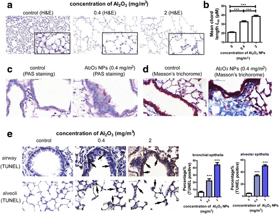
Exposure to Al2O3 NPs led to experimental COPD in a murine model. a Representative images of normal alveolar area and emphysema. The images on the right bottom of each panel are magnified (400) of area from the original one. b Lm was significantly increased in Al2O3 NPs-exposed mouse lungs (n = 36, *** P < 0.001). c Representative images of PAS staining, the PAS+ cells suggested hypersecretion of airway epithelial cells (shown by arrows). d Representative images of Masson’s Trichorome staining. Deposit of collagen around airway was stained purple. e Representative images of TUNEL staining and percentage of TUNEL+ cells. The TUNEL+ cells were shown by arrows. (n = 30, *** P < 0.001, compared with control group)
Fig. 3
Fig. 3
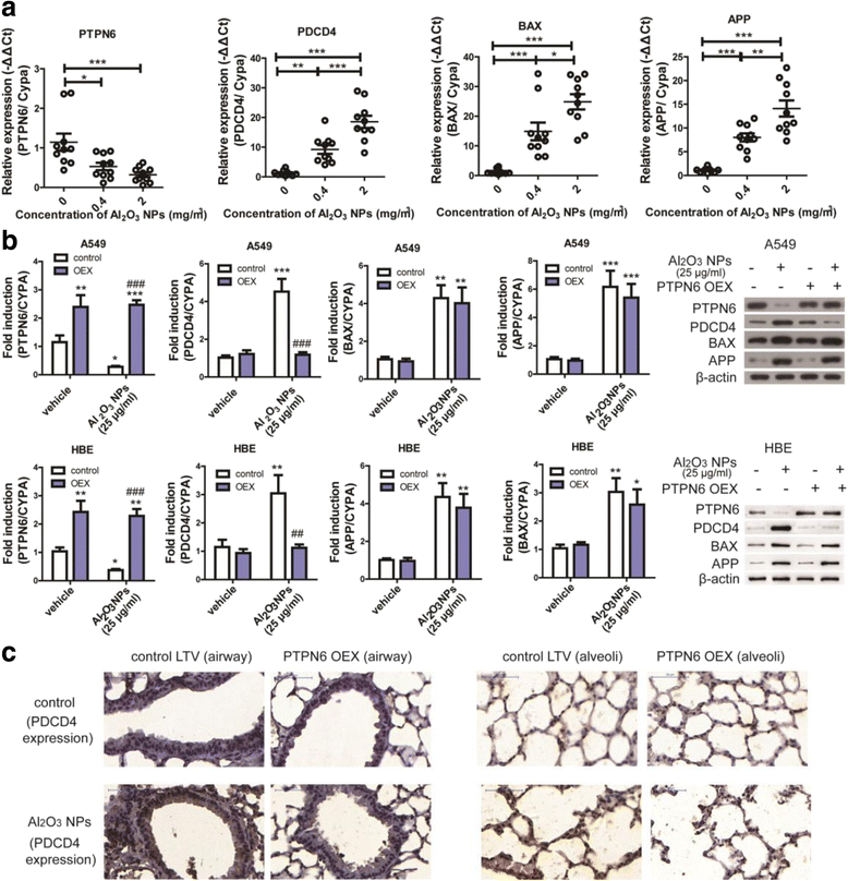
Overexpression of PTPN6 inhibited PDCD4 expression levels both in vitro and in vivo. a expression of PTPN6, PDCD4, BAX and APP in mouse lungs exposed to Al2O3 NPs (n = 10, * P < 0.05, ** P < 0.01, *** P < 0.001). b mRNA and protein expression levels of PTPN6, PDCD4, BAX and APP in A549 and HBE cells (n = 6, * P < 0.05, compared with vehicle-treated control, ** P < 0.01, compared with vehicle-treated control, *** P < 0.001, compared with vehicle-treated control, ## P < 0.01, compared with vehicle/Al2O3 NPs-exposed group, ### P < 0.001, compared with vehicle/Al2O3 NPs-exposed group). c Representative images of PDCD4 expression in mouse lung tissues. LTV: lentivirus; OEX: overexpression
Fig. 4
Fig. 4
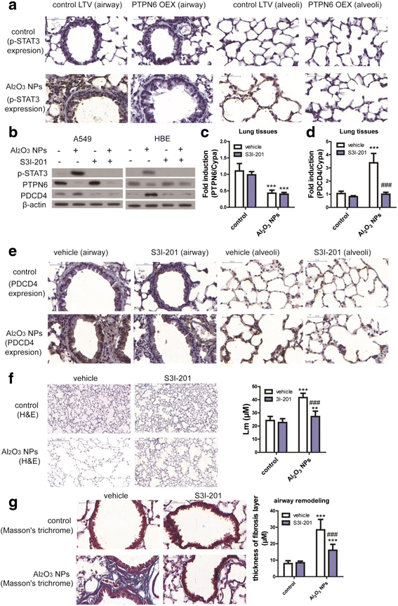
PTPN6 inhibited PDCD4 expression in a STAT3-dependent manner. a Representative images of p-STAT3 in mouse lung tissues. b The STAT3 inhibitor (S3I-201) inhibited activation of STAT3 and expression of PDCD4 in A549 and HBE cells. c The STAT3 inhibitor (S3I-201) did not inhibit expression of PTPN6 in mouse lung tissues (n = 6, *** P < 0.001, compared with vehicle control). d The STAT3 inhibitor (S3I-201) inhibited expression of PDCD4 in mouse lung tissues (n = 6, *** P < 0.001, compared with vehicle control, ### P < 0.001, compared with Al2O3 NPs-treated vehicle mice). e Representative images of PDCD4 expression in mouse lung tissues f Representative images of emphysema and Lm in mouse lungs (n = 36, ** P < 0.01, compared with vehicle control, *** P < 0.001, compared with vehicle control, ### P < 0.001, compared with Al2O3 NPs-exposed vehicle mice). g Representative images of airway remodeling and thickness of fibrosis layer around airway in mouse lungs (n = 36, *** P < 0.001, compared with vehicle control, ### P < 0.001, compared with Al2O3 NPs-exposed vehicle mice)
Fig. 5
Fig. 5
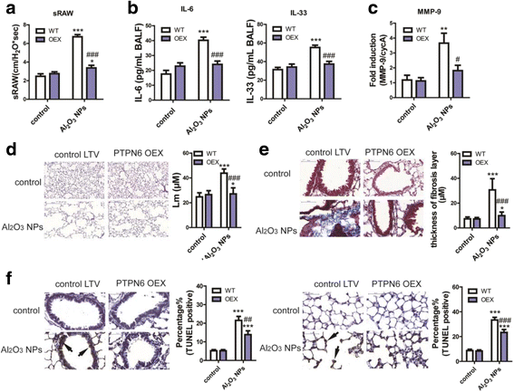
Overexpression of PTPN6 rescued experimental COPD in mouse model. a sRAW of conscious mice (n = 10, * P < 0.05, compared with WT control, *** P < 0.001, compared with WT control, ### P < 0.001, compared with Al2O3 NPs-exposed WT mice). b Levels of inflammatory mediators in BALF (n = 4, *** P < 0.001, compared with WT control, ### P < 0.001, compared with Al2O3 NPs-exposed WT mice). c Expression levels of MMP-9 in mouse lungs (n = 6, ** P < 0.01, compared with WT control, # P < 0.05, compared with Al2O3 NPs-exposed WT mice). d Representative images of emphysema and Lm in mouse lungs (n = 36, * P < 0.05, compared with WT control, *** P < 0.001, compared with WT control, ## P < 0.01, compared with Al2O3 NPs-exposed WT mice). e Representative images of airway remodeling and thickness of fibrosis layer around airway in mouse lungs (n = 36, * P < 0.05, compared with WT control, *** P < 0.001, compared with WT control, ### P < 0.001, compared with Al2O3 NPs-exposed WT mice). f Representative images of TUNEL staining and percentage of TUNEL+ cells in airway epithelia and alveolar epithelia (n = 30, *** P < 0.001, compared with WT control, ## P < 0.01, ### P < 0.001, compared with Al2O3 NPs-exposed WT mice)
Fig. 6
Fig. 6
Key molecular pathway involved in Al2O3 NPs-induced COPD-like lesions

Similar articles

Cited by

References

    1. Braakhuis HM, Cassee FR, Fokkens PH, de la Fonteyne LJ, Oomen AG, Krystek P, de Jong WH, van Loveren H, Park MV. Identification of the appropriate dose metric for pulmonary inflammation of silver nanoparticles in an inhalation toxicity study. Nanotoxicology. 2016;10:63–73. doi: 10.3109/17435390.2015.1127443. - DOI - PubMed
    1. Ho CC, Lee HL, Chen CY, Luo YH, Tsai MH, Tsai HT, Lin P. Involvement of the cytokine-IDO1-AhR loop in zinc oxide nanoparticle-induced acute pulmonary inflammation. Nanotoxicology. 2017;11:360–370. doi: 10.1080/17435390.2017.1306129. - DOI - PubMed
    1. Sager TM, Wolfarth M, Leonard SS, Morris AM, Porter DW, Castranova V, Holian A. Role of engineered metal oxide nanoparticle agglomeration in reactive oxygen species generation and cathepsin B release in NLRP3 inflammasome activation and pulmonary toxicity. Inhal Toxicol. 2016;28:686–697. doi: 10.1080/08958378.2016.1257664. - DOI - PMC - PubMed
    1. Willhite CC, Karyakina NA, Yokel RA, Yenugadhati N, Wisniewski TM, Arnold IM, Momoli F, Krewski D. Systematic review of potential health risks posed by pharmaceutical, occupational and consumer exposures to metallic and nanoscale aluminum, aluminum oxides, aluminum hydroxide and its soluble salts. Crit Rev Toxicol. 2014;44(Suppl 4):1–80. doi: 10.3109/10408444.2014.934439. - DOI - PMC - PubMed
    1. Chen L, Yokel RA, Hennig B, Toborek M. Manufactured aluminum oxide nanoparticles decrease expression of tight junction proteins in brain vasculature. J NeuroImmune Pharmacol. 2008;3:286–295. doi: 10.1007/s11481-008-9131-5. - DOI - PMC - PubMed

MeSH terms