Distinct cytokine patterns may regulate the severity of neonatal asphyxia-an observational study
- PMID: 29233180
- PMCID: PMC5727967
- DOI: 10.1186/s12974-017-1023-2
Distinct cytokine patterns may regulate the severity of neonatal asphyxia-an observational study
Abstract
Background: Neuroinflammation and a systemic inflammatory reaction are important features of perinatal asphyxia. Neuroinflammation may have dual aspects being a hindrance, but also a significant help in the recovery of the CNS. We aimed to assess intracellular cytokine levels of T-lymphocytes and plasma cytokine levels in moderate and severe asphyxia in order to identify players of the inflammatory response that may influence patient outcome.
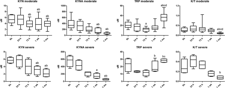
Methods: We analyzed the data of 28 term neonates requiring moderate systemic hypothermia in a single-center observational study. Blood samples were collected between 3 and 6 h of life, at 24 h, 72 h, 1 week, and 1 month of life. Neonates were divided into a moderate (n = 17) and a severe (n = 11) group based on neuroradiological and amplitude-integrated EEG characteristics. Peripheral blood mononuclear cells were assessed with flow cytometry. Cytokine plasma levels were measured using Bioplex immunoassays. Components of the kynurenine pathway were assessed by high-performance liquid chromatography.
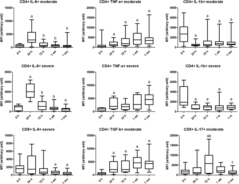
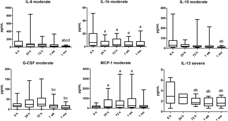
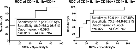
Results: The prevalence and extravasation of IL-1b + CD4 cells were higher in severe than in moderate asphyxia at 6 h. Based on Receiver operator curve analysis, the assessment of the prevalence of CD4+ IL-1β+ and CD4+ IL-1β+ CD49d+ cells at 6 h appears to be able to predict the severity of the insult at an early stage in asphyxia. Intracellular levels of TNF-α in CD4 cells were increased at all time points compared to 6 h in both groups. At 1 month, intracellular levels of TNF-α were higher in the severe group. Plasma IL-6 levels were higher at 1 week in the severe group and decreased by 1 month in the moderate group. Intracellular levels of IL-6 peaked at 24 h in both groups. Intracellular TGF-β levels were increased from 24 h onwards in the moderate group.
Conclusions: IL-1β and IL-6 appear to play a key role in the early events of the inflammatory response, while TNF-α seems to be responsible for prolonged neuroinflammation, potentially contributing to a worse outcome. The assessment of the prevalence of CD4+ IL-1β+ and CD4+ IL-1β+ CD49d+ cells at 6 h appears to be able to predict the severity of the insult at an early stage in asphyxia.
Keywords: Cytokine network; Hypoxic ischemic encephalopathy; Neuroinflammation; Perinatal asphyxia.
Conflict of interest statement
Authors’ information
VL is a professor of Neurology and an expert of the kynurenine system. TT is a professor of Pediatrics and an expert of neonatal development. MS is the head of the Neonatal Intensive Care Unit of the First Department of Pediatrics at Semmelweis University, Budapest (regional cooling centre), and an expert on neonatal asphyxia. GT is a consultant neonatologist and an expert of neonatal immunology.
Ethics approval and consent to participate
Our study was reviewed and approved by the Hungarian Medical Research Council (TUKEB 6578-0/2011-EKU), and written informed consent was obtained from parents of all participants. The study was adhered to the tenets of the most recent revision of the Declaration of Helsinki.
Consent for publication
Not applicable
Competing interests
The authors declare that they have no competing interests.
Publisher’s Note
Springer Nature remains neutral with regard to jurisdictional claims in published maps and institutional affiliations.
Figures


Similar articles
-
Cytokine production pattern of T lymphocytes in neonatal arterial ischemic stroke during the first month of life-a case study.J Neuroinflammation. 2018 Jun 22;15(1):191. doi: 10.1186/s12974-018-1229-y. J Neuroinflammation. 2018. PMID: 29933753 Free PMC article.
-
Cord Blood IL-16 Is Associated with 3-Year Neurodevelopmental Outcomes in Perinatal Asphyxia and Hypoxic-Ischaemic Encephalopathy.Dev Neurosci. 2017;39(1-4):59-65. doi: 10.1159/000471508. Epub 2017 May 11. Dev Neurosci. 2017. PMID: 28490023
-
Changes in serum cytokine and cortisol levels in normothermic and hypothermic term neonates after perinatal asphyxia.Inflamm Res. 2013 Jan;62(1):81-7. doi: 10.1007/s00011-012-0554-3. Epub 2012 Sep 18. Inflamm Res. 2013. PMID: 22986466
-
Long-term follow-up of term neonates with perinatal asphyxia.Clin Perinatol. 1993 Jun;20(2):483-500. Clin Perinatol. 1993. PMID: 7689432 Review.
-
[Cytokines in children with immunodeficiencies].Folia Med Cracov. 1999;40(1-2):5-97. Folia Med Cracov. 1999. PMID: 10909468 Review. Polish.
Cited by
-
Fas-ligand and interleukin-6 in the cerebrospinal fluid are early predictors of hypoxic-ischemic encephalopathy and long-term outcomes after birth asphyxia in term infants.J Neuroinflammation. 2018 Aug 8;15(1):223. doi: 10.1186/s12974-018-1253-y. J Neuroinflammation. 2018. PMID: 30089504 Free PMC article.
-
Interleukin-1 blockade attenuates white matter inflammation and oligodendrocyte loss after progressive systemic lipopolysaccharide exposure in near-term fetal sheep.J Neuroinflammation. 2021 Aug 31;18(1):189. doi: 10.1186/s12974-021-02238-4. J Neuroinflammation. 2021. PMID: 34465372 Free PMC article.
-
Low mucosal-associated invariant T-cell number in peripheral blood of patients with immune thrombocytopenia and their response to prednisolone.PLoS One. 2018 Nov 8;13(11):e0207149. doi: 10.1371/journal.pone.0207149. eCollection 2018. PLoS One. 2018. PMID: 30408105 Free PMC article.
-
Comparison of new biomarkers in the diagnosis of perinatal asphyxia.Iran J Child Neurol. 2023 Winter;17(1):99-110. doi: 10.22037/ijcn.v17i2.38561. Epub 2023 Jan 1. Iran J Child Neurol. 2023. PMID: 36721830 Free PMC article.
-
Differential Effects of Hypothermia and SZR72 on Cerebral Kynurenine and Kynurenic Acid in a Piglet Model of Hypoxic-Ischemic Encephalopathy.Int J Mol Sci. 2023 Sep 25;24(19):14522. doi: 10.3390/ijms241914522. Int J Mol Sci. 2023. PMID: 37833970 Free PMC article.
References
-
- Beckstrom AC, Humston EM, Snyder LR, Synovec RE, Juul SE. Application of comprehensive two-dimensional gas chromatography with time-of-flight mass spectrometry method to identify potential biomarkers of perinatal asphyxia in a non-human primate model. J Chromatogr A. 2011;1218(14):1899–1906. doi: 10.1016/j.chroma.2011.01.086. - DOI - PMC - PubMed
Publication types
MeSH terms
Substances
Grants and funding
LinkOut - more resources
Full Text Sources
Other Literature Sources
Research Materials

