. 2017 Dec 13;10(1):57.
doi: 10.1186/s13041-017-0338-3.

Restoring synaptic plasticity and memory in mouse models of Alzheimer's disease by PKR inhibition

Affiliations

Restoring synaptic plasticity and memory in mouse models of Alzheimer's disease by PKR inhibition

Kyoung-Doo Hwang et al. Mol Brain. .

Abstract

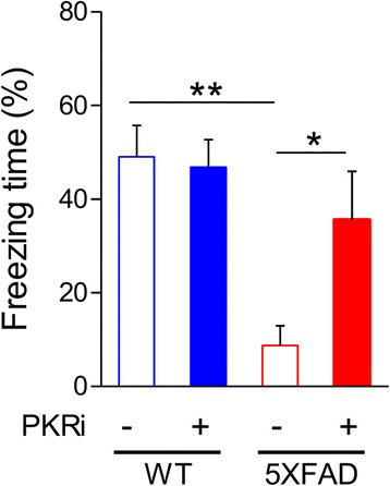
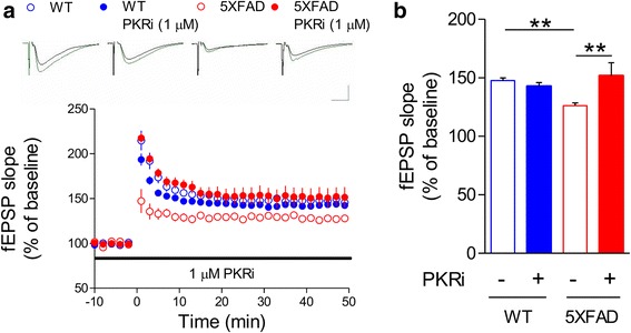
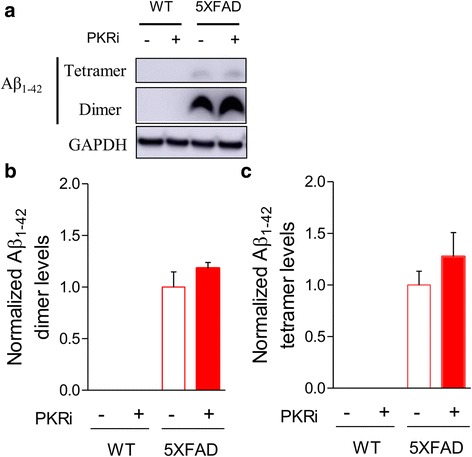
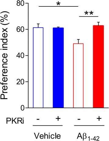
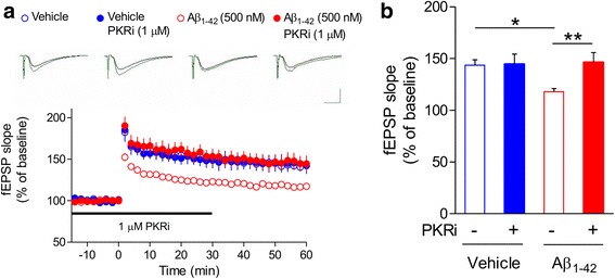
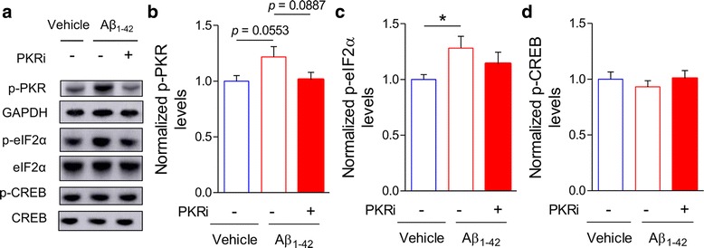
Alzheimer's disease (AD) is a neurodegenerative disorder associated with deficits in cognition and synaptic plasticity. While accumulation of amyloid β (Aβ) and hyper-phosphorylation of tau are parts of the etiology, AD can be caused by a large number of different genetic mutations and other unknown factors. Considering such a heterogeneous nature of AD, it would be desirable to develop treatment strategies that can improve memory irrespective of the individual causes. Reducing the phosphorylation of eukaryotic translation initiation factor 2α (eIF2α) was shown to enhance long-term memory and synaptic plasticity in naïve mice. Moreover, hyper-phosphorylation of eIF2α is observed in the brains of postmortem AD patients. Therefore, regulating eIF2α phosphorylation can be a plausible candidate for restoring memory in AD by targeting memory-enhancing mechanism. In this study, we examined whether PKR inhibition can rescue synaptic and learning deficits in two different AD mouse models; 5XFAD transgenic and Aβ1-42-injected mice. We found that the acute treatment of PKR inhibitor (PKRi) can restore the deficits in long-term memory and long-term potentiation (LTP) in both mouse models without affecting the Aβ load in the hippocampus. Our results prove the principle that targeting memory enhancing mechanisms can be a valid candidate for developing AD treatment.

Keywords: Alzheimer’s disease (AD); Amyloid β (Aβ); Contextual fear conditioning; Long-term potentiation (LTP); Object recognition memory; PKR inhibitor (PKRi).


Conflict of interest statement

Ethics approval

All the animal experiments were approved by the Seoul National University Institutional Animal Care and Use Committee (SNU IACUC) and the Chung-Ang University Institutional Animal Care and Use Committee (CAU IACUC).

Consent for publication

Not applicable.

Competing interests

The authors declare no conflict of interest.

Publisher’s Note

Springer Nature remains neutral with regard to jurisdictional claims in published maps and institutional affiliations.

Figures

Fig. 1
Fig. 1
PKRi treatment rescues fear memory deficit in 5XFAD mice. Eleven to twelve months old 5XFAD mice showed significant deficit in contextual fear memory, which was rescued by PKRi treatment (0.335 mg/kg) (% freezing: WT, 49.03 ± 6.67%, n = 9 mice; WT + PKRi, 46.78 ± 5.90%, n = 10 mice; 5XFAD, 8.74 ± 4.18%, n = 6 mice; 5XFAD + PKRi, 35.55 ± 10.38%, n = 7 mice; Two-way ANOVA, interaction between genotype and PKRi, p = 0.0558, Bonferroni post-tests, *p < 0.05, **p < 0.01). Bars represent as mean ± SEM
Fig. 2
Fig. 2
Inhibition of PKR restores LTP impairment in 5XFAD mice. a LTP in Schaffer-collateral-CA1 pathway was induced by theta burst stimulation (TBS). Field excitatory synaptic potential (fEPSP) slopes were normalized by the average of baseline recordings. Slices from 5XFAD mice showed significantly reduced LTP than WT, which can be restored by PKRi treatment (1 μM, 90 min). Representative traces were shown above. Black, baseline; Green, average between 40 and 50 min after TBS. Vertical bar, 1.0 mV; horizontal bar, 5 ms. b Cumulative data showing the average field excitatory synaptic potential (fEPSP) slope of 40–50 min after TBS (WT, 147.77 ± 2.19%, n = 6 slices from 4 mice; WT + PKRi, 142.83 ± 3.10%, n = 9 slices from 5 mice; 5XFAD, 126.22 ± 2.36%, n = 7 slices from 5 mice; 5XFAD and PKRi, 151.67 ± 11.20%, n = 5 slices from 3 mice; Two-way ANOVA, interaction between genotype and PKRi, *p < 0.05, Two-way ANOVA, Bonferroni post-tests, **p < 0.01). Bars represent as mean ± SEM
Fig. 3
Fig. 3
PKRi treatment does not decrease Aβ1–42 in the hippocampus of 5XFAD mice. a Representative immunoblots of protein extracts from the hippocampi 1 h after PKRi injection (0.335 mg/kg) in WT and 5XFAD mice. b, c Quantification of the of Aβ1–42 oligomers such as dimers and tetramers showing that PKRi treatment did not affect Aβ1–42 oligomers in 5XFAD mice (dimer levels normalized by that of 5XFAD; vehicle, 0; vehicle + PKRi, 0; Aβ1–42, 1.00 ± 0.15; Aβ1–42 + PKRi, 1.18 ± 0.06; unpaired t-test, 5XFAD vs 5XFAD + PKRi, p = 0.2674; tetramer levels normalized by that of 5XFAD; vehicle, 0; vehicle + PKRi, 0; Aβ1–42, 1.00 ± 0.13; Aβ1–42 + PKRi, 1.28 ± 0.23; unpaired t-test, 5XFAD vs 5XFAD + PKRi, p = 0.3243; 6 hippocampi from 3 mice per group). Bars represent as mean ± SEM
Fig. 4
Fig. 4
PKRi treatment rescues memory deficit in novel object recognition (NOR) in Aβ1–42–injected mice. Injection of Aβ1–42 oligomers (3 μg/mouse) induced NOR memory deficit, which was rescued by PKRi treatment. PKRi (0.335 mg/kg) was intraperitoneally injected 20 min before NOR training (Preference index for the novel object: Vehicle, 61.33 ± 2.85%; PKRi, 60.92 ± 0.83%; Aβ, 49.09 ± 3.21%; Aβ1–42 and PKRi, 62.7 ± 2.79%; Two-way ANOVA, interaction between Aβ1–42 and PKRi, *p < 0.05; Bonferroni post-tests, *p < 0.05, **p < 0.01, n = 6 mice for each group). Bars represent as mean ± SEM
Fig. 5
Fig. 5
Inhibition of PKR restores Aβ1–42–induced LTP impairment in hippocampus. a PKRi treatment rescued the LTP deficit in Aβ1–42-treated slices. Aβ1–42 (500 nM) was treated for 2 h before recording and PKRi (1 μM) was applied for 1 h (30 min before/after LTP induction). Representative traces were shown above. Black, baseline; Green, average between 40 and 50 min after HFS. Vertical bar, 1.0 mV; horizontal bar, 5 ms. b Cumulative data showing the average field excitatory synaptic potential (fEPSP) slope of 50–60 min after LTP induction (2X HFS) (Vehicle, 143.52 ± 5.22%, n = 7 slices from 6 mice; PKRi, 144.48 ± 9.73%, n = 7 slices from 5 mice; Aβ1–42, 118.00 ± 2.99%, n = 12 slices from 8 mice; Aβ1–42 and PKRi, 146.28 ± 9.45%, n = 7 slices from 7 mice; Two-way ANOVA, interaction between Aβ1–42 and PKRi, *p < 0.05, Two-way ANOVA, Bonferroni post-tests, *p < 0.05, **p < 0.01). Bars represent as mean ± SEM
Fig. 6
Fig. 6
PKRi treatment has a trend to reverse Aβ1–42-mediated changes in PKR signaling. a Representative immunoblots of protein extracts from hippocampi 30 min after PKRi injection (0.335 mg/kg) in Aβ1–42-treated mice. b PKRi treatment showed a trend to decrease eIF2α phosphorylation in Aβ1–42-treated mice, but the effect was not statistically significant (normalized p-PKR, vehicle, 1.00 ± 0.05, 14, 14 hippocampi from 11 mice; Aβ1–42, 1.22 ± 0.09, 15 hippocampi from 13 mice; Aβ1–42 + PKRi, 1.01 ± 0.06, 14 hippocampi from 11 mice; unpaired t-test, vehicle vs Aβ1–42, p = 0.055; Aβ1–42 vs Aβ1–42 + PKRi, p = 0.089). c PKRi treatment showed a trend to decrease eIF2α phosphorylation in Aβ1–42-treated mice, but the effect was not statistically significant (normalized p-eIF2α, vehicle, 1.00 ± 0.04, 17 hippocampi from 11 mice; Aβ1–42, 1.28 ± 0.11, 19 hippocampi from 13 mice; Aβ1–42 + PKRi, 1.14 ± 0.10, 18 hippocampi from 12 mice; unpaired t-test, vehicle vs Aβ1–42, *p < 0.05; Aβ1–42 vs Aβ1–42 + PKRi, p = 0.354). (D) CREB phosphorylation was slightly reduced by Aβ1–42 and was rescued by PKRi treatment although it was not statistically significant (normalized p-CREB, vehicle, 1.00 ± 0.07, 15 hippocampi from 9 mice; Aβ1–42, 0.93 ± 0.05, 15 hippocampi from 9 mice; Aβ1–42 + PKRi, 1.01 ± 0.07, 16 hippocampi from 10 mice; unpaired t-test, vehicle vs Aβ1–42, p = 0.426; Aβ1–42 vs Aβ1–42 + PKRi, p = 0.390). Bars represent as mean ± SEM

Similar articles

Cited by

References

    1. Goate A, Chartier-Harlin MC, Mullan M, Brown J, Crawford F, Fidani L, Giuffra L, Haynes A, Irving N, James L, et al. Segregation of a missense mutation in the amyloid precursor protein gene with familial Alzheimer's disease. Nature. 1991;349:704–706. doi: 10.1038/349704a0. - DOI - PubMed
    1. Schellenberg GD, Bird TD, Wijsman EM, Orr HT, Anderson L, Nemens E, White JA, Bonnycastle L, Weber JL, Alonso ME, et al. Genetic linkage evidence for a familial Alzheimer's disease locus on chromosome 14. Science. 1992;258:668–671. doi: 10.1126/science.1411576. - DOI - PubMed
    1. Levy-Lahad E, Wasco W, Poorkaj P, Romano DM, Oshima J, Pettingell WH, CE Y, Jondro PD, Schmidt SD, Wang K, et al. Candidate gene for the chromosome 1 familial Alzheimer's disease locus. Science. 1995;269:973–977. doi: 10.1126/science.7638622. - DOI - PubMed
    1. Hardy J, Higgins G. Alzheimer's disease: the amyloid cascade hypothesis. Science. 1992;256:184–185. doi: 10.1126/science.1566067. - DOI - PubMed
    1. Duyckaerts C, Delatour B, Potier MC. Classification and basic pathology of Alzheimer disease. Acta Neuropathol. 2009;118:5–36. doi: 10.1007/s00401-009-0532-1. - DOI - PubMed