

Effects of Angelicae Pubescentis and Loranthi Decotion on repairing knee joint cartilages in rats
- PMID: 29233184
- PMCID: PMC5727648
- DOI: 10.1186/s13018-017-0679-8
Effects of Angelicae Pubescentis and Loranthi Decotion on repairing knee joint cartilages in rats
Abstract
Background: Knee osteoarthritis (KOA) is a common orthopedics disease and its pathological changes at early stage are the damage and loss of articular cartilage. Traditional Chinese medicine (TCM) prescription contains multiple components and has the unique advantages of the diversity of targets.We compared the traditional Chinese medical formulae (Angelicae Pubescentis and Loranthi decotion, APLD, or Duhuo Jisheng) with a western medicine (glucosamine sulfate, GS) to treat the rat arthritis models, and tracked the outcomes.
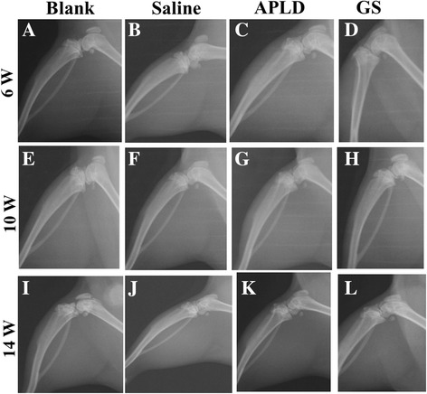
Methods: Thirty-two Wistar rats (weight 180 ± 10 g, 6-week-old) were randomly divided into four groups (eight for each): group A as normal control group (no surgery and no drug treatment), group B as SIA (surgery-induced arthritis) model control without drug treatment, group C as SIA model + APLD, and group D as SIA model + GS. Anterior cruciate ligament in the knee joint of both hind legs from each rat in groups B, C, and D was shown and cut off to establish the SIA model. After 6 weeks of the surgery, rats in group C or D were treated with APLD or GS, respectively, for 8 weeks. Bone X-ray examination, histological images, and determination of genes of collagen II and aggrecan were performed. At week 14, both knee joint gap and bone structure disappeared in rats of group B, but they were visible in rats of groups A, C, and D.
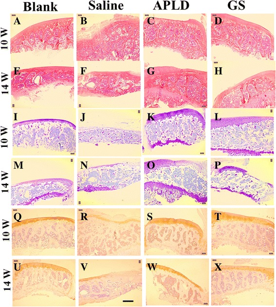
Results: Histological images revealed that the structure and composition of the knee joint cartilage were significantly degenerated in group B and improved in group C. Genes of collagen II and aggrecan were significantly increased in both group C and D.
Conclusion: APLD or GS gavage treatment for knee osteoarthritis (KOA) rat models was effective on the proliferation of cartilage chondrocytes and the damaged knee joint tissue repairing, and the APLD showed slightly superior in general.
Keywords: Angelicae Pubescentis and Loranthi decotion; Chondrocyte; Duhuo; Knee osteoarthritis; Serum.
Conflict of interest statement
Ethics approval
The study protocol was approved by the Ethics Committee of the Shanghai University of Traditional Chinese Medicine.
Consent for publication
The current research did not involve human participants, this information is not applicable.
Competing interests
The authors declare that they have no competing interests.
Publisher’s Note
Springer Nature remains neutral with regard to jurisdictional claims in published maps and institutional affiliations.
Figures
Similar articles
-
Effect of Ermiao Recipe with medicinal guide Angelicae Pubescentis Radix on promoting the homing of bone marrow stem cells to treat cartilage damage in osteoarthritis rats.Chin J Integr Med. 2014 Aug;20(8):600-9. doi: 10.1007/s11655-014-1761-2. Epub 2014 Aug 5. Chin J Integr Med. 2014. PMID: 25087604
-
Intra-Articular Injection of Cross-Linked Hyaluronic Acid-Dexamethasone Hydrogel Attenuates Osteoarthritis: An Experimental Study in a Rat Model of Osteoarthritis.Int J Mol Sci. 2016 Apr 15;17(4):411. doi: 10.3390/ijms17040411. Int J Mol Sci. 2016. PMID: 27092487 Free PMC article.
-
Glucosamine sulfate reduces experimental osteoarthritis and nociception in rats: association with changes of mitogen-activated protein kinase in chondrocytes.Osteoarthritis Cartilage. 2010 Sep;18(9):1192-202. doi: 10.1016/j.joca.2010.05.012. Epub 2010 May 25. Osteoarthritis Cartilage. 2010. PMID: 20510383
-
Knee osteoarthritis: A review of animal models and intervention of traditional Chinese medicine.Animal Model Exp Med. 2024 Apr;7(2):114-126. doi: 10.1002/ame2.12389. Epub 2024 Feb 26. Animal Model Exp Med. 2024. PMID: 38409942 Free PMC article. Review.
-
Joint structure modification in osteoarthritis: development of SMOAD drugs.Curr Rheumatol Rep. 2001 Dec;3(6):506-12. doi: 10.1007/s11926-001-0065-7. Curr Rheumatol Rep. 2001. PMID: 11709113 Review.
Cited by
-
Anti-inflammatory activity of Radix Angelicae biseratae in the treatment of osteoarthritis determined by systematic pharmacology and in vitro experiments.Exp Ther Med. 2021 Jan;21(1):5. doi: 10.3892/etm.2020.9437. Epub 2020 Nov 2. Exp Ther Med. 2021. PMID: 33235614 Free PMC article.
-
Duhuo Jisheng Decoction inhibits SDF-1-induced inflammation and matrix degradation in human degenerative nucleus pulposus cells in vitro through the CXCR4/NF-κB pathway.Acta Pharmacol Sin. 2018 Jun;39(6):912-922. doi: 10.1038/aps.2018.36. Epub 2018 May 24. Acta Pharmacol Sin. 2018. PMID: 29795361 Free PMC article.
-
Integrated Network and Experimental Pharmacology for Deciphering the Medicinal Substances and Multiple Mechanisms of Duhuo Jisheng Decoction in Osteoarthritis Therapy.Evid Based Complement Alternat Med. 2020 Nov 2;2020:7275057. doi: 10.1155/2020/7275057. eCollection 2020. Evid Based Complement Alternat Med. 2020. PMID: 33204290 Free PMC article.
-
Melatonin ameliorates osteoarthritis rat cartilage injury by inhibiting matrix metalloproteinases and JAK2/STAT3 signaling pathway.Inflammopharmacology. 2023 Feb;31(1):359-368. doi: 10.1007/s10787-022-01102-y. Epub 2022 Nov 24. Inflammopharmacology. 2023. PMID: 36427113
-
Efficacy and safety of combined Chinese and Western medicine in the treatment of knee osteoarthritis: a prospective, multicenter cohort study.Front Pharmacol. 2023 Aug 28;14:1176980. doi: 10.3389/fphar.2023.1176980. eCollection 2023. Front Pharmacol. 2023. PMID: 37701040 Free PMC article.
References
-
- Zhang G-D, Yang C, Yang G, Qi X. Application and development of kinematical alighment during total knee arthroplasty. Zhongguo Gu Shang. 2015;28:1162–1165. - PubMed
MeSH terms
Substances
LinkOut - more resources
Full Text Sources
Other Literature Sources
Miscellaneous
