Review
. 2018 Jan;24(1):7-17.
doi: 10.1016/j.molmed.2017.11.004. Epub 2017 Dec 7.

Monitoring Neurodegeneration in Glaucoma: Therapeutic Implications

Affiliations
Review

Monitoring Neurodegeneration in Glaucoma: Therapeutic Implications

Norimitsu Ban et al. Trends Mol Med. 2018 Jan.

Abstract

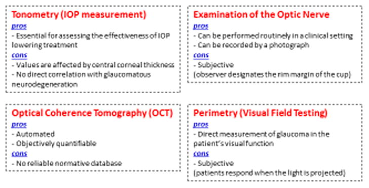
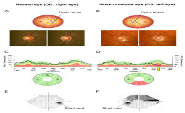
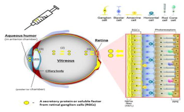
Glaucoma is one of the leading causes of blindness globally, and is characterized by loss of retinal ganglion cells (RGCs). Because vision loss in glaucoma is not reversible, therapeutic interventions early in disease are highly desirable. However, owing to the current limitations in evaluating glaucomatous neurodegeneration, it is challenging to monitor the disease severity and progression objectively, and to design rational therapeutic strategies accordingly. Therefore, there is a clear need to identify quantifiable molecular biomarkers of glaucomatous neurodegeneration. As such, in our opinion, molecular biomarker(s) that specifically reflect stress or death of RGCs, and which correlate with disease severity, progression, and response to therapy, are highly desirable.

Keywords: aqueous humor; biomarker; glaucoma; growth differentiation factor 15 (GDF15); neurodegeneration; retinal ganglion cell.


Figures

Figure 1
Figure 1
Current methods of detecting/monitoring glaucomatous neurodegeneration: pros and cons.
Figure 2
Figure 2. Biometric tools to assess the presence and severity of glaucoma in humans
(A–B) Cupping (excavation) of the optic nerve head (ONH) of the (A) normal and (B) glaucomatous eye. Loss of retinal ganglion cells is associated with enlargement of the optic cup. Solid circle represents the outline of the ONH, and dashed circle represents the contour of the cup within the ONH. Note that the glaucomatous eye has a larger cup than the normal eye. There is also a hemorrhage at the ONH rim (white arrow), which is another characteristic of glaucoma. Average diameter of ONH is approximately 1.5 mm. (C–D) Representative graphs of retinal nerve fiber layer (RNFL) thickness of the (C) normal and (D) glaucomatous eye measured by optical coherence tomography (OCT) are shown. Note that the inferior RNFL of the glaucomatous eye is thinner (yellow arrow) than normative database. S or SUP: superior. T or TMP: temporal. I or INF: inferior. N or NAS: nasal refer to the thickness of the RNFL in selected quadrants. (E–F) Perimetry of the (E) normal and (F) glaucomatous eye. Dark region in the normal visual field of the right eye indicates “physiological” blind spot corresponding to the reflection of the optic nerve in the temporal visual field. In contrast, perimetry of the glaucomatous eye demonstrates a superior arcuate visual field defect corresponding to loss of RNFL in the inferior quadrant.
Figure 3 (Key figure)
Figure 3 (Key figure). Structure of the neurosensory retina and access to molecular biomarkers of retinal ganglion cell health
The retina is composed of various types of neurons (red box inset); photoreceptors (rods and cones) in the outer retina, intermediate neurons (bipolar, amacrine, and horizontal cells), and ganglion cells in the inner retina. Axons of retinal ganglion cell (RGC) come together to form the optic nerve that extends processes that synapse in the lateral geniculate nucleus, ultimately sending axons to the occipital (visual) cortex of the brain. Retinal pigmented epithelium (RPE), which is located external to the neurosensory retina, performs essential specialized functions such as phagocytosis of photoreceptor outer segments and recycling photopigments in the visual cycle. Secreted proteins or soluble factors are released from RGCs into vitreous (1) and can diffuse throughout the vitreous body (2). In addition, they can be detected in the aqueous humor (AH) in the posterior chamber (3), and in the anterior chamber (4) of the eye. AH in anterior chamber is collected by a micro-invasive procedure called paracentesis through the peripheral cornea.

References

    1. Resnikoff S, et al. Global data on visual impairment in the year 2002. Bulletin of the World Health Organization. 2004;82:844–851. - PMC - PubMed
    1. Kingman S. Glaucoma is second leading cause of blindness globally. Bulletin of the World Health Organization. 2004;82:887–888. - PMC - PubMed
    1. Quigley HA, et al. Blockade of rapid axonal transport. Effect of intraocular pressure elevation in primate optic nerve. Archives of ophthalmology. 1979;97:525–531. - PubMed
    1. Takihara Y, et al. In vivo imaging of axonal transport of mitochondria in the diseased and aged mammalian CNS. Proceedings of the National Academy of Sciences of the United States of America. 2015;112:10515–10520. - PMC - PubMed
    1. The Advanced Glaucoma Intervention Study (AGIS): 7. The relationship between control of intraocular pressure and visual field deterioration. The AGIS Investigators. American journal of ophthalmology. 2000;130:429–440. - PubMed

Publication types