. 2018 Jan 9;10(1):58-72.
doi: 10.1016/j.stemcr.2017.11.005. Epub 2017 Dec 7.

Human Neural Stem Cell Transplantation Rescues Functional Deficits in R6/2 and Q140 Huntington's Disease Mice

Affiliations

Human Neural Stem Cell Transplantation Rescues Functional Deficits in R6/2 and Q140 Huntington's Disease Mice

Jack C Reidling et al. Stem Cell Reports. .

Abstract

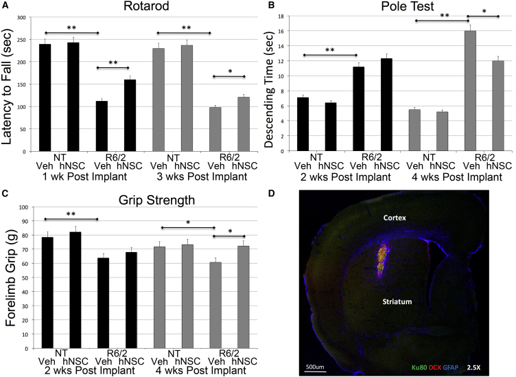
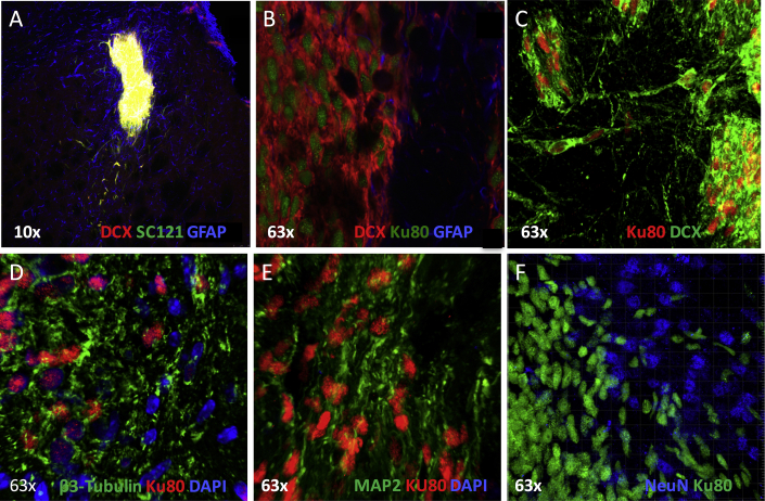
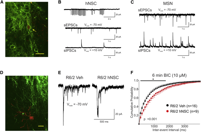
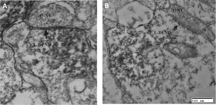
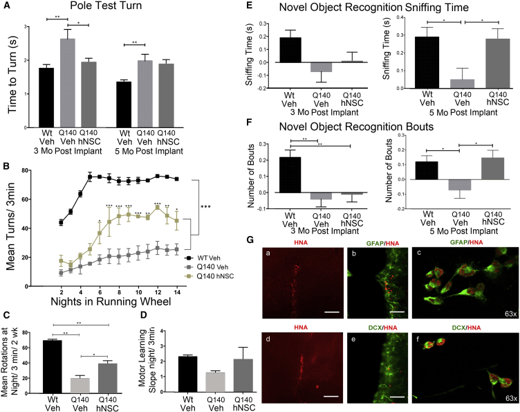
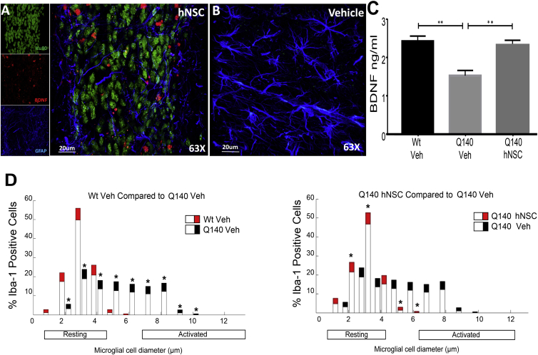
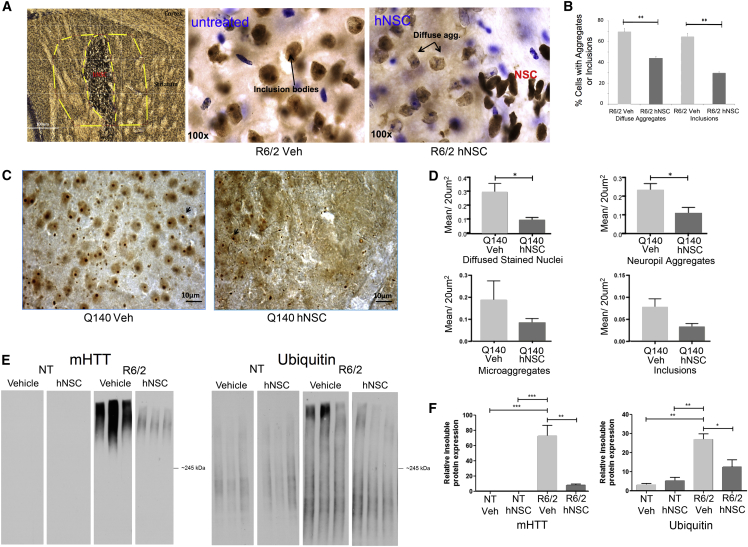
Huntington's disease (HD) is an inherited neurodegenerative disorder with no disease-modifying treatment. Expansion of the glutamine-encoding repeat in the Huntingtin (HTT) gene causes broad effects that are a challenge for single treatment strategies. Strategies based on human stem cells offer a promising option. We evaluated efficacy of transplanting a good manufacturing practice (GMP)-grade human embryonic stem cell-derived neural stem cell (hNSC) line into striatum of HD modeled mice. In HD fragment model R6/2 mice, transplants improve motor deficits, rescue synaptic alterations, and are contacted by nerve terminals from mouse cells. Furthermore, implanted hNSCs are electrophysiologically active. hNSCs also improved motor and late-stage cognitive impairment in a second HD model, Q140 knockin mice. Disease-modifying activity is suggested by the reduction of aberrant accumulation of mutant HTT protein and expression of brain-derived neurotrophic factor (BDNF) in both models. These findings hold promise for future development of stem cell-based therapies.

Keywords: Huntington's disease; Q140 mice; R6/2 mice; embryonic stem cells; neural stem cell; transplantation.


Figures

Figure 1
Figure 1
ESI-017 hNSCs Implanted in R6/2 Mice Improve Behavior and Exhibit Evidence of Differentiation into Immature Neurons and Astrocytes (A) Rotarod task demonstrates a deficit in R6/2 mice compared with non-transgenic littermates (NT), and hNSC-treated R6/2 mice have increased average latency to fall 1 week (black bars) and 3 weeks (gray bars) after implantation compared with vehicle-treated (Veh) mice. (B) Pole test demonstrates a deficit with R6/2 mice compared with NT. hNSC-treated R6/2 mice descend faster than Veh mice 4 weeks after implantation (gray bars) but not 2 weeks after implantation (black bars). (C) Grip strength demonstrates a deficit in R6/2 mice compared with NT. hNSC-treated R6/2 mice have greater grams of strength after 4 weeks compared with Veh mice (black bars) but not after 2 weeks (gray bars). (D) Immunohistochemistry (IHC). hNSCs (human marker SC121, green) implanted in striatum of R6/2 mice co-localize (yellow) with marker for neuron-restricted progenitors (doublecortin [DCX], red) and astrocytes (SC121 and GFAP, blue). One-way ANOVA followed by Tukey’s HSD test with Scheffé, Bonferroni, and Holm multiple comparison calculation performed post hoc. p < 0.05, ∗∗p < 0.01 (n = 15). Graphs show means ± SEM.
Figure 2
Figure 2
IHC Shows that ESI-017 hNSCs Implanted in R6/2 Mice Differentiate (A) hNSCs (SC121, green) implanted in R6/2 mice differentiate into neuron-restricted progenitors (doublecortin [DCX], red) and astrocytes (SC121 and GFAP, blue). (B) High magnification (63×) showing differentiation: hNSCs (human nuclear marker Ku80, green) implanted in R6/2 mice differentiate into neuron-restricted progenitors (DCX, red) and some astrocytes (Ku80 and GFAP, blue). (C) hNSCs (Ku80, red) and neuron-restricted progenitors (DCX, green). (D) hNSCs (Ku80, red) and neuron-restricted progenitors (βIII-tubulin, green); mouse cell nuclei shown with DAPI in blue. (E) hNSCs (Ku80, red) and neuron-restricted progenitors (MAP-2, green); mouse cell nuclei shown with DAPI in blue. (F) hNSCs (Ku80, green) do not co-localize with differentiated post-mitotic neuronal cell marker (NeuN, blue).
Figure 3
Figure 3
Implantation of ESI-017 hNSCs Reduces Corticostriatal Hyperexcitability in R6/2 Mice (A) Biocytin-filled (red, yellow arrow) hNSC that was recorded in the striatum and IHC with SC121 (green). Scale bar, 20 μm. (B) Top trace: cell-attached recording of spontaneously firing hNSC. Bottom traces: sEPSCs and sIPSCs from hNSC. Recordings illustrate spontaneous inward and outward synaptic currents in the hNSC. (C) sEPSCs and sIPSCs recorded in MSN. (D) Biocytin-filled MSN (red) near a cluster of hNSCs (SC121 green). Scale bar, 20 μm. (E) Recordings of sEPSCs in a subpopulation of R6/2 MSNs show “epileptiform” activity after the addition of the GABAA receptor antagonist, bicuculline (10 μM) (first trace). These large-amplitude excitatory events are usually followed by high-frequency small-amplitude sEPSCs. In mice with hNSC implants these events were markedly reduced in frequency (second trace). (F) In cells with “epileptiform” activity (6–8 min after BIC), there was a rightward shift in the cumulative inter-event interval probability distributions for the hNSC-implanted R6/2 group compared with vehicle, corresponding to a significant decrease in high-frequency spontaneous events (p < 0.001, two-way repeated-measures ANOVA followed by Bonferroni post hoc analysis; p < 0.05).
Figure 4
Figure 4
Nerve Terminals from the Host Make Synaptic Contact with the Implanted hNSCs (A) Unlabeled nerve terminal (U-NT), containing synaptic vesicles, making a synaptic-like contact (arrow) with an underlying labeled (SC121) hNSC dendrite (L-DEND). The connection may be symmetrical. (B) Unlabeled nerve terminal (U-NT), containing synaptic vesicles, making an asymmetrical synaptic contact (arrow) with an underlying labeled (SC121) hNSC dendrite (L-DEND). This asymmetrical contact suggests an excitatory synaptic contact.
Figure 5
Figure 5
ESI-017 hNSCs Implanted in Q140 Mice Improve Behavior and Exhibit Evidence of Differentiation into Immature Neurons and Astrocytes (A) Transient improvement in motor coordination (pole task) 3 months after cell injection. WT Veh (n = 20), Q140 Veh (n = 18), Q140 hNSC (n = 18). One-way ANOVA with Bonferroni post hoc test: p < 0.05, ∗∗p < 0.01. (B–D) Persistent improvement of running wheel deficits 5.5 months post treatment (n = 5 per group). (B) Graph showing mean running wheel rotations/3 min/night over 2 weeks, in 7.5-month-old male WT or Q140 mice 5.5 months post treatment. Comparison by two-way ANOVA: group effect F = 52.93, p < 0.0001; night in running wheel effect F = 17, p < 0.0001. Bonferroni post hoc test: p < 0.01, ∗∗p < 0.001, and ∗∗∗p < 0.0001 compared with Q140 Veh. (C) Total average running wheel turns at night over 2 weeks. Two-way ANOVA with Bonferroni post hoc test: p < 0.01, ∗∗p < 0.001. (D) Slope of motor learning not significant between the three groups. (E and F) Novel object recognition. hNSCs prevented the deficit in Q140 mice 5 months post treatment but not at 3 months in the discrimination index of sniffing time (E) or number of bouts (F). WT Veh n = 18, Q140 Veh n = 18, and Q140 hNSC n = 19. One-way ANOVA with Bonferroni post hoc test: p < 0.05, ∗∗p < 0.01. (G) Survival and differentiation of hNSCs in Q140 mice by staining with the human specific antibody (HNA, red; a and d) co-expressing with astrocytes (GFAP, green; b and c) or neuron-restricted progenitors (DCX, green; e and f). Scale bar, 20 μm. All graphs show mean ± SEM.
Figure 6
Figure 6
ESI-017 hNSCs Implanted in HD Mice Increase Expression of BDNF (A) ESI-017 hNSCs (Ku80, green) show co-localization with BDNF (red); astrocytes are shown as GFAP positive (blue). (B) Veh-treated mice show no BDNF or hNSCs but have GFAP (blue). (C) BDNF levels by ELISA in striatum of Q140 or WT mice 6 months post implant. (D) hNSC treatment in Q140 mice decreased microglial activation. Data are presented as the mean + 95% confidence interval (n = 5 per group). Bars represent percentage of cells of each diameter and the colored portion represents the confidence interval. Significant striatal microglial activation observed in Q140 Veh compared with WT Veh. Q140 hNSC mice showed significant reduction of microglial activation in striatum compared with Q140 Veh mice. p < 0.05 and ∗∗p < 0.01 by one-way ANOVA with Bonferroni post hoc test. Graphs show means ± SEM.
Figure 7
Figure 7
ESI-017 hNSCs Implanted in R6/2 Mice Cause Decreases in Diffuse Aggregates and Inclusions and Reduce Huntingtin Aggregates in Q140 Mice (A and B) ESI-017 hNSCs cause decreases in diffuse aggregates and inclusions (arrows in A) in R6/2 mice. (A) Image of Ku80 with nickel, HTT marker EM48, and cresyl violet for non-hNSC nuclear staining. Stereological assessment performed using StereoInvestigator. Contour tracing under 5× objective (dashed lines, example in left panel) and counting at 100×. Every third section was counted (40-μm coronal sections) for 6 sections throughout the striatum where Ku80 could be seen between bregma 0.5 mm and bregma −0.34 mm. (B) Graph depicting percentage of cells with aggregates or inclusions (n = 4/group) ∗∗p < 0.01 by one-way ANOVA with Bonferroni post hoc test. (C and D) ESI-017 hNSCs reduce Huntingtin aggregates in Q140 mice. (C) Images of HTT marker EM48 (arrows indicate inclusions). (D) HTT-stained nuclei and aggregates were analyzed with StereoInvestigator for quantification of aggregate type/section. Data are shown as mean ± SEM (n = 5/group). p < 0.05 by one-way ANOVA with Bonferroni post hoc test. (E and F) hNSC transplantation modulates insoluble protein accumulation in R6/2 mice. Western blot of striatal lysates separated into detergent-soluble and detergent-insoluble fractions. (E) R6/2 enriched in insoluble accumulated mHTT compared with NT. hNSC transplantation in R6/2 results in a significant reduction of insoluble HMW accumulated HTT compared with veh-treated animals. R6/2 striatum is also enriched in insoluble ubiquitin-conjugated proteins compared with NT. hNSC transplantation in R6/2 mice results in a significant reduction of ubiquitin-modified insoluble conjugated proteins compared with veh treatment with no significant effect in NT compared with veh controls. (F) Quantitation of the relative protein expression for mHTT and ubiquitin. Values represent means ± SEM. Statistical significance for relative insoluble accumulated mHTT and ubiquitin-conjugated protein expression in R6/2 was determined with a one-way ANOVA followed by Bonferroni post hoc test (n = 3/treatment). p < 0.05, ∗∗p < 0.01, ∗∗∗p < 0.001. Graphs show means ± SEM.

References

    1. Ager R.R., Davis J.L., Agazaryan A., Benavente F., Poon W.W., LaFerla F.M., Blurton-Jones M. Human neural stem cells improve cognition and promote synaptic growth in two complementary transgenic models of Alzheimer's disease and neuronal loss. Hippocampus. 2015;25:813–826. - PMC - PubMed
    1. An M.C., Zhang N., Scott G., Montoro D., Wittkop T., Mooney S., Melov S., Ellerby L.M. Genetic correction of Huntington's disease phenotypes in induced pluripotent stem cells. Cell Stem Cell. 2012;11:253–263. - PMC - PubMed
    1. Andre V.M., Cepeda C., Fisher Y.E., Huynh M., Bardakjian N., Singh S., Yang X.W., Levine M.S. Differential electrophysiological changes in striatal output neurons in Huntington's disease. J. Neurosci. 2011;31:1170–1182. - PMC - PubMed
    1. Benraiss A., Wang S., Herrlinger S., Li X., Chandler-Militello D., Mauceri J., Burm H.B., Toner M., Osipovitch M., Jim Xu Q. Human glia can both induce and rescue aspects of disease phenotype in Huntington disease. Nat. Commun. 2016;7:11758. - PMC - PubMed
    1. Blurton-Jones M., Kitazawa M., Martinez-Coria H., Castello N.A., Muller F.J., Loring J.F., Yamasaki T.R., Poon W.W., Green K.N., LaFerla F.M. Neural stem cells improve cognition via BDNF in a transgenic model of Alzheimer disease. Proc. Natl. Acad. Sci. USA. 2009;106:13594–13599. - PMC - PubMed

Publication types