. 2018 Apr 17;137(16):1712-1730.
doi: 10.1161/CIRCULATIONAHA.117.030785. Epub 2017 Dec 12.

Large Cardiac Muscle Patches Engineered From Human Induced-Pluripotent Stem Cell-Derived Cardiac Cells Improve Recovery From Myocardial Infarction in Swine

Affiliations

Large Cardiac Muscle Patches Engineered From Human Induced-Pluripotent Stem Cell-Derived Cardiac Cells Improve Recovery From Myocardial Infarction in Swine

Ling Gao et al. Circulation. .

Abstract

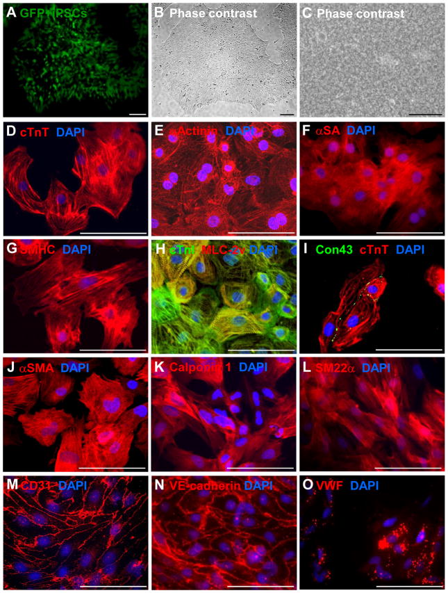
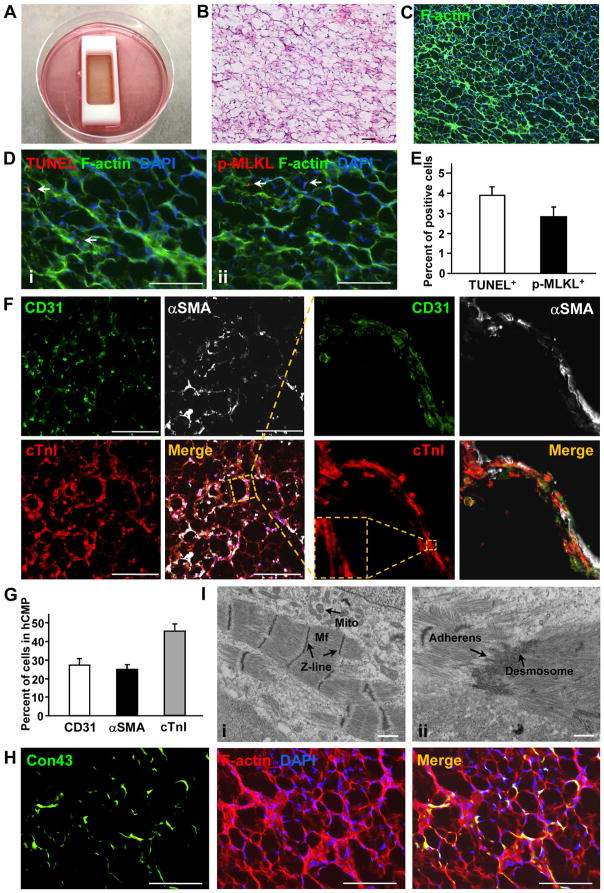
Background: Here, we generated human cardiac muscle patches (hCMPs) of clinically relevant dimensions (4 cm × 2 cm × 1.25 mm) by suspending cardiomyocytes, smooth muscle cells, and endothelial cells that had been differentiated from human induced-pluripotent stem cells in a fibrin scaffold and then culturing the construct on a dynamic (rocking) platform.

Methods: In vitro assessments of hCMPs suggest maturation in response to dynamic culture stimulation. In vivo assessments were conducted in a porcine model of myocardial infarction (MI). Animal groups included: MI hearts treated with 2 hCMPs (MI+hCMP, n=13), MI hearts treated with 2 cell-free open fibrin patches (n=14), or MI hearts with neither experimental patch (n=15); a fourth group of animals underwent sham surgery (Sham, n=8). Cardiac function and infarct size were evaluated by MRI, arrhythmia incidence by implanted loop recorders, and the engraftment rate by calculation of quantitative polymerase chain reaction measurements of expression of the human Y chromosome. Additional studies examined the myocardial protein expression profile changes and potential mechanisms of action that related to exosomes from the cell patch.

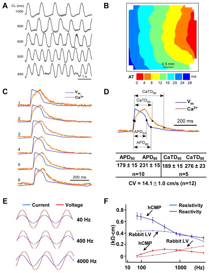
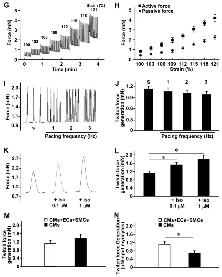
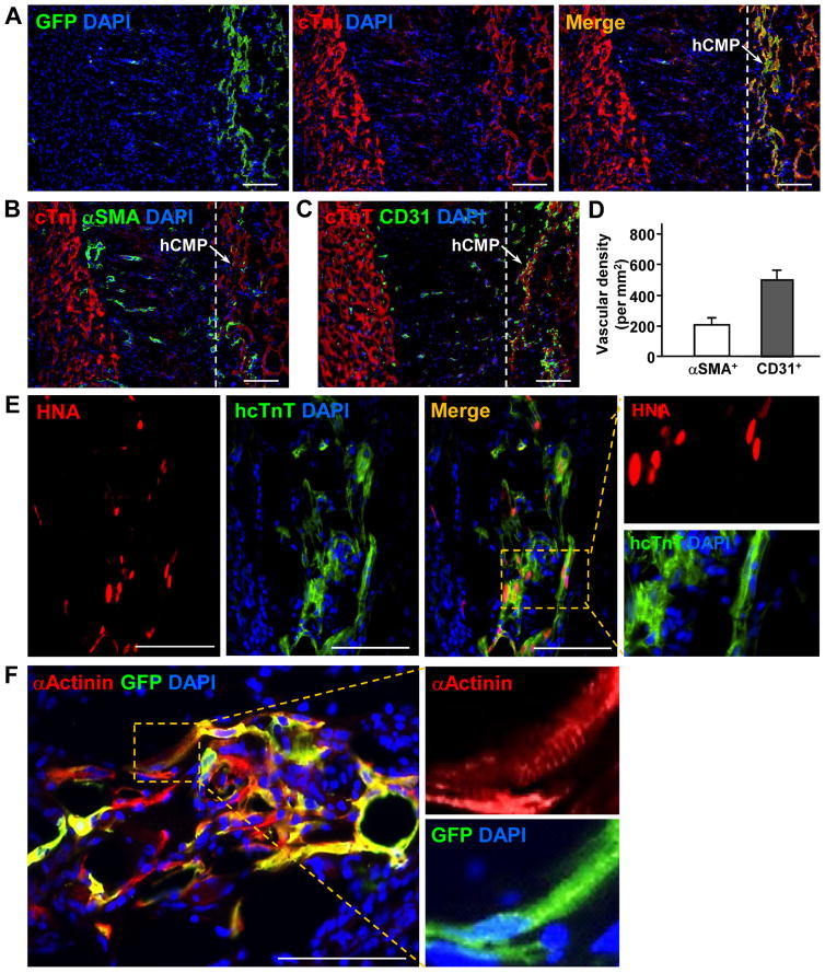
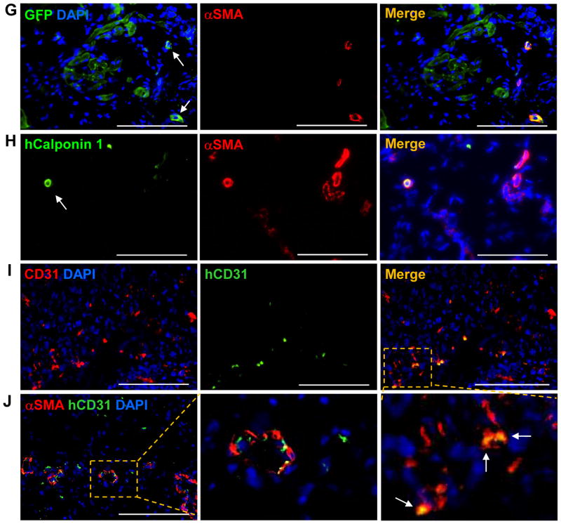
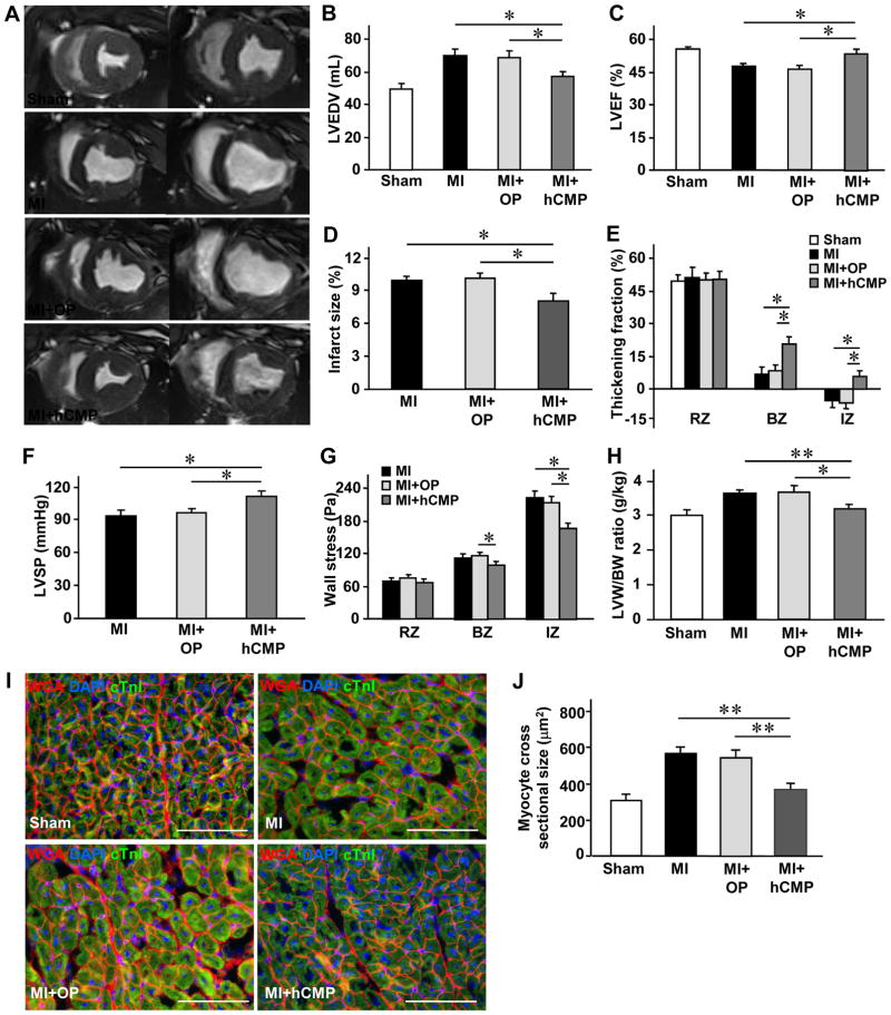
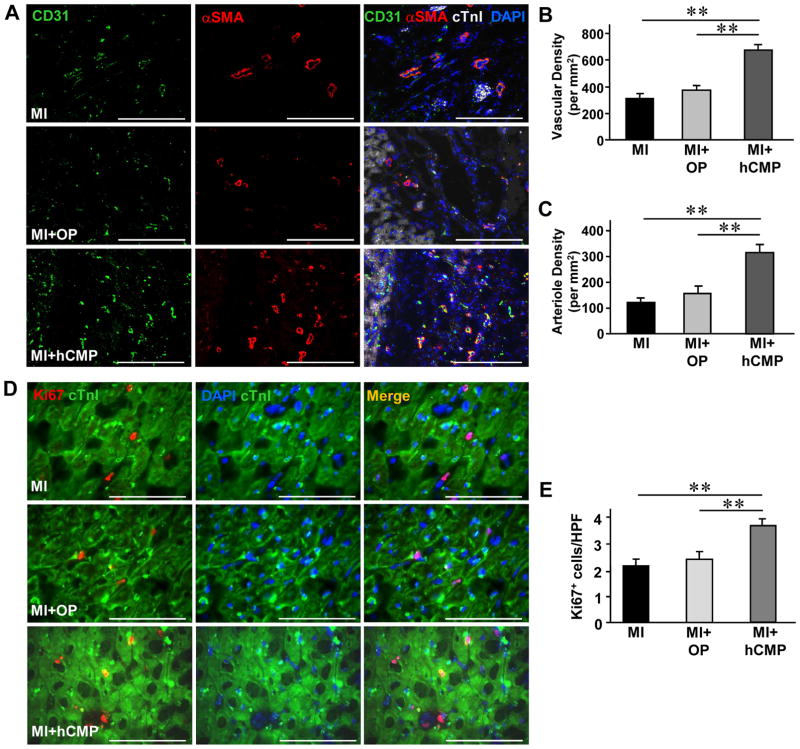
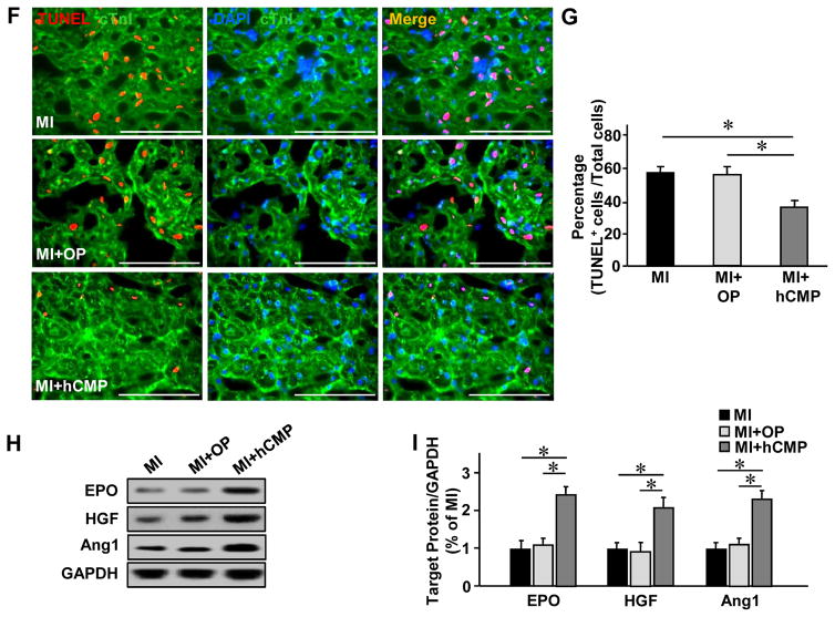
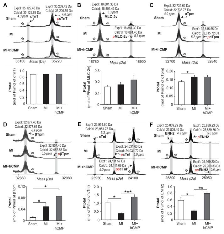
Results: The hCMPs began to beat synchronously within 1 day of fabrication, and after 7 days of dynamic culture stimulation, in vitro assessments indicated the mechanisms related to the improvements in electronic mechanical coupling, calcium-handling, and force generation, suggesting a maturation process during the dynamic culture. The engraftment rate was 10.9±1.8% at 4 weeks after the transplantation. The hCMP transplantation was associated with significant improvements in left ventricular function, infarct size, myocardial wall stress, myocardial hypertrophy, and reduced apoptosis in the periscar boarder zone myocardium. hCMP transplantation also reversed some MI-associated changes in sarcomeric regulatory protein phosphorylation. The exosomes released from the hCMP appeared to have cytoprotective properties that improved cardiomyocyte survival.

Conclusions: We have fabricated a clinically relevant size of hCMP with trilineage cardiac cells derived from human induced-pluripotent stem cells. The hCMP matures in vitro during 7 days of dynamic culture. Transplantation of this type of hCMP results in significantly reduced infarct size and improvements in cardiac function that are associated with reduction in left ventricular wall stress. The hCMP treatment is not associated with significant changes in arrhythmogenicity.

Keywords: heart; models, animal; myocardial infarction; pluripotent stem cells; tissue engineering.


Figures

Figure 1
Figure 1. Characterization of human induced-pluripotent stem cells (hiPSCs) and hiPSC-derived cardiac cells
The hiPSCs used for this investigation were reprogrammed from human left atrial fibroblasts and (A) engineered to express green fluorescent protein (GFP). When cultured as a monolayer with Matrigel, (B) the cells grew to form flat, compact colonies with distinct cell borders (magnification: 40×) and displayed the morphological characteristics of hiPSCs, including (C) prominent nuclei and a high nucleus-to-cytoplasm ratio (magnification: 100×). (D–I) hiPSC-derived cardiomyocytes (hiPSC-CMs) were characterized via immunofluorescent analyses of (D) cardiac troponin T (cTnT), (E) α-sarcomeric actinin (αActinin), (F) α-sarcomeric actin (αSA), (G) slow myosin heavy chain (SMHC), (H) cardiac troponin I (cTnI) and myosin light chain 2v (MLC-2v), and (I) connexin43 (Con43) and cTnT expression. (J–L) hiPSC-derived smooth muscle cells (hiPSC-SMCs) were characterized via immunofluorescent analyses of (J) α-smooth muscle actin (αSMA), (K) calponin 1, and (L) smooth muscle 22 alpha (SM22α) expression. (M–O) hiPSC-derived endothelial cells (hiPSC-ECs) were characterized via immunofluorescent analyses of (M) CD31, (N) vascular endothelial cadherin (VE-cadherin), and (O) von Willebrand factor (VWF) expressions. Nuclei were counterstained with 4′,6-Diamidino-2-Phenylindole (DAPI). Bar = 100 μm.
Figure 2
Figure 2. Characterization of the structure and cellular composition of the human cardiac muscle patch (hCMP)
(A) Large hCMPs (4 cm× 2 cm× 1.25 mm) were fabricated by suspending 4 million hiPSC-CMs, 2 million hiPSC-ECs, and 2 million hiPSC-SMCs in a fibrinogen solution, mixing the cell-containing fibrinogen solution with a thrombin solution, quickly pouring the mixture into a mold (internal dimensions: 4 cm × 2 cm× 1 cm), and then culturing the cells for one week. (BC) The internal structure of the hCMP was evaluated via (B) hematoxylin/eosin (HE) staining and (C) phalloidin staining to identify the presence of F-actin (bar = 100 μm). (D) Apoptotic and necrotic cells were identified via (Di) terminal deoxynucleotidyl transferase dUTP nick end labeling (TUNEL) and (Dii) immunofluorescence staining for phosphorylated mixed lineage kinase domain like pseudokinase (p-MLKL), respectively (bar = 100 μm), and then (E) quantified as the percentage of cells that were positive for each stain (n=4 hCMPs). (F) ECs, SMCs, and CMs were identified in the hCMP via immunofluorescence staining for the presence of CD31, αSMA, and cTnI, respectively (bar = 300 μm); then, (G) the percentage of cells that stained positively for each marker was calculated (n=4 hCMPs). (H) Gap-junctions between adjacent cardiac cells were identified in hCMPs stained for the presence of F-actin and Con43 (bar = 100 μm). (I) The ultrastructure of the hCMP was analyzed by transmission electron microscope to identify (Ii) myofibrils (Mf), Z-lines, mitochondria (Mito) and (Iii) primitive intercalated disc-like structures with fascia adherens junctions and desmosomes. Scale bar = 1 μm.
Figure 3
Figure 3. Characterization of hCMP electrophysiology and function
(AD) Action potential (AP) propagation and Ca2+ transient kinetics were evaluated in hCMPs 7 days after manufacture by staining hCMPs with a voltage-sensitive dye (RH-237, for AP assessments) or a Ca2+-sensitive dye (Cal-520FF, for Ca2+ transient measurements) and then measuring the intensity of transmitted light. (A) Traces (i.e., light intensity) were recorded at the indicated cycle lengths (CL). (B) Isochronal maps of AP propagation (AT: activation time) and (C) optical traces of membrane potential (Vm, blue) and Ca2+ transient traces (red) were recorded during pacing at CL=800 ms and used to determine (D) the conduction velocity (CV), duration of the action potential until 50% and 80% repolarization (APD50 and APD80, respectively), and the duration of the Ca2+ transients until 50% and 80% relaxation (CaTD50 and CaTD80, respectively). (E) Voltage (red) and current (blue) recordings were obtained in hCMPs during stimulation at 40 Hz, 400 Hz, and 4000 Hz; recordings were windowed to enable the current and voltage traces to be compared across 1.5 cycles for each frequency. (F) Tissue resistivity and reactivity in the hCMPs and in rabbit ventricular myocardium (from a previous report) were summarized as a function of stimulation frequency. (GL) hCMP force-generation measurements were determined 7 days after hCMP generation. (G) The relationship between force generation and tensile strain was evaluated by recording force traces as the hCMP was stretched from 100% to 121% of slack length over a four-minute period; (H) active and passive force generation was summarized as a function of hCMP length. (I) hCMPs were stretched to 110% of slack length, and force traces were recorded as the hCMPs beat spontaneously (s) or in response electronic pacing at frequencies of 1, 2, and 3 Hz; (J) twitch force was summarized as a function of pacing frequency. (K) hCMPs were stretched to 110% of slack length, and force traces were recorded as the hCMPs beat in the presence of increasing concentrations (0, 0.1 μM, 1 μM) of isoproterenol; (L) twitch force was summarized as a function of isoproterenol concentration. (MN) Twitch-force was measured at 110% of slack length in hCMPs and in patches that lacked hiPSC-SMCs and ECs but were otherwise identical to the hCMPs; then, force-generation was calculated (M) for the entire hCMP or patch and (N) per cardiomyocyte in the hCMP or the patch. CM: cardiomyocyte; EC: endothelial cell; SMC: smooth muscle cell. *p<0.05. n=4–5 in each group.
Figure 4
Figure 4. hCMPs engraft and survive after transplantation onto infarcted swine hearts
Ischemia-reperfusion injury was surgically induced in swine hearts, and then two hCMPs were sutured over the site of infarction. Sections taken at week 4 from the region of patch application were immufluorescently stained for the presence of cTnI, cTnT, GFP, human specific nuclear antigen (HNA), human specific TnT (hcTnT), α-sarcomeric actinin (αActinin), αSMA, human specific calponin 1 (hCalponin 1), CD31, and the human isoform of CD31 (hCD31); nuclei were counterstained with DAPI. (A) Engrafted hiPSC-CMs were identified by the co-expression of GFP and cTnI; the transplanted hCMP is located to the right of the dashed line in the merged image. (B–D) Vascular structures were identified via the presence of (B) αSMA or (C) CD31 expression; then (D) vessel density in the hCMP was quantified by calculating the number of αSMA+ or CD31+ structures per unit area. (E) Engrafted hiPSC-CMs were further identified by the co-expression of HNA or hcTnT. (F) Evidence of maturing sarcomeric structure (insets) was visible in sections stained for the co-expression of GFP and αActinin. (G–H) hiPSC-SMCs were identified as evidence of arterioles within the engrafted patch via the co-expression of (G) αSMA and GFP, or (H) hCalponin 1 and αSMA. (I–J) hiPSC-ECs were identified in (I) the vasculature of the engrafted patch via the expression of CD31 and hCD31; and in (J) arterioles within the engrafted patch via the expression of hCD31 and αSMA. These data indicate that the majority of neovascularization vessels are from angiogenesis, with small portion of the total spouting arterials or arterioles that are involved by vascular genesis. Scale bar = 100 μm.
Figure 5
Figure 5. hCMP transplantation improves the recovery of cardiac function and limits adverse remodeling in infarcted pig hearts
MI was surgically induced in swine hearts by occluding the coronary artery for 60 minutes; then, two hCMPs were sutured over the site of infarction in animals from the MI+hCMP group, two large fibrin patches lacking the hiPSC-derived cardiac cells were sutured over the injury site in animals from the MI+OP group, and animals in the MI group recovered without either experimental treatment. Animals in the Sham group underwent all surgical procedures for MI induction except the occlusion step. (AE) Four weeks after MI or Sham surgery, (A) magnetic resonance images (left: end systole, right: end diastole) were obtained and used to measure (B) left ventricular end-diastolic volumes (LVEDV), (C) left ventricular ejection fractions (LVEF), (D) infarct sizes, and (E) systolic thickening fractions in the infarcted zone (IZ) of the LV wall, in the border zone (BZ) of the infarct, and in a remote (i.e., noninfarcted) zone (RZ). n=8–10 per experimental group. (F) Hemodynamic measurements. (G) end-systolic LV wall stress in the IZ, BZ and RZ. n=5–7 per experimental group. (HJ) Animals were sacrificed at week 4; then, (H) the LV weight to body weight (LVW/BW) ratios were determined (n=8–11 per experimental group), and (IJ) sections from the border-zone of the infarct were collected and stained with (I) wheat germ agglutinin (WGA) and cTnI to visualize cardiomyocytes (n=6–8 per experimental group, scale bar = 100 μm). Nuclei were counterstained with DAPI, and (J) cardiomyocyte cross-sectional surface areas were measured. *p<0.05, **p<0.01.
Figure 6
Figure 6. Characteristics and cytoprotective effects of hCMP-secreted exosomes
Exosomes were isolated from the hCMP culture medium; then, (A) exosome size was evaluated via nanoparticle tracking analysis, (B) exosome morphology was evaluated via electron microscopy (bar = 100 nm), and (C) the presence of exosome marker proteins (ALG-2-interacting protein X [Alix], tumor susceptibility gene 101 protein [TSG101], CD81, CD63, CD9) was evaluated via Western blot. (D) Cardiomyocytes were incubated for 30 minutes or 24 hours with PKH26-labeled hCMP-secreted exosomes that had been pretreated with or without the exosome uptake inhibitor annexin V (2 μg/mL); then, the cardiomyocytes were fixed and immunofluorescently stained for α-actinin, nuclei were counterstained with DAPI, and exosomes that had been taken up by the cardiomyocytes were identified by PKH26 fluorescence (bar = 100 μm). (EH) Cardiomyocytes were cultured under hypoxic conditions in serum free Dulbecco’s Modified Eagle’s medium (DMEM) for 48 h with phosphate-buffered saline (PBS) or with exosomes from hCMPs (hCMP-Exo) that had been treated with or without an exosome-release inhibitor (GW4869, 10 μM) or an exosome-internalization inhibitor (annexin V, 2 μg/mL). (E) The intensity of lactate dehydrogenase (LDH) fluorescence observed in the media was measured and expressed as a percentage of the intensity observed in PBS-cultured cells. (F) Cell viability was measured with a colorimetric assay that detected the reduction of a tetrazolium compound into a colored formazan product (the conversion occurs through the metabolic activity of living cells), and expressed as a percentage of the measurement in PBS group. (G) Cardiomyocytes were fixed, immunofluorescently stained for cTnT expression and TUNEL stained; nuclei were counter-stained with DAPI (bar = 100 μm). (H) Apoptosis was quantified as the percentage of cells that were TUNEL-positive. (IJ) Cardiomyocytes were cultured under hypoxic conditions for 48 h in serum-free DMEM medium and treated with PBS or with exosomes collected from monolayer cultures of ECs (EC-Exo), SMCs (SMC-Exo), or cardiomyocytes (CM-Exo), from a co-culture of ECs and SMCs (EC/SMC-Exo), or from a co-culture of all three cell types (EC/SMC/CM-Exo). Cytotoxicity was evaluated via (I) measurements of cardiomyocytes LDH leakage and (J) cell viability. *p<0.05, **p<0.01. n=4–5 experiments.
Figure 7
Figure 7. hCMP transplantation enhances the vasculogenic response, promotes cell proliferation, and reduces apoptosis after MI
Sections were collected from the border-zone of the infarct in MI, MI+OP, and MI+hCMP animals four weeks after MI induction. (A) Sections were stained with fluorescent antibodies against CD31, αSMA, and cTnI (bar = 200 μm), and nuclei were counterstained with DAPI; then, (B) vascular density was determined by quantifying the number of structures that expressed CD31, and (C) arteriole density was determined by quantifying the number of structures that expressed both CD31 and αSMA. (D) Sections were stained for expression of the cell proliferation marker Ki67, muscle fibers were visualized by fluorescent immunostaining for cTnI, and nuclei were counterstained with DAPI (bar = 100 μm); then, (E) cell proliferation was quantified as the number of Ki67-positive cells per high-power field (HPF) (n=6–8 per experimental group). (F) Sections taken from the border zone of infarction at week 1 after MI were stained with antibodies against cTnI, apoptotic cells were identified via a TUNEL staining, and nuclei were counterstained with DAPI (bar = 100 μm); then (G) apoptosis was quantified as the percentage of cells that were positive for TUNEL staining (n=3–4 in each group). (H) Expression of the prosurvival proteins erythropoietin (EPO), hepatocyte growth factor (HGF), and angiopoietin 1 (Ang1) at week 1 after MI were evaluated in tissues from the border zone of infarction via Western blot. Glyceraldehyde phosphate dehydrogenase (GAPDH) protein levels were evaluated to serve as a control for unequal loading. (I) EPO, HGF, and Ang1 protein levels were quantified via densitometry analysis and normalized to GAPDH protein levels (n=3 in each group). *P<0.05, **P<0.01.
Figure 8
Figure 8. MI-induced changes in sarcomeric protein phosphorylation are partially prevented or reversed by hCMP transplantation
Four weeks after MI induction, phosphorylation of the sarcomeric proteins (A) cTnT, (B) MLC-2v, (C) α-tropomyosin (αTpm), (D) βTpm, (E) cTnI, and (F) enigma homolog isoform 2 (ENH2), was quantified in myocardium from the border zone of infarction in animals from the MI and MI+hCMP groups and from the corresponding region of hearts in Sham animals via top-down proteomics and mass spectroscopy. Expt’l: experimentally determined most abundant molecular mass, Calc’d: calculated most abundant molecular mass based on the DNA-predicted protein sequence. The star in the cTnT/pcTnT spectrum identifies a peak caused by the loss of H3PO4, diamonds identify peaks caused by the loss of NH3, and ovals identify peaks caused by oxidation of the protein. *p<0.05, **p<0.01, ***p<0.001. n=6 per experimental group.

References

    1. Sayed N, Liu C, Wu JC. Translation of Human-Induced Pluripotent Stem Cells: From Clinical Trial in a Dish to Precision Medicine. J Am Coll Cardiol. 2016;67:2161–76. doi: 10.1016/j.jacc.2016.01.083. - DOI - PMC - PubMed
    1. Shiba Y, Gomibuchi T, Seto T, Wada Y, Ichimura H, Tanaka Y, Ogasawara T, Okada K, Shiba N, Sakamoto K, Ido D, Shiina T, Ohkura M, Nakai J, Uno N, Kazuki Y, Oshimura M, Minami I, Ikeda U. Allogeneic transplantation of iPS cell-derived cardiomyocytes regenerates primate hearts. Nature. 2016;538:388–391. doi: 10.1038/nature19815. - DOI - PubMed
    1. Ye L, Chang YH, Xiong Q, Zhang P, Zhang L, Somasundaram P, Lepley M, Swingen C, Su L, Wendel JS, Guo J, Jang A, Rosenbush D, Greder L, Dutton JR, Zhang J, Kamp TJ, Kaufman DS, Ge Y, Zhang J. Cardiac repair in a porcine model of acute myocardial infarction with human induced pluripotent stem cell-derived cardiovascular cells. Cell Stem Cell. 2014;15:750–61. doi: 10.1016/j.stem.2014.11.009. - DOI - PMC - PubMed
    1. Riegler J, Tiburcy M, Ebert A, Tzatzalos E, Raaz U, Abilez OJ, Shen Q, Kooreman NG, Neofytou E, Chen VC, Wang M, Meyer T, Tsao PS, Connolly AJ, Couture LA, Gold JD, Zimmermann WH, Wu JC. Human Engineered Heart Muscles Engraft and Survive Long Term in a Rodent Myocardial Infarction Model. Circulation research. 2015;117:720–30. doi: 10.1161/CIRCRESAHA.115.306985. - DOI - PMC - PubMed
    1. Chong JJ, Yang X, Don CW, Minami E, Liu YW, Weyers JJ, Mahoney WM, Van Biber B, Cook SM, Palpant NJ, Gantz JA, Fugate JA, Muskheli V, Gough GM, Vogel KW, Astley CA, Hotchkiss CE, Baldessari A, Pabon L, Reinecke H, Gill EA, Nelson V, Kiem HP, Laflamme MA, Murry CE. Human embryonic-stem-cell-derived cardiomyocytes regenerate non-human primate hearts. Nature. 2014;510:273–7. doi: 10.1038/nature13233. - DOI - PMC - PubMed

Publication types

MeSH terms