. 2018 Jan 19;122(2):231-245.
doi: 10.1161/CIRCRESAHA.117.312392. Epub 2017 Dec 12.

VE-Cadherin-Mediated Epigenetic Regulation of Endothelial Gene Expression

Affiliations

VE-Cadherin-Mediated Epigenetic Regulation of Endothelial Gene Expression

Marco F Morini et al. Circ Res. .

Abstract

Rationale: The mechanistic foundation of vascular maturation is still largely unknown. Several human pathologies are characterized by deregulated angiogenesis and unstable blood vessels. Solid tumors, for instance, get their nourishment from newly formed structurally abnormal vessels which present wide and irregular interendothelial junctions. Expression and clustering of the main endothelial-specific adherens junction protein, VEC (vascular endothelial cadherin), upregulate genes with key roles in endothelial differentiation and stability.

Objective: We aim at understanding the molecular mechanisms through which VEC triggers the expression of a set of genes involved in endothelial differentiation and vascular stabilization.

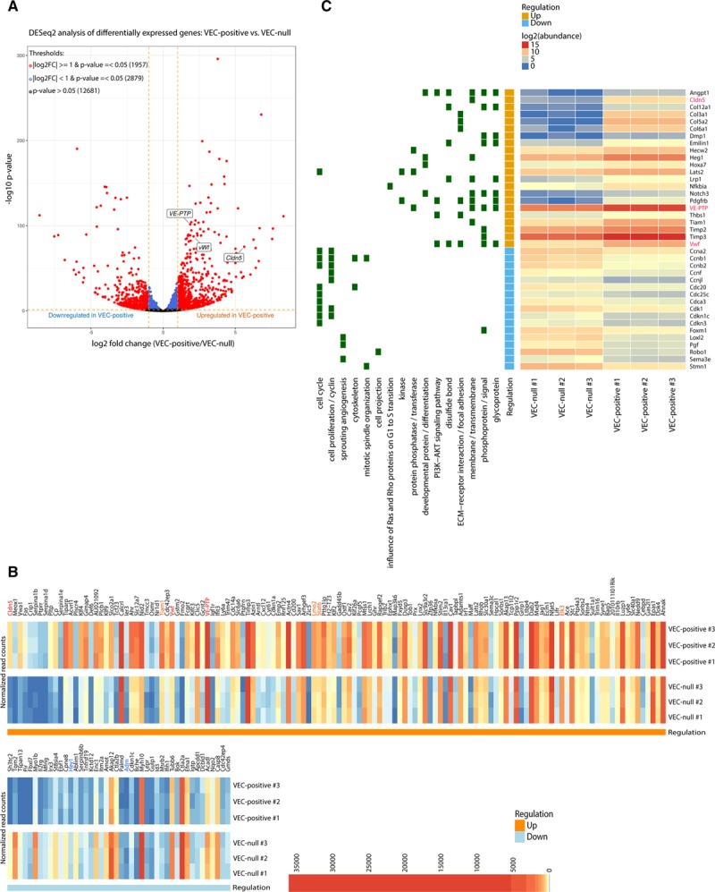
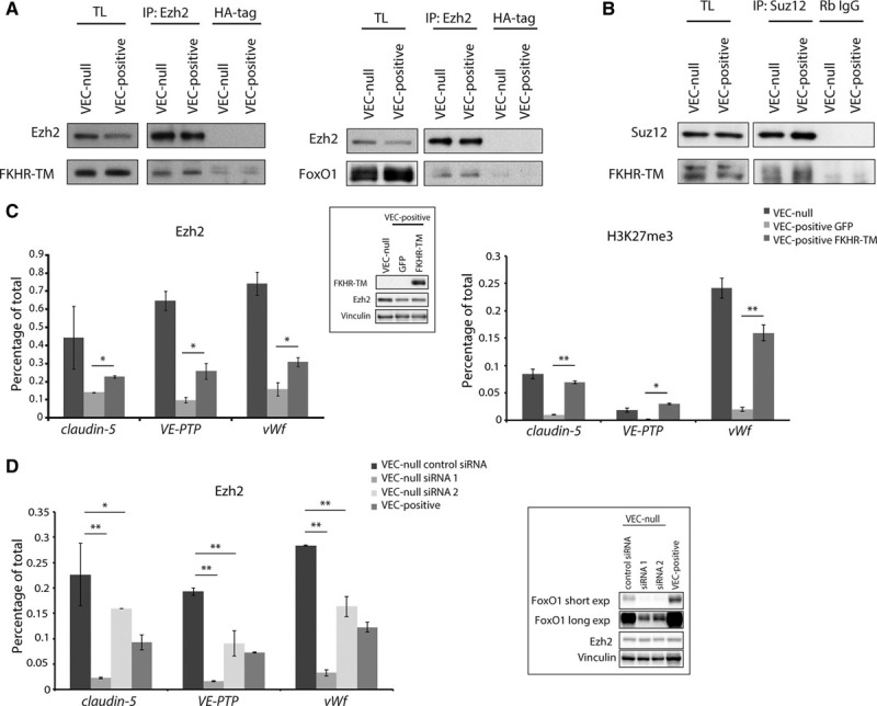
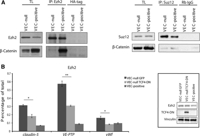
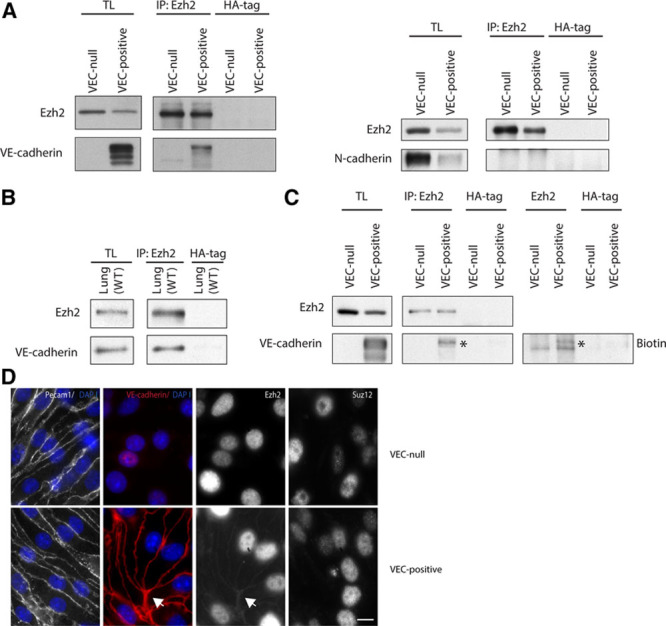
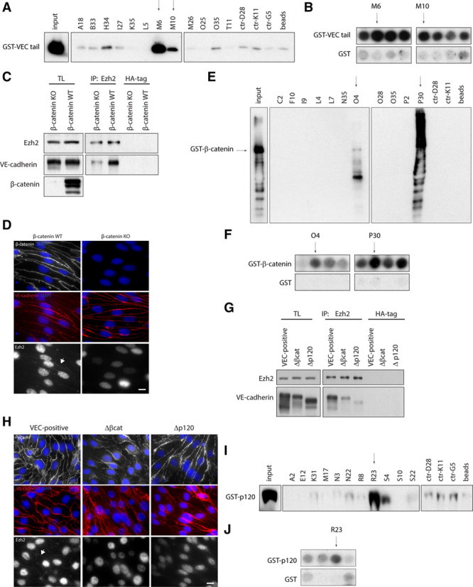
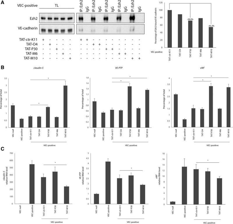
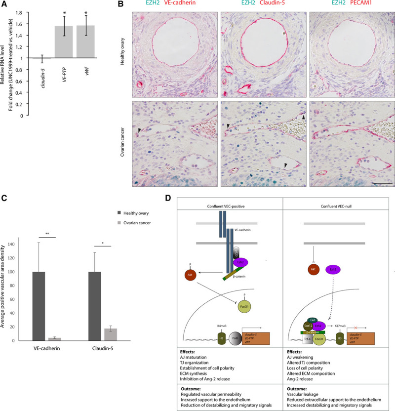
Methods and results: We compared a VEC-null cell line with the same line reconstituted with VEC wild-type cDNA. VEC expression and clustering upregulated endothelial-specific genes with key roles in vascular stabilization including claudin-5, vascular endothelial-protein tyrosine phosphatase (VE-PTP), and von Willebrand factor (vWf). Mechanistically, VEC exerts this effect by inhibiting polycomb protein activity on the specific gene promoters. This is achieved by preventing nuclear translocation of FoxO1 (Forkhead box protein O1) and β-catenin, which contribute to PRC2 (polycomb repressive complex-2) binding to promoter regions of claudin-5, VE-PTP, and vWf. VEC/β-catenin complex also sequesters a core subunit of PRC2 (Ezh2 [enhancer of zeste homolog 2]) at the cell membrane, preventing its nuclear translocation. Inhibition of Ezh2/VEC association increases Ezh2 recruitment to claudin-5, VE-PTP, and vWf promoters, causing gene downregulation. RNA sequencing comparison of VEC-null and VEC-positive cells suggested a more general role of VEC in activating endothelial genes and triggering a vascular stability-related gene expression program. In pathological angiogenesis of human ovarian carcinomas, reduced VEC expression paralleled decreased levels of claudin-5 and VE-PTP.

Conclusions: These data extend the knowledge of polycomb-mediated regulation of gene expression to endothelial cell differentiation and vessel maturation. The identified mechanism opens novel therapeutic opportunities to modulate endothelial gene expression and induce vascular normalization through pharmacological inhibition of the polycomb-mediated repression system.

Keywords: blood vessels; cadherin; cell differentiation; endothelial cells; polycomb-group proteins.


Figures

Figure 1.
Figure 1.
Transcriptome profile determined by VEC (vascular endothelial cadherin) expression and clustering. A, Volcano plot showing the magnitude of differential expression between VEC-positive and VEC-null endothelial cells (ECs). Each dot represents 1 gene with detectable expression in both cell types. The horizontal dashed line (orange) together with the vertical lines (orange) mark thresholds used (P value ≤0.05 and |log2FC| ≥1) to define a gene as differentially regulated in VEC-positive (red). Genes that only passed threshold P value ≤0.05 are depicted in blue. Dots representing claudin-5, vascular endothelial-protein tyrosine phosphatase (VE-PTP), and von Willebrand factor (vWF) are labeled in the figure. B, Heat-map showing the expression pattern of significantly differentially expressed endothelial genes (P value ≤0.05 and |log2FC| ≥1) within and between biological replicates. Endothelial genes upregulated (red/orange) or downregulated (blue) in VEC-positive cells which were further investigated in this study are highlighted in the figure. Genes are displayed in decreasing |log2FC| order (left to right). C, Heat-map showing normalized abundance of significantly changing genes across all samples. Genes belonging to selected functionally enriched terms are highlighted in green on the left of the plot. Claudin-5, VE-PTP, and vWf genes are highlighted in red. In (B) and (C), VEC-positive numbers 1/2/3 represent biological replicates in VEC-positive cells, whereas VEC-null numbers 1/2/3 represent biological replicates in VEC-null cells.
Figure 2.
Figure 2.
Claudin-5, vascular endothelial-protein tyrosine phosphatase (VE-PTP), and von Willebrand factor (vWf) are polycomb targets. A, Quantitative real-time polymerase chain reaction (qRT-PCR) for the transcription start site (TSS) of claudin-5, VE-PTP, and vWf performed on endogenous Ezh (enhancer of zeste homolog)2-, Suz (suppressor of zeste)12-, Bmi1 (B lymphoma Mo-MLV insertion region 1)-, and H3K27me3 (histone H3 trimethylated on lysine 27)-bound chromatin immunoprecipitated from confluent VEC (vascular endothelial cadherin)-null and VEC-positive endothelial cells (ECs). B, qRT-PCR for the TSS of claudin-5, VE-PTP, and vWf performed on endogenous H3K4me3-bound and RNA polymerase II (p-PolII) Ser5–bound chromatin immunoprecipitated from confluent VEC-null and VEC-positive ECs. C, Western blot (WB) analysis of indicated proteins in extracts of confluent VEC-null and VEC-positive ECs upon Suz12 overexpression. D, Quantification of WB in (C). Suz12 and Ezh2 levels were normalized to tubulin. Columns are means±SEM of 3 independent experiments. E, qRT-PCR analysis of claudin-5, VE-PTP, and vWf expression in confluent VEC-null and VEC-positive ECs upon Suz12 overexpression. F, WB analysis of indicated proteins in extracts of confluent VEC-null ECs upon Suz12 knockdown (sh-Suz12). G, qRT-PCR for the TSS of claudin-5, VE-PTP, and vWf performed on endogenous Suz12- and H3K27me3-bound chromatin immunoprecipitated from confluent VEC-positive, VEC-null-sh-Empty, and VEC-null-sh-Suz12 ECs. H, qRT-PCR analysis of claudin-5, VE-PTP, and vWf expression in confluent VEC-null-sh-Empty and VEC-null-sh-Suz12 ECs. A, B, G, Levels of DNA are normalized to input, columns are means±SD of triplicates from a representative experiment. C, F, Tubulin and vinculin are the loading controls. E, H, Levels of mRNA are normalized to 18S; columns are means±SEM of triplicates from a representative experiment. In (A) and (B), *P<0.05; **P<0.01, t test VEC-null vs VEC-positive. In (G), *P<0.05; **P<0.01, t test VEC-null Sh-Empty vs VEC-null Sh-Suz12. In (D), (E), and (H), *P<0.05; **P<0.01, t test. nd indicates not detectable.
Figure 3.
Figure 3.
FoxO1 (Forkhead box protein O1) enhances PcG (polycomb group) protein association to claudin-5, vascular endothelial-protein tyrosine phosphatase (VE-PTP), and von Willebrand factor (vWf) promoters. A, B, Coimmunoprecipitation and Western blot (WB) of endogenous Ezh (enhancer of zeste homolog)2 or Suz (suppressor of zeste)12 and endogenous FoxO1 or FKHR-TM (Forkhead transcription factor triple mutant) from extracts of confluent VEC (vascular endothelial cadherin)-null and VEC-positive endothelial cells (ECs) or the same cells types expressing FKHR-TM (myc-tagged). C, Quantitative real-time polymerase chain reaction (qRT-PCR) for the transcription start site (TSS) of claudin-5, VE-PTP, and vWf performed on endogenous Ezh2- and H3K27me3 (histone H3 trimethylated on lysine 27)-bound chromatin immunoprecipitated from confluent VEC-null and VEC-positive ECs expressing either FKHR-TM or GFP (green fluorescent protein; negative control). Inset: WB analysis of FKHR-TM and Ezh2. D, qRT-PCR for the TSS of claudin-5, VE-PTP, and vWf performed on endogenous Ezh2-bound chromatin immunoprecipitated from confluent VEC-positive or VEC-null ECs transfected with control siRNA or with 2 siRNAs targeting FoxO1 mRNA. Inset: WB analysis of FoxO1 and Ezh2. Two different film exposure timings are shown for FoxO1. C, D, Vinculin is the loading control. Levels of DNA are normalized to input, columns are means±SD of triplicates from a representative experiment. In (C), *P<0.05; **P<0.01, t test VEC-positive GFP vs VEC-positive FKHR-TM. In (D), *P<0.05; **P<0.01, t test VEC-null control siRNA vs VEC-null siRNA1 or VEC-null siRNA2. IP indicates immunoprecipitation; and TL, total cell lysate.
Figure 4.
Figure 4.
β-Catenin stabilizes polycomb/DNA interaction on claudin-5, vascular endothelial-protein tyrosine phosphatase (VE-PTP), and von Willebrand factor (vWf) promoters. A, Coimmunoprecipitation and Western blot (WB) analysis of endogenous Ezh (enhancer of zeste homolog)2 or Suz (suppressor of zeste)12 and β-catenin from extracts of confluent VEC (vascular endothelial cadherin)-null and VEC-positive endothelial cells (ECs). B, Quantitative real-time polymerase chain reaction (qRT-PCR) for the TSS of claudin-5, VE-PTP, and vWf performed on endogenous Ezh2-bound chromatin immunoprecipitated from confluent VEC-positive and VEC-null ECs expressing dominant negative form of Tcf4 (TCF4-DN) or GFP (green fluorescent protein; negative control). Inset: WB analysis of TCF4-DN and Ezh2 in extracts from confluent VEC-positive and VEC-null ECs expressing TCF4-DN or control GFP. Vinculin is the loading control. *P<0.05; **P<0.01, t test VEC-null GFP vs VEC-null TCF4-DN. IP indicates immunoprecipitation; and TL, total cell lysate.
Figure 5.
Figure 5.
VEC (vascular endothelial cadherin) sequesters Ezh2 (enhancer of zeste homolog 2) at the plasma membrane. A, Coimmunoprecipitation and WB of endogenous Ezh2 and VEC or N-cadherin from extracts of confluent VEC-null and VEC-positive endothelial cells (ECs). B, Coimmunoprecipitation and Western blot (WB) of endogenous Ezh2 and VEC from wild-type (WT) murine whole lung extracts. C, Coimmunoprecipitation and WB of endogenous Ezh2 and VEC from extracts of confluent VEC-null and VEC-positive ECs after biotinylation of cell surface proteins. Asterisk highlights Ezh2-associated total and surface VEC bands. D, Immunofluorescence analysis of Ezh2 junctional localization (arrow) in confluent VEC-null and VEC-positive ECs. Junctional Suz (suppressor of zeste)12 was not detected. Platelet/endothelial cell adhesion molecule-1 (Pecam1) and VEC were used as junctional markers. Scale bar: 10 μm. IP indicates immunoprecipitation; and TL, total cell lysate.
Figure 6.
Figure 6.
Analysis of Ezh2 (enhancer of zeste homolog 2) interaction with VEC (vascular endothelial cadherin) junctional complex. A, Streptavidin pull-down of selected biotinylated Ezh2 peptides and GST-tagged VEC cytoplasmic tail. GST-VEC cytoplasmic tail (400 ng) was loaded as input. Peptides displaying no interaction in peptide array were used as controls. Arrows indicate peptides showing positive signal. B, Regions of peptide array in the Online Figure IXA and IXB corresponding to selected peptides in (A). C, Coimmunoprecipitation and Western blot (WB) of Ezh2 and VEC from extracts of confluent β-catenin knockout (KO) and β-catenin wild-type (WT) endothelial cells (ECs). D, Immunofluorescence analysis of Ezh2 (arrow) and β-catenin junctional localization in confluent β-catenin KO and β-catenin WT ECs. VE-cadherin (red) was used as junctional marker. E, Streptavidin pull-down of selected biotinylated Ezh2 peptides and GST-tagged β-catenin. GST-β-catenin (400 ng) was loaded as input. Peptides displaying no interaction in peptide array were used as controls. Arrows indicate peptides showing positive signal. F, Regions of peptide array in the Online Figure IXA and IXC corresponding to selected peptides in (E). G, Coimmunoprecipitation and WB of Ezh2 and VEC from extracts of confluent VEC-positive, Δβcat, and Δp120 ECs. H, Immunofluorescence analysis of Ezh2 junctional localization (arrow) in confluent VEC-positive, Δβcat, and Δp120 ECs. VEC and platelet/endothelial cell adhesion molecule-1 (Pecam1) were used as junctional markers. I, Streptavidin pull-down of selected biotinylated Ezh2 peptides and GST-tagged p120-catenin. GST-p120-catenin (300 ng) was loaded as input. Peptides displaying no interaction in peptide array were used as controls. Arrow indicates peptide showing positive signal. J, Regions of peptide array in the Online Figure IXA and IXD corresponding to selected peptides in (I). In (D) and (H), scale bar: 10 μm. GST indicates glutathione S-transferase; HA, human influenza hemagglutinin; IP, immunoprecipitation; and TL, total cell lysate.
Figure 7.
Figure 7.
Inhibition of Ezh2 (enhancer of zeste homolog 2)/VEC (vascular endothelial cadherin) interaction causes claudin-5, vascular endothelial-protein tyrosine phosphatase (VE-PTP), and von Willebrand factor (vWf) downregulation. A, Coimmunoprecipitation and Western blot (WB) of endogenous Ezh2 and VEC from extracts of VEC-positive endothelial cells (ECs) treated with VEC-binding transactivator of transcription (TAT)-M6 and TAT-M10 peptides, β-catenin–binding TAT-O4 and TAT-P30 peptides or nonbinding TAT-ctr-K11 peptide as control (left). Quantification of coprecipitated VEC protein normalized on precipitated Ezh2 level (right). B, Quantitative real-time polymerase chain reaction (qRT-PCR) for the transcription start site (TSS) of claudin-5, VE-PTP, and vWf performed on endogenous Ezh2-bound chromatin immunoprecipitated from VEC-positive ECs treated with VEC-binding TAT-M6 and TAT-M10 peptides, β-catenin–binding TAT-O4 and TAT-P30 peptides or nonbinding TAT-ctr-K11 peptide as control. Levels of DNA are normalized to input; columns are means±SD of triplicates from a representative experiment. *P<0.01, t test TAT-ctr-K11 vs TAT-P30 or TAT-M10 treatment. C, qRT-PCR analysis of claudin-5, VE-PTP, and vWf expression in VEC-positive ECs treated with VEC-binding TAT-M10 peptide, β-catenin–binding TAT-P30 peptide or nonbinding TAT-ctr-K11 peptide as control. Levels of mRNA are normalized to GAPDH; columns are means±SEM of triplicates from a representative experiment. *P<0.01, t test TAT-ctr-K11 vs. TAT-M10 treatment. IP indicates immunoprecipitation; and TL, total cell lysate.
Figure 8.
Figure 8.
Ezh2 (enhancer of zeste homolog 2) activity correlates with claudin-5, vascular endothelial-protein tyrosine phosphatase (VE-PTP), and von Willebrand factor (vWf) repression in vivo. A, Quantitative real-time polymerase chain reaction (qRT-PCR) analysis of claudin-5, VE-PTP, and vWf expression in lungs of vehicle- or UNC1999-treated pups (P6). Data are represented as fold change of UNC1999 treated vs vehicle and are means±SD from at least 4 mice per group. Gene expression was normalized to VEC (vascular endothelial cadherin) expression. B, Immunohistochemistry (IHC) staining of VEC, Claudin-5, platelet/endothelial cell adhesion molecule-1 (PECAM1; red) and EZH2 (green) expression in serial sections of human healthy ovary (upper) or serous surface papillary ovarian carcinoma (lower). Black arrowheads point to tumor vessel endothelial cells (ECs) expressing high levels of EZH2. Scale bar: 50 μm. C, Quantification of IHC stainings in (B). For VEC and claudin-5, areas of specific signal, divided by the total measured area, were normalized to the corresponding values of PECAM1 staining. Columns are means±SEM (n=3 healthy ovaries; 4 ovarian carcinomas; at least 3 fields per sample). D, Suggested model for the regulation of claudin-5, VE-PTP, and vWf genes. Clustered VEC recruits β-catenin and activates Akt leading to FoxO1 (Forkhead box protein O1) phosphorylation and inhibition. Furthermore, Ezh2 is sequestered at the cell membrane by association with VEC cytoplasmic tail (left). These mechanisms allow gene activation by impeding the recruitment of PcG (polycomb group) proteins to gene promoters. Claudin-5 expression allows the correct organization of tight junctions (TJs) and regulation of vessel permeability., VE-PTP regulates adherens junction (AJ) maturation and VEGFR2 (vascular endothelial growth factor receptor-2) activity,, whereas vWf contributes to extracellular matrix (ECM) formation and inhibits Ang (angiopoietin)-2 release., These effects are likely to contribute to vessel stabilization and prevent vascular leakage. In (A) and (C), *P<0.05; **P<0.01, t test.

References

    1. Carmeliet P, Jain RK. Principles and mechanisms of vessel normalization for cancer and other angiogenic diseases. Nat Rev Drug Discov. 2011;10:417–427. doi: 10.1038/nrd3455. - PubMed
    1. Baluk P, Morikawa S, Haskell A, Mancuso M, McDonald DM. Abnormalities of basement membrane on blood vessels and endothelial sprouts in tumors. Am J Pathol. 2003;163:1801–1815. doi: 10.1016/S0002-9440(10)63540-7. - PMC - PubMed
    1. Giannotta M, Trani M, Dejana E. VE-cadherin and endothelial adherens junctions: active guardians of vascular integrity. Dev Cell. 2013;26:441–454. doi: 10.1016/j.devcel.2013.08.020. - PubMed
    1. Dejana E, Giampietro C. Vascular endothelial-cadherin and vascular stability. Curr Opin Hematol. 2012;19:218–223. doi: 10.1097/MOH.0b013e3283523e1c. - PubMed
    1. Taddei A, Giampietro C, Conti A, Orsenigo F, Breviario F, Pirazzoli V, Potente M, Daly C, Dimmeler S, Dejana E. Endothelial adherens junctions control tight junctions by VE-cadherin-mediated upregulation of claudin-5. Nat Cell Biol. 2008;10:923–934. doi: 10.1038/ncb1752. - PubMed

Publication types