Review
. 2017 Dec 12;61(6):733-749.
doi: 10.1042/EBC20170055. Print 2017 Dec 12.

Dysregulation of autophagy as a common mechanism in lysosomal storage diseases

Affiliations
Review

Dysregulation of autophagy as a common mechanism in lysosomal storage diseases

Elena Seranova et al. Essays Biochem. .

Abstract

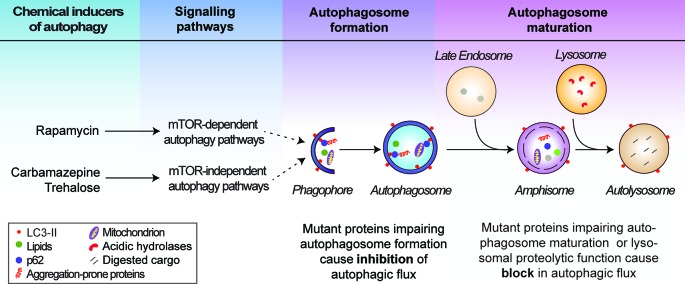
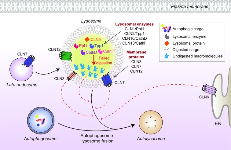
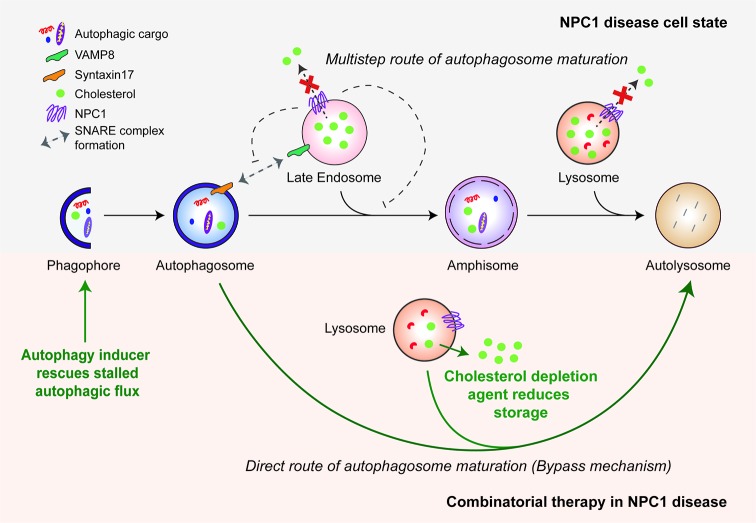
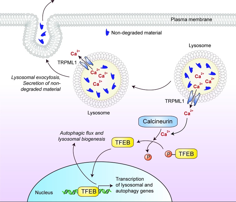
The lysosome plays a pivotal role between catabolic and anabolic processes as the nexus for signalling pathways responsive to a variety of factors, such as growth, nutrient availability, energetic status and cellular stressors. Lysosomes are also the terminal degradative organelles for autophagy through which macromolecules and damaged cellular components and organelles are degraded. Autophagy acts as a cellular homeostatic pathway that is essential for organismal physiology. Decline in autophagy during ageing or in many diseases, including late-onset forms of neurodegeneration is considered a major contributing factor to the pathology. Multiple lines of evidence indicate that impairment in autophagy is also a central mechanism underlying several lysosomal storage disorders (LSDs). LSDs are a class of rare, inherited disorders whose histopathological hallmark is the accumulation of undegraded materials in the lysosomes due to abnormal lysosomal function. Inefficient degradative capability of the lysosomes has negative impact on the flux through the autophagic pathway, and therefore dysregulated autophagy in LSDs is emerging as a relevant disease mechanism. Pathology in the LSDs is generally early-onset, severe and life-limiting but current therapies are limited or absent; recognizing common autophagy defects in the LSDs raises new possibilities for therapy. In this review, we describe the mechanisms by which LSDs occur, focusing on perturbations in the autophagy pathway and present the latest data supporting the development of novel therapeutic approaches related to the modulation of autophagy.

Keywords: Autophagy; Glycogenoses; Lysosomal storage disorders; Lysosomes; Neuronal ceroid lipofuscinoses; Sphingolipidoses.


Conflict of interest statement

The authors declare that there are no competing interests associated with the manuscript.

Figures

Figure 1
Figure 1. Schematic representation of the autophagy pathway
Autophagy initiates by the de novo synthesis and elongation of phagophores, which engulf cytosolic materials (autophagic cargo) to form autophagosomes. Autophagosomes predominantly fuse with the late endosomes to form amphisomes and subsequently with the lysosomes to form autolysosomes where the autophagic cargo is degraded by the lysosomal hydrolases. Autophagy can be stimulated by chemical inducers acting via the mTOR-dependent and mTOR-independent pathways regulating autophagy. Defects in autophagic flux at the autophagosome formation and maturation stages are indicated.
Figure 2
Figure 2. CLN protein distribution and their link to autophagy defects in neuronal ceroid lipofuscinoses
Many CLN proteins reside in the lysosomal matrix (CLN1, 2, 5, 10, 13) or at the lysosomal membrane (CLN3, 7), while others localize to different cellular compartments such as the ER membrane (CLN6). Disease-causing mutations in some of the CLN proteins inhibit autophagosome maturation (dashed red lines) and block autophagic flux, but the underlying mechanisms are unknown. Mutated lysosomal hydrolases (CLN1, 2, 10, 13) are unable to degrade autophagic cargo, which subsequently accumulate and impair lysosomal function.
Figure 3
Figure 3. Autophagy defects in NPC1 disease and the bypass mechanism of autophagosome maturation for restoring autophagic flux
Mutant NPC1 protein prevents cholesterol efflux from the endo-lysosomal compartments and impairs autophagosome maturation in the multi-step route due to failure in the SNARE machinery. Induction of autophagy by chemical inducers bypasses this block and restores autophagic flux via direct autophagosome–lysosome fusion. A combinatorial treatment strategy is shown with cholesterol depletion agents. The green arrows indicate therapeutic effects of autophagy induction and cholesterol depletion.
Figure 4
Figure 4. Cellular effects of TFEB that might be of therapeutic benefit in lysosomal storage disorders
Lysosomal Ca2+ efflux through TRPML1 activates the Ca2+-dependent phosphatase calcineurin, which mediates dephosphorylation-dependent nuclear translocation of TFEB. Nuclear TFEB up-regulates the transcription of genes involved in lysosome biogenesis and autophagy, thereby enhancing autophagic flux. In addition, Ca2+ efflux from peripheral lysosomes promotes lysosomal exocytosis and the secretion of non-degraded materials.

References

    1. Luzio J.P., Pryor P.R. and Bright N.A. (2007) Lysosomes: fusion and function. Nat. Rev. Mol. Cell Biol. 8, 622–632 - PubMed
    1. Perera R.M. and Zoncu R. (2016) The lysosome as a regulatory hub. Annu. Rev. Cell Dev. Biol. 32, 223–253 - PMC - PubMed
    1. Xu H. and Ren D. (2015) Lysosomal physiology. Annu. Rev. Physiol. 77, 57–80 - PMC - PubMed
    1. Futerman A.H. and van Meer G. (2004) The cell biology of lysosomal storage disorders. Nat. Rev. Mol. Cell Biol. 5, 554–565 - PubMed
    1. Platt F.M., Boland B. and van der Spoel A.C. (2012) The cell biology of disease: lysosomal storage disorders: the cellular impact of lysosomal dysfunction. J. Cell Biol. 199, 723–734 - PMC - PubMed

Publication types

LinkOut - more resources