. 2017 Dec 12;8(6):e01964-17.
doi: 10.1128/mBio.01964-17.

Prophages and Growth Dynamics Confound Experimental Results with Antibiotic-Tolerant Persister Cells

Affiliations

Prophages and Growth Dynamics Confound Experimental Results with Antibiotic-Tolerant Persister Cells

Alexander Harms et al. mBio. .

Abstract

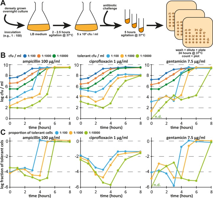
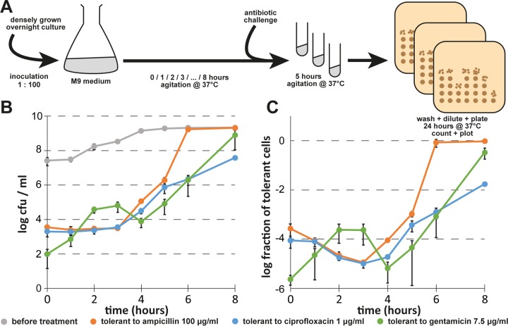
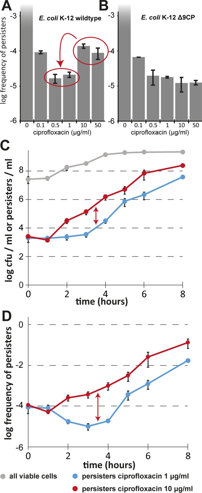
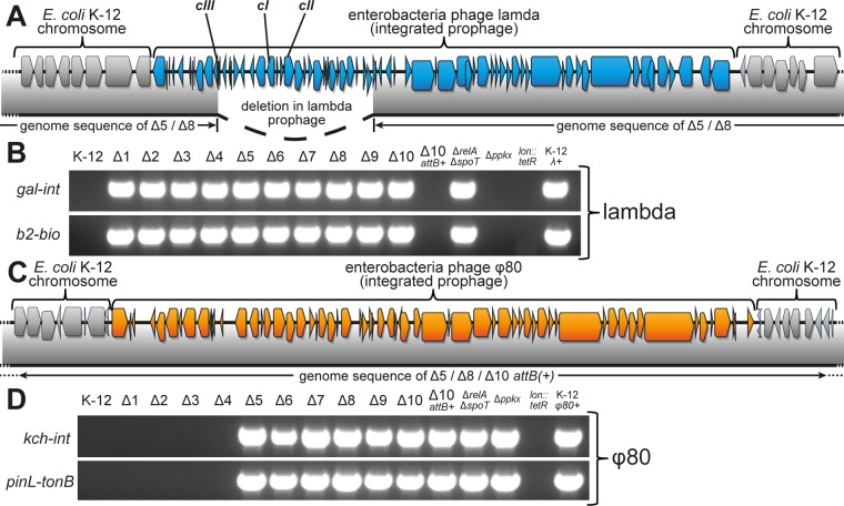
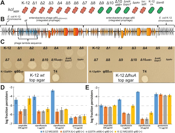
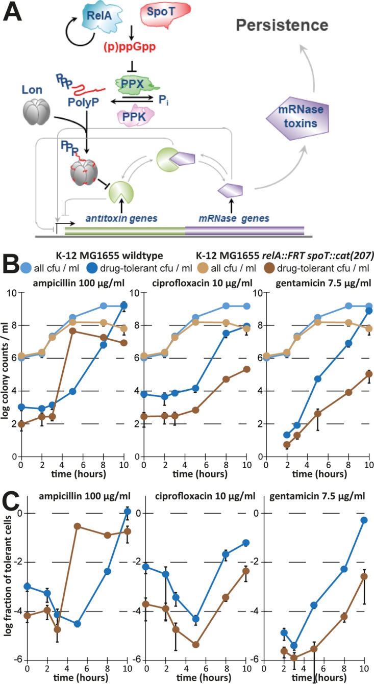
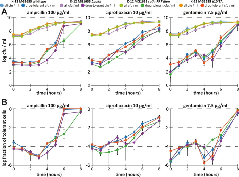
Bacterial persisters are phenotypic variants that survive antibiotic treatment in a dormant state and can be formed by multiple pathways. We recently proposed that the second messenger (p)ppGpp drives Escherichia coli persister formation through protease Lon and activation of toxin-antitoxin (TA) modules. This model found considerable support among researchers studying persisters but also generated controversy as part of recent debates in the field. In this study, we therefore used our previous work as a model to critically examine common experimental procedures to understand and overcome the inconsistencies often observed between results of different laboratories. Our results show that seemingly simple antibiotic killing assays are very sensitive to variations in culture conditions and bacterial growth phase. Additionally, we found that some assay conditions cause the killing of antibiotic-tolerant persisters via induction of cryptic prophages. Similarly, the inadvertent infection of mutant strains with bacteriophage ϕ80, a notorious laboratory contaminant, apparently caused several of the phenotypes that we reported in our previous studies. We therefore reconstructed all infected mutants and probed the validity of our model of persister formation in a refined assay setup that uses robust culture conditions and unravels the dynamics of persister cells through all bacterial growth stages. Our results confirm the importance of (p)ppGpp and Lon but no longer support a role of TA modules in E. coli persister formation under unstressed conditions. We anticipate that the results and approaches reported in our study will lay the ground for future work in the field.IMPORTANCE The recalcitrance of antibiotic-tolerant persister cells is thought to cause relapsing infections and antibiotic treatment failure in various clinical setups. Previous studies identified multiple genetic pathways involved in persister formation but also revealed reproducibility problems that sparked controversies about adequate tools to study persister cells. In this study, we unraveled how typical antibiotic killing assays often fail to capture the biology of persisters and instead give widely differing results based on poorly controlled experimental parameters and artifacts caused by cryptic as well as contaminant prophages. We therefore established a new, robust assay that enabled us to follow the dynamics of persister cells through all growth stages of bacterial cultures without distortions by bacteriophages. This system also favored adequate comparisons of mutant strains with aberrant growth phenotypes. We anticipate that our results will contribute to a robust, common basis for future studies on the formation and eradication of antibiotic-tolerant persisters.

Keywords: (p)ppGpp; antibiotic tolerance; bacteriophage genetics; persistence; toxin-antitoxin modules.


Figures

FIG 1
FIG 1
Persister assays are affected by inoculum and growth phase. (A) Scheme illustrating the setup of a persister assay as it is commonly performed in the field. (B) Cultures of E. coli K-12 MG1655 were grown in LB medium after inoculation at a dilution of 1:100 (blue), 1:1,000 (orange), or 1:10,000 (green) from dense overnight cultures, and levels of all CFUs as well as levels of antibiotic-tolerant CFUs were determined at each time point. (C) Fraction of antibiotic-tolerant cells for each data point reported as described for panel B. Data points are shown for one representative experiment, because the absolute numbers of antibiotic-tolerant cells (but not their dynamics) were affected considerably by batch-to-batch variations of the LB medium (see Fig. S1). n.d., not detected (no gentamicin-tolerant bacteria recovered at t = 0 h from inoculation at a dilution of 1:10,000).
FIG 2
FIG 2
Persister formation of E. coli K-12 MG1655 in M9 medium. (A) Scheme illustrating how the dynamics of persister formation were determined. (B) Bacterial growth and the dynamics of antibiotic-tolerant cells were determined for cultures of E. coli K-12 MG1655 in M9 minimal medium as described for the experiment whose results are shown in Fig. 1 after inoculation at a dilution of 1:100 from a dense overnight culture. (C) Fraction of antibiotic-tolerant cells for each data point shown in panel B. We generated biphasic kill curves to verify that the antibiotic-tolerant cells present during exponential growth (3 h after inoculation) represented persisters (Fig. S2). Data points represent means of results from at least three independent experiments, and error bars indicate standard deviations.
FIG 3
FIG 3
Induction of cryptic prophages distorts persister measurements of E. coli K-12. Exponentially growing cultures of E. coli K-12 strain BW25113 (A) and its derivative lacking all nine cryptic prophages (B; Δ9CP) created by Wang et al. (28) were treated with different concentrations of ciprofloxacin in M9 medium, and the level of surviving persister cells was determined. Note that the level of survivors for the wild-type strain increased at high ciprofloxacin concentrations, while no such effect can be observed in the Δ9CP mutant. The latter strain generally exhibits a lower level of persister formation than its ancestor, possibly due to the roles of prophage-encoded factors in persistence and general stress tolerance (28). (C) Bacterial growth and the dynamics of antibiotic-tolerant cells were determined for cultures of E. coli K-12 MG1655 in M9 minimal medium as described for Fig. 2B, and the results of treatment with 1 µg/ml and 10 µg/ml ciprofloxacin were compared. (D) Data representing the fraction of antibiotic-tolerant cells at each time point are shown. Data points represent means of results from at least three independent experiments, and error bars indicate standard deviations.
FIG 4
FIG 4
Lambda and ϕ80 infection of strains from our previous studies. (A) The insertion of a lambda prophage (blue gene arrows) in the E. coli K-12 chromosome (gray gene arrows) is shown together with coverage of the prophage insertion in the genomes of the Δ5TA and Δ8TA mutants (gray bar below). The 13-kilobase deletion comprising cI, cII, and cIII repressors spans a sequence from the gam nuclease inhibitor gene to the endolysin gene R. (B) Diagnostic PCR analyses of the two junctions between the lambda prophage and the gal-bio region of the E. coli chromosome were performed to determine the extent of lambda infection in important strains from our previous studies (8, 9). (C) Insertion of a ϕ80 prophage (orange gene arrows) in the E. coli K-12 chromosome (gray gene arrows) in the genomes of Δ5TA, Δ8TA, and Δ10TA attB(+) mutants. (D) Diagnostic PCR analyses of the two junctions between the ϕ80 prophage and the yciI locus in the E. coli K-12 chromosome were performed to determine the prevalence of ϕ80 infections among important strains from our previous work (8, 9).
FIG 5
FIG 5
Lambda immunity and active ϕ80 prophages and their effect on persistence. (A) The sensitivity of different E. coli strains to the lambda cIb221 mutant was determined by streaking across lines of phage stock on agar plates. Red, no growth/sensitive; green, growth/immune. (B) Illustration of neighboring ϕ80 and ϕ80h(80)imm(λ) integration in the chromosome of E. coli Δ10TA as deduced from the genome sequence. The E. coli chromosome backbone is shown in gray; genes of ϕ80 and ϕ80h(80)imm(λ) are shown in orange and dark orange, respectively; and the lambda region of the ϕ80h(80)imm(λ) mutant is highlighted in blue. Note that the genome sequence does not unambiguously tell which of the prophages is upstream and which is downstream in the integration site. (C) A plaque assay with culture supernatants and control phage stocks was performed to determine the infectivity of prophage carriers. wt, wild type. (D and E) Exponentially growing E. coli K-12 MG1655 and different mutant derivatives/lysogens were challenged with ampicillin, ciprofloxacin, or gentamicin in LB medium (D) or M9 medium (E), and the fractions of surviving persisters were calculated. Data points represent averages of results from three independent experiments, and error bars represent standard deviations.
FIG 6
FIG 6
The model of (p)ppGpp-dependent persister formation through TA modules. (A) The illustration (adapted from Germain et al. [54]) shows our previously published model of persister formation initiated by stochastic bursts of (p)ppGpp that induce the production of polyphosphate which stimulates Lon to degrade TA module antitoxins. Consequently, the activation of mRNA interferase toxins would induce bacterial persistence (8). (B and C) In order to verify the most upstream element of the model, we created a new (p)ppGpp-deficient mutant of E. coli K-12 MG1655 (relA spoT) and assayed the dynamics of antibiotic tolerance seen in the experiment described for Fig. 2 with minor modifications (see Materials and Methods).
FIG 7
FIG 7
Protease Lon, but not mRNA endonuclease TA modules, contributes to persistence. We studied the dynamics of antibiotic tolerance for cultures of newly constructed E. coli K-12 Δppkx, sulA::FRT Δlon, and Δ10TA mutants in comparison to the parental wild type similarly to the experiment whose results are shown in Fig. 2. Changes in overall and antibiotic-tolerant CFU per milliliter are plotted in panel A, and the fractions of antibiotic-tolerant cells are plotted in panel B. While the sulA::FRT Δlon mutant showed a clear defect in persister formation or survival during exponential growth (around 3 h after inoculation; see also Fig. S2), the parental sulA::FRT strain had no such phenotype (Fig. S3), confirming that the defect was due to the lack of Lon.

References

    1. Lewis K. 2010. Persister cells. Annu Rev Microbiol 64:357–372. doi: 10.1146/annurev.micro.112408.134306. - DOI - PubMed
    1. Harms A, Maisonneuve E, Gerdes K. 2016. Mechanisms of bacterial persistence during stress and antibiotic exposure. Science 354. doi: 10.1126/science.aaf4268. - DOI - PubMed
    1. Dörr T, Vulić M, Lewis K. 2010. Ciprofloxacin causes persister formation by inducing the TisB toxin in Escherichia coli. PLoS Biol 8:e1000317. doi: 10.1371/journal.pbio.1000317. - DOI - PMC - PubMed
    1. Verstraeten N, Knapen WJ, Kint CI, Liebens V, Van den Bergh B, Dewachter L, Michiels JE, Fu Q, David CC, Fierro AC, Marchal K, Beirlant J, Versées W, Hofkens J, Jansen M, Fauvart M, Michiels J. 2015. Obg and membrane depolarization are part of a microbial bet-hedging strategy that leads to antibiotic tolerance. Mol Cell 59:9–21. doi: 10.1016/j.molcel.2015.05.011. - DOI - PubMed
    1. Shan Y, Brown Gandt A, Rowe SE, Deisinger JP, Conlon BP, Lewis K. 2017. ATP-dependent persister formation in Escherichia coli. mBio 8:e02267-16. doi: 10.1128/mBio.02267-16. - DOI - PMC - PubMed

MeSH terms

Substances

LinkOut - more resources