. 2017 Dec 12;8(12):3204.
doi: 10.1038/s41419-017-0001-z.

Implication of 4E-BP1 protein dephosphorylation and accumulation in pancreatic cancer cell death induced by combined gemcitabine and TRAIL

Affiliations

Implication of 4E-BP1 protein dephosphorylation and accumulation in pancreatic cancer cell death induced by combined gemcitabine and TRAIL

Androulla Elia et al. Cell Death Dis. .

Abstract

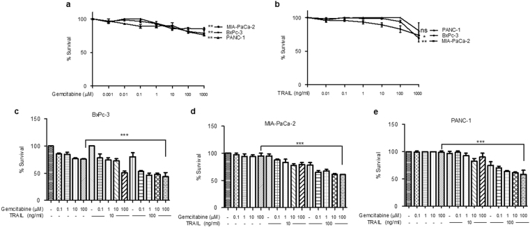
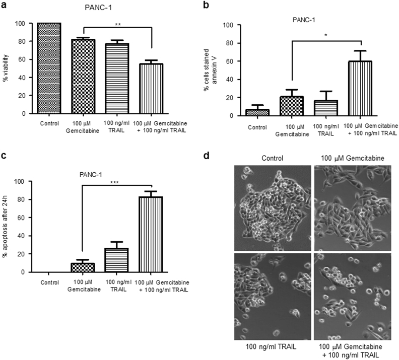
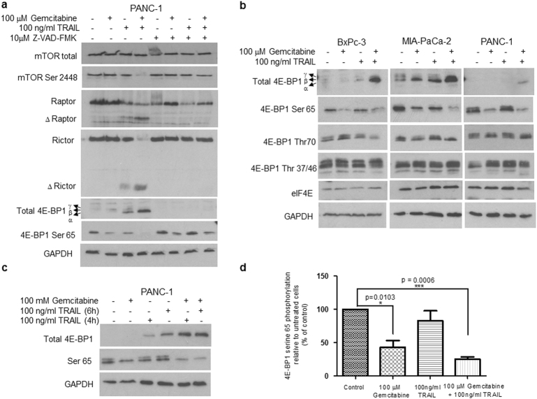
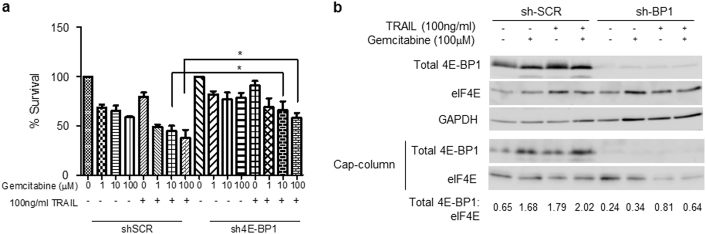
Pancreatic cancer cells show varying sensitivity to the anticancer effects of gemcitabine. However, as a chemotherapeutic agent, gemcitabine can cause intolerably high levels of toxicity and patients often develop resistance to the beneficial effects of this drug. Combination studies show that use of gemcitabine with the pro-apoptotic cytokine TRAIL can enhance the inhibition of survival and induction of apoptosis of pancreatic cancer cells. Additionally, following combination treatment there is a dramatic increase in the level of the hypophosphorylated form of the tumour suppressor protein 4E-BP1. This is associated with inhibition of mTOR activity, resulting from caspase-mediated cleavage of the Raptor and Rictor components of mTOR. Use of the pan-caspase inhibitor Z-VAD-FMK indicates that the increase in level of 4E-BP1 is also caspase-mediated. ShRNA-silencing of 4E-BP1 expression renders cells more resistant to cell death induced by the combination treatment. Since the levels of 4E-BP1 are relatively low in untreated pancreatic cancer cells these results suggest that combined therapy with gemcitabine and TRAIL could improve the responsiveness of tumours to treatment by elevating the expression of 4E-BP1.


Conflict of interest statement

The authors declare that they have no conflict of interest.

Figures

Fig. 1
Fig. 1. Effect of gemcitabine and/or TRAIL on PDAC survival. BxPC-3, MIA PaCa-2 and PANC-1 cells were seeded in 96-well plates at a cell seeding density of 3x10⁴ cells/cm2
a Sensitivity of cells to gemcitabine was assessed by MTT assay. Cells were treated with increasing amounts of gemcitabine (0.001–1000 μM) for 24 h (n = 4). b Sensitivity of cells to TRAIL was assessed by MTT assay. Cells were treated with increasing amounts of TRAIL (0.001–1000 ng/ml) for 4 h (n = 4). ce Sensitivity of cells to gemcitabine and TRAIL combination treatment was assessed by MTT assay. Cells were treated with increasing amounts of gemcitabine (0.1–100 μM) for 24 h (n = 4) and/or 10 or 100 ng/ml TRAIL for 4 h for BxPC-3 and MIA PaCa-2 cells and 6 h for PANC-1 cells (n = 4). All experiments were repeated three times and data are provided as means ± SEM (one representative experiment is shown). P-values were calculated using Student’s t-test to determine the statistical significance of the difference between a, b untreated cells and cells treated with either 1000 μM gemcitabine or 1000 ng/ml TRAIL, respectively (ns: P > 0.05, *P < 0.05, **P < 0.01) and ce cells treated with 100 μM gemcitabine and cells treated with 100 μM gemcitabine plus 100 ng/ml TRAIL (***P < 0.001)
Fig. 2
Fig. 2. Combination treatment induces apoptosis
a PANC-1 cells were seeded in triplicate in 12-well plates at a cell seeding density of 3×10⁴ cells/cm2 and left to attach overnight. Cells were treated with 100 μM gemcitabine for 24 h and/or 100 ng/ml TRAIL for 6 h. The viability of the cells was assessed by trypan blue exclusion assay. Quadruplicate cell counts were used to calculate each cell density. These were performed for three independently seeded wells and percentage viability was determined. b A total of 1×10⁶ PANC-1 cells were treated with 100 μM gemcitabine for 24 h and/or 100 ng/ml TRAIL for 6 h. Induction of early apoptosis in PANC-1 cells was assessed using flow cytometry following staining with FITC Annexin V. The data represent means ± SEM of three experiments performed in triplicate. c, d PANC-1 cells were seeded in triplicate in 12-well plates at a cell seeding density of 3×10⁴ cells/cm2 and left to attach overnight. Cells were treated with 100 μM gemcitabine for 24 h and/or 100 ng/ml TRAIL for 6 h and monitored by time-lapse microscopy. c The appearance of a pre-apoptotic morphology was scored and the % apoptotic cells after 24 h determined. The data are the means ± SEM from three independent experiments. d Phase contrast microscopy images of cells treated as indicated. ac All experiments were repeated three times and data are provided as means ± SEM (one representative experiment is shown). P-values were calculated using Student’s t-test to determine the statistical significance of the difference between cells treated with 100 μM gemcitabine and cells treated with 100 μM gemcitabine plus 100 ng/ml TRAIL (*P < 0.05, **P < 0.01, ***P < 0.001)
Fig. 3
Fig. 3. Combination treatment induces caspase-dependent apoptosis. BxPC-3, MIA PaCa-2 and PANC-1 cells were seeded in 96-well plates at a cell seeding density of 3×10⁴ cells/cm2
a, b Caspase-mediated cleavage of caspase-8 and PARP was assessed by western blotting in cells treated with 100 μΜ gemcitabine for 24 h and/or 100 ng/ml TRAIL for 4 h for BxPC-3 and MIA PaCa-2 cells, and 4 and 6 h for PANC-1 cells (n = 3). One representative experiment is shown. Lysates were prepared and equal amounts (15 μg total protein) were subjected to SDS–PAGE, transferred to PVDF membranes and then immunoblotted with antibodies directed against a PARP (top panel), caspase-8 (middle panel) or GAPDH (bottom panel). b Caspase-mediated cleavages of caspase-8, PARP and BID in the presence or absence of the pan-caspase inhibitor Z-VAD-FMK (10 μM) were assessed by western blotting in cells treated as described above. Membranes were immunoblotted with antibodies directed against caspase-8, PARP and BID. GAPDH was used as a loading control. c The inhibition of cell survival following combination treatment was assessed in the presence or absence of the pan-caspase inhibitor Z-VAD-FMK. PANC-1 cells were seeded in 96-well plates at a cell seeding density of 3×10⁴ cells/cm2. Cells were treated with 100 μM gemcitabine for 24 h and/or 100 ng/ml TRAIL for 6 h in the presence or absence of 10 μM Z-VAD-FMK. Cell survival was assessed using the MTT assay. All experiments were repeated three times and data are provided as means ± SEM (one representative experiment is shown). P-values were calculated using Student’s t-test to determine the statistical significance of the difference between cells treated with 100 μM gemcitabine and those treated with both 100 μM gemcitabine and 100 ng/ml TRAIL (*P < 0.05, **P < 0.01, ***P < 0.001)
Fig. 4
Fig. 4. Combination treatment targets the mTOR pathway and alters the phosphorylation of 4E-BP1 in PDAC cells
BxPC-3, MIA PaCa-2 cells and PANC-1 cells were treated with 100 μΜ gemcitabine for 24 h and/or 100 ng/ml TRAIL for 4 h. A unit of 15 μg of total protein lysate was analysed using western blotting. a PANC-1 cell lysates were analysed with antibodies directed against total mTOR, mTOR Ser2448, Raptor, Rictor, total 4E-BP1, 4E-BP1 Ser65 and GAPDH. b BxPC-3, MIA PaCA-2 and PANC-1 lysates were analysed to look at the effect on levels and phosphorylation of 4E-BP1 at residues Ser65, Thr 37/46 and Thr70 as well as levels of eIF4E. GAPDH was used as a loading control. c The change in phosphorylation of 4E-BP1 at Ser65 in PANC-1 cells following combination treatment using TRAIL treatment for either 4 or 6 h was assessed by western blotting. PVDF membranes were immunoblotted with antibodies directed against total 4E-BP1 and 4E-BP1 residue Ser65. d The relative levels of phosphorylation of 4E-BP1 at Ser65 were quantified by scanning densitometry using ImageJ and the data are shown on the histogram as % of the values for untreated cells. All experiments were repeated three times and data are provided as means ± SEM. P-values were calculated using Student’s t-test to determine the statistical significance of the difference between untreated cells and cells treated with either gemcitabine or gemcitabine plus TRAIL (*P < 0.05 and ***P < 0.001)
Fig. 5
Fig. 5. 4E-BP1 is involved in the regulation of cell survival following gemcitabine and TRAIL treatment
a, b MIA PaCa-2 cells expressing a small hairpin RNA (shRNA) directed against 4E-BP1 and control cells expressing a scrambled shRNA were seeded in 96-well plates at a cell seeding density of 3×10⁴ cells/cm2. a The sensitivity of cells to gemcitabine and TRAIL combination treatment was assessed by MTT assay. Cells were treated with increasing amounts of gemcitabine (0.1–100 μM) for 24 h (n = 4) and/or 100 ng/ml TRAIL for 24 h (n = 4). All experiments were repeated three times and data are provided as means ± SEM. One representative experiment is shown. P-values were calculated using Student’s t-test to determine the statistical significance of the difference between cells expressing a scrambled shRNA and cells expressing a shRNA directed against 4E-BP1, both cell lines having been treated with 10 or 100 μM gemcitabine and 100 ng/ml TRAIL (*P < 0.05). b Lysates made from cells treated as in a were used to purify eIF4E using chromatography on m7GTP-Sepharose beads as described in Methods. The levels of eIF4E and of the 4E-BP1 associated with it were determined by SDS gel electrophoresis and immunoblotting. Total cell lysates were analysed in parallel. Quantification was carried out by densitometry using ImageJ and the ratios of 4E-BP1 to eIF4E in the m7GTP-purified samples (in arbitrary units) are indicated

References

    1. Kamisawa T, Wood LD, Itoi T, Takaori K. Pancreatic cancer. Lancet. 2016;388:73–85. doi: 10.1016/S0140-6736(16)00141-0. - DOI - PubMed
    1. Elshaer, M., Gravante, G., Kosmin, M., Riaz, A., Al-Bahrani, A. A systematic review of the prognostic value of lymph node ratio, number of positive nodes and total nodes examined in pancreatic ductal adenocarcinoma. Ann. R. Coll. Surg. Engl. 99, 1–6 (2016). - PMC - PubMed
    1. Cid-Arregui A, Juarez V. Perspectives in the treatment of pancreatic adenocarcinoma. World J. Gastroenterol. 2015;21:9297–9316. doi: 10.3748/wjg.v21.i31.9297. - DOI - PMC - PubMed
    1. Zabernigg A, et al. Quality of life across chemotherapy lines in patients with cancers of the pancreas and biliary tract. BMC Cancer. 2012;12:390. doi: 10.1186/1471-2407-12-390. - DOI - PMC - PubMed
    1. Rathos MJ, Joshi K, Khanwalkar H, Manohar SM, Joshi KS. Molecular evidence for increased antitumor activity of gemcitabine in combination with a cyclin-dependent kinase inhibitor, P276-00 in pancreatic cancers. J. Transl. Med. 2012;10:161. doi: 10.1186/1479-5876-10-161. - DOI - PMC - PubMed

MeSH terms

LinkOut - more resources