. 2017 Dec 12;8(1):2073.
doi: 10.1038/s41467-017-02228-2.

Structure of outer membrane protein G in lipid bilayers

Affiliations

Structure of outer membrane protein G in lipid bilayers

Joren S Retel et al. Nat Commun. .

Abstract

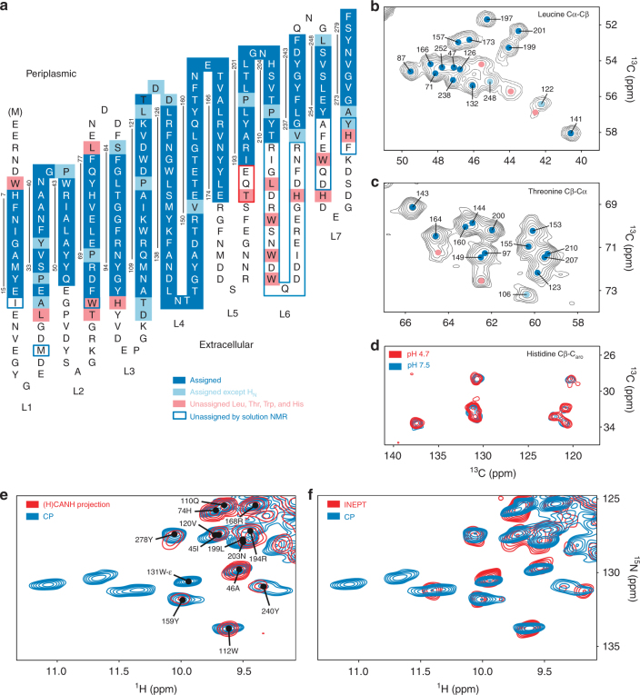
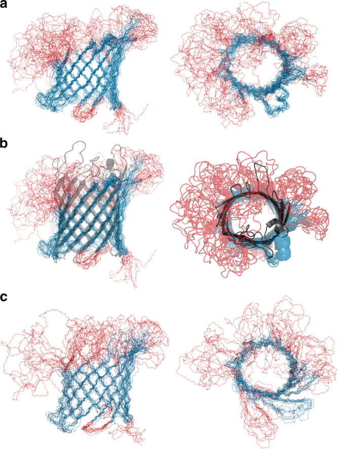
β-barrel proteins mediate nutrient uptake in bacteria and serve vital functions in cell signaling and adhesion. For the 14-strand outer membrane protein G of Escherichia coli, opening and closing is pH-dependent. Different roles of the extracellular loops in this process were proposed, and X-ray and solution NMR studies were divergent. Here, we report the structure of outer membrane protein G investigated in bilayers of E. coli lipid extracts by magic-angle-spinning NMR. In total, 1847 inter-residue 1H-1H and 13C-13C distance restraints, 256 torsion angles, but no hydrogen bond restraints are used to calculate the structure. The length of β-strands is found to vary beyond the membrane boundary, with strands 6-8 being the longest and the extracellular loops 3 and 4 well ordered. The site of barrel closure at strands 1 and 14 is more disordered than most remaining strands, with the flexibility decreasing toward loops 3 and 4. Loop 4 presents a well-defined helix.


Conflict of interest statement

The authors declare no competing financial interests.

Figures

Fig. 1
Fig. 1
Resonance assignment and OmpG topology. Assigned residues are indicated in blue. a For residues in light blue, the 1HN shift is unknown but partial carbon assignment was obtained. Pink indicates unassigned residues as discussed in the text. Residues in blue frames do not show signals in solution NMR spectra and residues in the red frame were assigned by solution NMR but not solid-state NMR, see text. Vertical lines indicate the β-strands with residue numbers. bd Spectral regions of 13C–13C correlation spectra comprising Cα–Cβ peaks of b leucine in the GAVLS(W) sample (20 ms DARR), c threonine in a DARR spectrum of the 1,3-TEMPQANDSG sample (50 ms mixing), and d histidine in a 50 ms DARR spectrum of the GANDSH(LV) sample. For the peaks indicated by pink dots in these 13C–13C spectra, no strip could be found in the 1H-detected 3D spectra. e, f Overlays of a CP-based 1H–15N-correlation (blue) comprising the region of Trp side chain cross peaks with the projection of the CANH spectrum (e) and an INEPT-based HSQC (f)
Fig. 2
Fig. 2
Set of two planes from the 3D (H)NHH and (H)N(HH)NH spectra. Strips taken at the 15N chemical shifts of Y75 (left) and L87 (right) from the (H)N(HH)NH and (H)NHH spectra, respectively. The proton–proton cross-peak pattern is indicative of cross-strand hydrogen bonding between the backbone amide and carbonyl groups of tyrosine 75 and leucine 87. Red lines correspond to the 1H and 15N chemical shifts of L87. Blue lines correspond to the 1H and 15N chemical shifts of Y75. A total of four cross peaks are present at the intersections of red and blue lines. Dotted circles indicate positions of potential sequential cross peaks (see text)
Fig. 3
Fig. 3
Solid-state NMR structure of OmpG in lipid bilayers and comparison to X-ray and solution NMR structures. a Regular secondary structure is shown in blue, loop regions in red. The structures to the right are turned by 90°. b Overlay of solid-state (blue and red) and X-ray structure (dark gray). The beta-sheet is extended further in the model derived by X-ray crystallography (2IWV), see left edge. c Same views of the solution NMR structure 2JQY obtained from OmpG solutions in dodecylphosphocholine. Figure generated using pymol

References

    1. Fairman JW, Noinaj N, Buchanan SK. The structural biology of beta-barrel membrane proteins: a summary of recent reports. Curr. Opin. Struct. Biol. 2011;21:523–531. doi: 10.1016/j.sbi.2011.05.005. - DOI - PMC - PubMed
    1. Wimley WC. The versatile beta-barrel membrane protein. Curr. Opin. Struct. Biol. 2003;13:404–411. doi: 10.1016/S0959-440X(03)00099-X. - DOI - PubMed
    1. Fajardo DA, et al. Biochemistry and regulation of a novel Escherichia coli K-12 porin protein, OmpG, which produces unusually large channels. J. Bacteriol. 1998;180:4452–4459. - PMC - PubMed
    1. Misra R, Benson SA. A novel mutation, cog, which results in production of a new porin protein (OmpG) of Escherichia coli K-12. J. Bacteriol. 1989;171:4105–4111. doi: 10.1128/jb.171.8.4105-4111.1989. - DOI - PMC - PubMed
    1. Conlan S, Zhang Y, Cheley S, Bayley H. Biochemical and biophysical characterization of OmpG: a monomeric porin. Biochemistry. 2000;39:11845–11854. doi: 10.1021/bi001065h. - DOI - PubMed

Publication types

MeSH terms