. 2017 Dec 12;8(1):2076.
doi: 10.1038/s41467-017-02145-4.

Global unleashing of transcription elongation waves in response to genotoxic stress restricts somatic mutation rate

Affiliations

Global unleashing of transcription elongation waves in response to genotoxic stress restricts somatic mutation rate

Matthieu D Lavigne et al. Nat Commun. .

Abstract

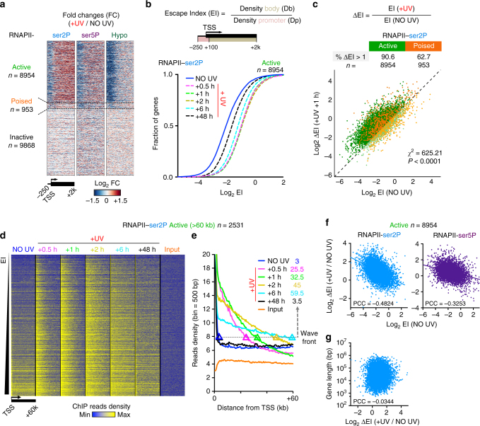
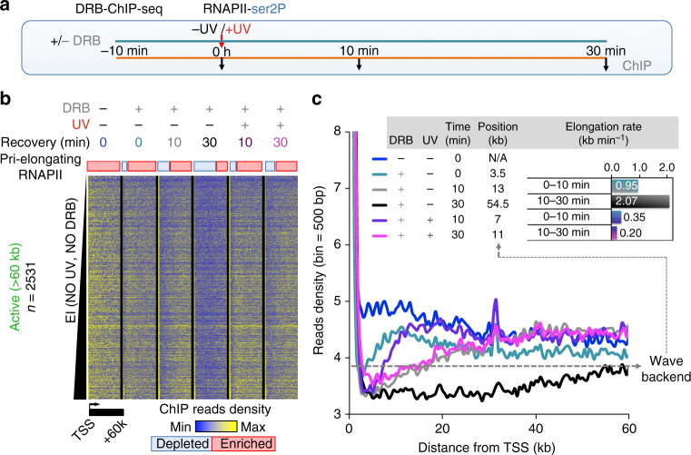
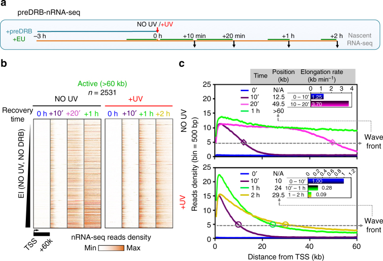
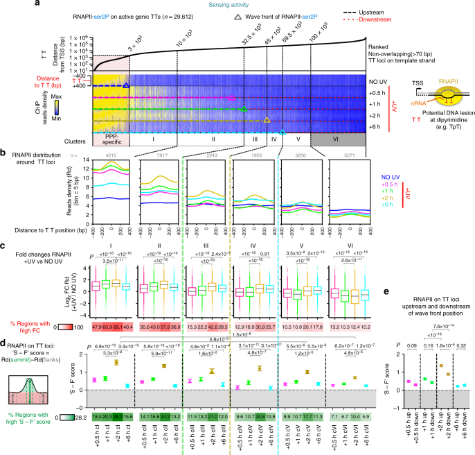
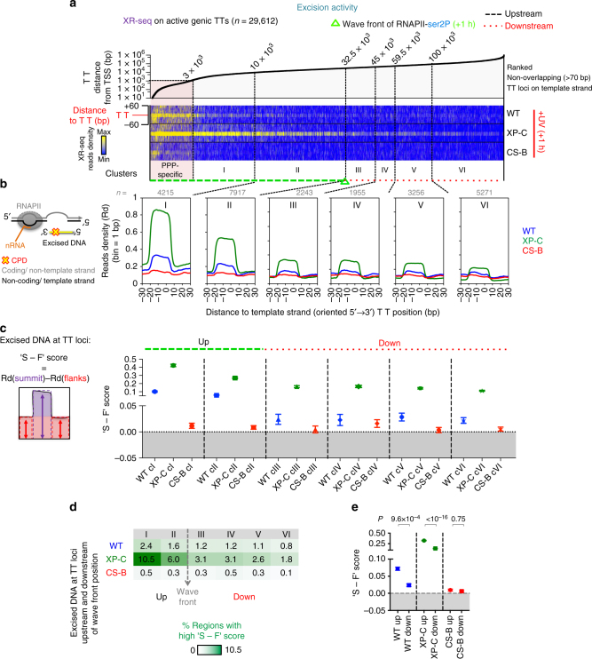
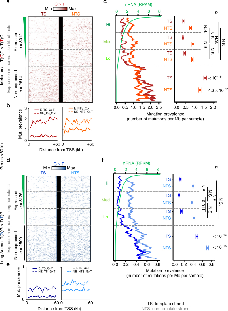
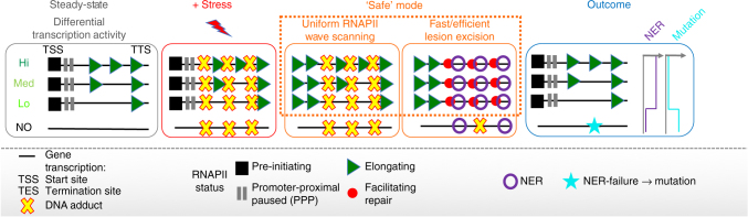
Complex molecular responses preserve gene expression accuracy and genome integrity in the face of environmental perturbations. Here we report that, in response to UV irradiation, RNA polymerase II (RNAPII) molecules are dynamically and synchronously released from promoter-proximal regions into elongation to promote uniform and accelerated surveillance of the whole transcribed genome. The maximised influx of de novo released RNAPII correlates with increased damage-sensing, as confirmed by RNAPII progressive accumulation at dipyrimidine sites and by the average slow-down of elongation rates in gene bodies. In turn, this transcription elongation 'safe' mode guarantees efficient DNA repair regardless of damage location, gene size and transcription level. Accordingly, we detect low and homogenous rates of mutational signatures associated with UV exposure or cigarette smoke across all active genes. Our study reveals a novel advantage for transcription regulation at the promoter-proximal level and provides unanticipated insights into how active transcription shapes the mutagenic landscape of cancer genomes.


Conflict of interest statement

The authors declare no competing financial interests.

Figures

Fig. 1
Fig. 1
Global triggering of transcription waves on virtually all active genes in response to UV irradiation. a Heatmaps illustrating the Log2 fold change (FC) of main RNAPII isoforms comparing reads density (Rd) between UV irradiation (8 J m−2) and steady-state (FC = (Rd+UV)/(RdNO UV), as aligned at individual (rows) regions (−250 bp to +2 kb relative to TSS) and categorised by gene expression status (see Supplementary Fig. 1). b RNAPII Escape Indexes from promoter regions (EI) before and after UV plotted as empirical cumulative distribution of fraction (ECDF) of active (labelled in solid green throughout the paper) genes. EI represents the ratio of reads density in gene body over reads density in promoter. c Individual comparison of EI before and after UV for all active and poised genes. Percentages of genes with increased escape after UV (ΔEI > 1, dark green dots) are shown. Chi-square test (χ 2) determines whether observed number of genes with ΔEI > 1 differs from expected value purely by chance. P is indicated. d Heatmaps illustrating RNAPII-ser2P and Input read densities from TSS to TSS + 60 kb before and after UV, as aligned at individual (rows) active genes larger than 60 kb and ranked by increasing EI (as determined before UV in b). e Average plots of read densities shown in d indicating progression of wave front position (kb) at arbitrary threshold representing the transition state (enriched to steady-state). f Correlation plot between constitutive EI (NO UV) for RNAPII-ser2P (left) or RNAPII-ser5P (right) and EI change after UV (ΔEI(+UV vs NO UV)) for all active genes. PCC scores are indicated. g Same as in f for RNAPII-ser2P ΔEI (+UV vs NO UV) plotted against gene length (bp)
Fig. 2
Fig. 2
Inhibition of RNAPII transition into elongation unmasks the kinetics of RNAPII molecules elongating prior to UV stress (pri-elongating). a Scheme of DRB-inhibition methodology (DRB-ChIP-seq), UV irradiation was performed at 20 J m−2, see also Methods. b Heatmaps illustrating RNAPII-ser2P read densities from TSS to TSS + 60 kb, for samples defined in a, as aligned at individual (rows) active genes larger than 60 kb and ranked by increasing steady-state EI (see Fig. 1d). c Average plots of read densities for RNAPII-ser2P derived from b highlighting the pri-elongating RNAPII wave backend positions at arbitrary threshold (dashed line) representing the transition state (depleted to enriched for RNAPII-ser2P). Insert shows corresponding average (n = 2531) elongation rates calculated from differences between wave backend positions in the considered time interval. Note: gradients of colours reflect the transition between indicative time points
Fig. 3
Fig. 3
Stress-triggered de novo transcription influx decelerates progressively. a Scheme of DRB-release methodology (preDRB-nRNA-seq), UV irradiation was performed at 20 J m−2 see also Methods. b Heatmaps illustrating nascent RNA read densities from TSS to TSS + 60 kb, for samples defined in a, as aligned at individual (rows) active genes larger than 60 kb and ranked by increasing steady-state EI (Fig. 1d). c Average plots of read densities for nascent RNA derived from b, highlighting de novo wave-release of RNAPII. Differences in wave front positions at an arbitrary threshold (dashed line representing the transition from enriched levels to steady-state levels) were used to calculate average (n = 2531) elongation rates in the indicated time intervals (inserts)
Fig. 4
Fig. 4
RNAPII-dependent sensing of DNA lesions is enhanced by the de novo wave-release. a Heatmaps illustrating the distribution of RNAPII-ser2P reads aligned around TT loci localized in active genes (as defined in Supplementary Fig. 8a) before (NO UV) and after UV irradiation (+UV, 8 J m−2) and sorted from left to right by increasing distance relative to TSS. TT loci were clustered (upstream (i.e. Clusters I, II, III for +2 h) or downstream (i.e. Clusters IV, V, VI for +2 h)) relatively to RNAPII-ser2P wave front positions, which indicate the transition state from de novo-enriched to pri-elongating only population of RNAPII (see Fig. 1e, Supplementary Fig. 8d and Methods for details). TT loci near PPP-specific RNAPII signal were not considered for analysis. b Average plots of read densities (Rd) as mapped in a for individual clusters (n is indicated). c Top; box plots showing the changes in Rd in regions analysed in b and normalised by Rd of NO UV (P value based on two-sided t-test using the Benjaminin–Hoechberg (BH) adjustment). Boxes refer to the first quartile, the median and the third quartile. Whiskers refer to the 10–90% interquantile range. Bottom; heatmap representing the proportion of regions (per cluster) with high fold changes (FC > 2) in Rd. d Top; plot showing the average (± SEM) of the difference between Rd at summit and Rd at flanks (defined as ‘S - F’ score) of all regions analysed in b (P-value based on two-sided Wilcoxon rank-sum test the BH adjustment). Bottom; heatmap representing the proportion of regions (per cluster) with high ‘S - F’ scores (> threshold = average[S - F]exon start + 3×SD[S - F]exon start, see Supplementary Fig. 9 and Methods). e Plot showing the comparison of average (± SEM) ‘S - F’ score for all regions upstream (Up) and downstream (Down) of the respective wave front position, for each time of recovery (P-value based on two-sided Wilcoxon rank-sum test using BH adjustment)
Fig. 5
Fig. 5
NER of DNA lesions is enhanced by the de novo wave-release of RNAPII upon genotoxic stress. a Heatmaps illustrating the distribution of excised DNA fragments representative of repair activity at 1 h of recovery in cell lines proficient for TC-NER activity (WT, XP-C), or not (CS-B) (reads from XR-seq study) (see Methods for details). XR-seq reads were aligned around TT loci localised in active genes (Supplementary Fig. 8a) and sorted from left to right by increasing distances relative to TSS. TT loci were clustered (Upstream; clusters I and II, or Downstream; clusters III–VI) relatively to RNAPII-ser2P wave front position at +1 h of recovery (as in Fig. 4a). TT loci near PPP-specific RNAPII signal were not considered for analysis. b Average plots of Read densities (Rd) mapped in a for individual clusters (n is indicated). c Plot showing the average (± SEM) of the difference between Rd at summit and Rd at flanks (‘S - F’ score) of all regions analysed in b (P-value based on two-sided Wilcoxon rank-sum test). d Heatmap representing the proportion of regions (per cluster) with high ‘S - F’ scores (> threshold = average ‘S - F’ + 3 × SD, as calculated for the control exon start regions, see Methods). e Plot showing the comparison of average (± SEM) ‘S - F’ score for all regions upstream (Up) and downstream (Down) of the respective wave front position, for each cell line (P-value based on two-sided Wilcoxon rank-sum test using BH adjustment)
Fig. 6
Fig. 6
Low and uniform mutation rate is detected in all expressed genes in environmentally exposed tumour genomes NER. a, d Heatmaps showing representative density and location (TSS to +60 kb) of most frequent substitutions associated with a UV (C > T) in skin melanoma tumour genomes (extracted from WES data) and d cigarette smoking (G > T) in lung adenocarcinoma tumour genomes (extracted from WGS data), for TS and NTS strand separately (see Supplementary Fig. 12a–c and Alexandrov et al.). Genes are stratified by expression levels, as determined by nRNA levels of normal skin and lung fibroblast cell lines, respectively (see Methods). Expressed is labelled as E and non-expressed as NE. b, e Average mutation prevalence profiles across gene bodies (number of mutations per Mb per sample was also corrected by exon density in the case of WES), for expressed (plain) or not-expressed (dashed) genes. c, f Left panel: moving average (across expression ranks, n = 200) of mutation prevalence (per gene) in a and d. Corresponding nRNA levels are shown in green. Right panel: comparison of average (± SEM) mutation prevalence (top: pairwise between Hi, Med, Lo expression categories, bottom: between all E and all NE genes, see also Supplementary Fig. 12e, h) for TS and NTS. N.S. indicate non-significant P-value (>0.05) (two-sided Wilcoxon rank-sum test using BH adjustment)
Fig. 7
Fig. 7
Model describing the proposed ‘safe’ mode of elongation. Upon genotoxic stress, steady-state nascent transcription levels of virtually all expressed genes are adjusted via a transient and uniform de novo wave-release of scanning RNAPII (green triangles) from PPP. This mechanism maximises both speed and probability of the sensing (red dots) and removal (purple rings) of NER-dependent DNA lesions (crosses) throughout the whole transcribed genome. In turn, environmentally exposed genomes are characterised by low and uniform mutation landscape across all active genes in both strands, which sharply contrasts the mutation rates observed in constitutively non-expressed genes. In case NER fails or is not recruited efficiently during stress recovery, unrepaired lesions can provoke error-prone DNA synthesis and result in mutations (turquoise star) during replication

Similar articles

Cited by

References

    1. Jackson SP, Bartek J. The DNA-damage response in human biology and disease. Nature. 2009;461:1071–1078. doi: 10.1038/nature08467. - DOI - PMC - PubMed
    1. Finkel T, Serrano M, Blasco MA. The common biology of cancer and ageing. Nature. 2007;448:767–774. doi: 10.1038/nature05985. - DOI - PubMed
    1. Lee TI, Young RA. Transcriptional regulation and its misregulation in disease. Cell. 2013;152:1237–1251. doi: 10.1016/j.cell.2013.02.014. - DOI - PMC - PubMed
    1. Smith E, Shilatifard A. Transcriptional elongation checkpoint control in development and disease. Genes Dev. 2013;27:1079–1088. doi: 10.1101/gad.215137.113. - DOI - PMC - PubMed
    1. Curtin NJ. DNA repair dysregulation from cancer driver to therapeutic target. Nat. Rev. Cancer. 2012;12:801–817. doi: 10.1038/nrc3399. - DOI - PubMed

Publication types

MeSH terms

Substances