. 2017 Dec 12;7(1):17428.
doi: 10.1038/s41598-017-17393-z.

Adeno-associated virus serotype rh10 is a useful gene transfer vector for sensory nerves that innervate bone in immunodeficient mice

Affiliations

Adeno-associated virus serotype rh10 is a useful gene transfer vector for sensory nerves that innervate bone in immunodeficient mice

Sun H Park et al. Sci Rep. .

Abstract

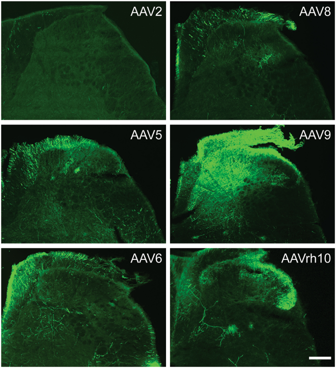
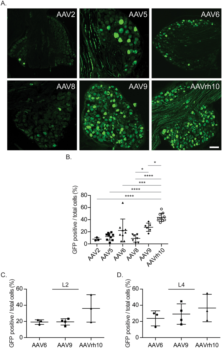
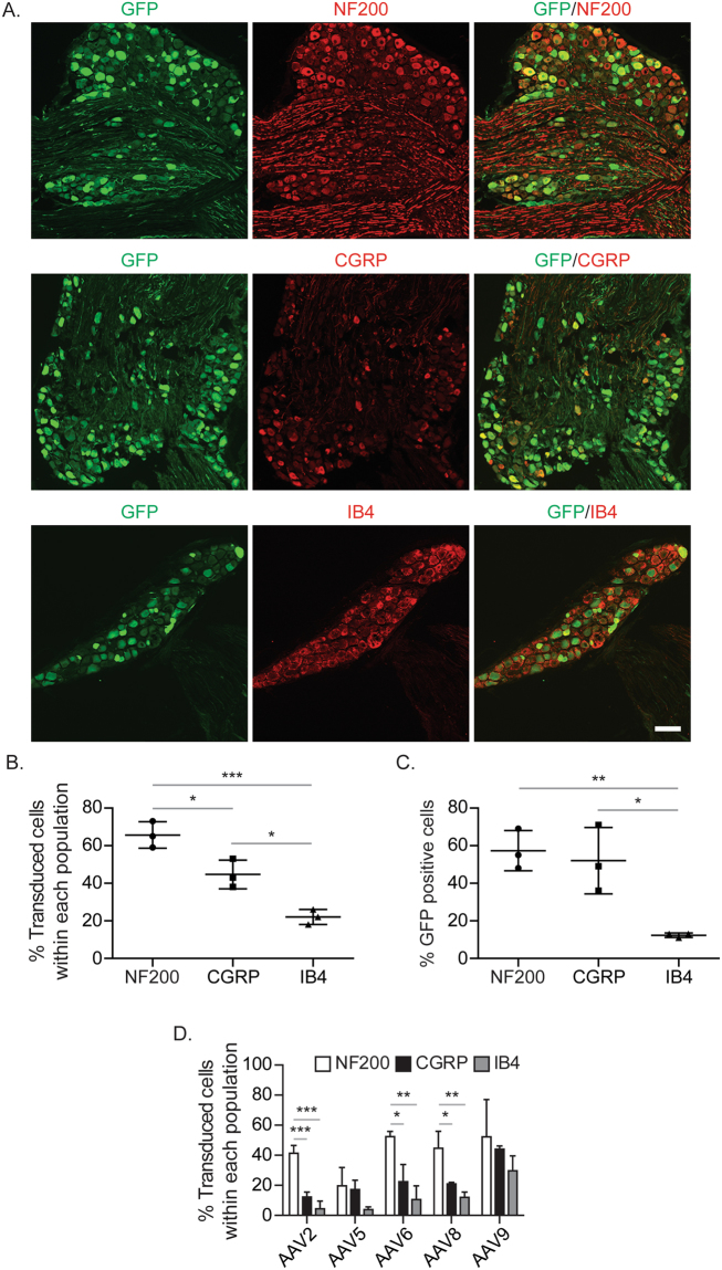
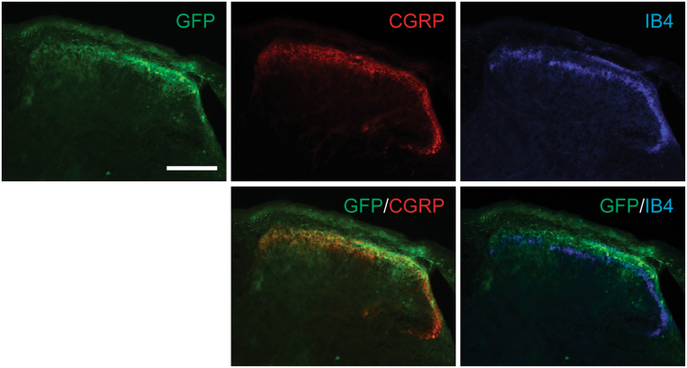
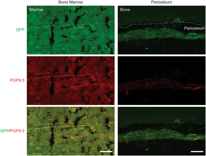
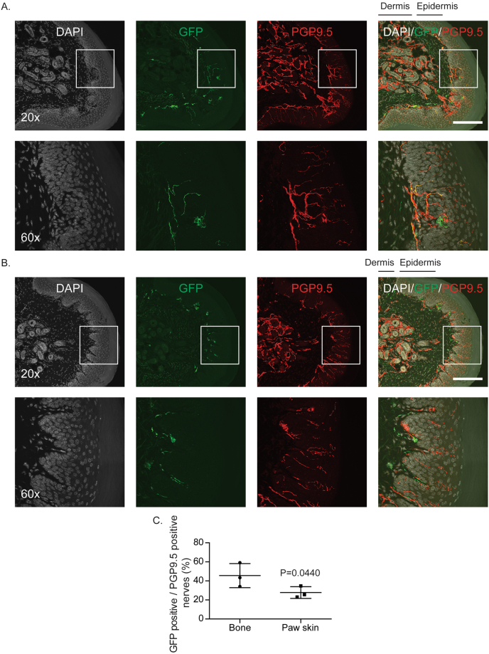
Adeno-associated virus (AAV) is frequently used to manipulate gene expression in the sensory nervous system for the study of pain mechanisms. Although some serotypes of AAV are known to have nerve tropism, whether AAV can distribute to sensory nerves that innervate the bone or skeletal tissue has not been shown. This information is crucial, since bone pain, including cancer-induced bone pain, is an area of high importance in pain biology. In this study, we found that AAVrh10 transduces neurons in the spinal cord and dorsal root ganglia of immunodeficient mice with higher efficacy than AAV2, 5, 6, 8, and 9 when injected intrathecally. Additionally, AAVrh10 has tropism towards sensory neurons in skeletal tissue, such as bone marrow and periosteum, while it occasionally reaches the sensory nerve fibers in the mouse footpad. Moreover, AAVrh10 has higher tropic affinity to large myelinated and small peptidergic sensory neurons that innervate bone, compared to small non-peptidergic sensory neurons that rarely innervate bone. Taken together, these results suggest that AAVrh10 is a useful gene delivery vector to target the sensory nerves innervating bone. This finding may lead to a greater understanding of the molecular mechanisms of chronic bone pain and cancer-induced bone pain.


Conflict of interest statement

The authors declare that they have no competing interests.

Figures

Figure 1
Figure 1
Distribution of AAV vectors in spinal cord. GFP-tagged AAV vectors (AAV2, 5, 6, 8, 9, and rh10) were placed intrathecally between the L4/5 vertebrae of immunodeficient mice by percutaneous lumbar puncture. At 4 weeks, spinal cords were collected for immunofluorescence analyses for GFP positivity to determine AAV distribution. Representative image of spinal cords obtained from AAV injected mice. Magnification, ×20. Scale Bar = 100 µm.
Figure 2
Figure 2
Distribution of AAV vectors in DRG. GFP-tagged AAV vectors (AAV2, 5, 6, 8, 9, and rh10) were placed intrathecally between the L4/5 vertebrae of immunodeficient mice by percutaneous lumbar puncture. At 4 weeks, DRGs were collected for immunofluorescence analyses for GFP positivity to determine AAV distribution. (A) Representative image of L3 DRGs obtained from AAV injected mice. Magnification, × 20. Scale Bar = 100 µm. (B) Quantifications of (A). (C,D) Quantifications of GFP positivity of (C) L2 and (D) L4 DRGs obtained from AAV injected mice. Presented as mean ± standard deviation, Significant differences were determined by one-way ANOVA with Tukey’s multiple comparisons. *p ≤ 0.05, ***p ≤ 0.0001, ****p ≤ 0.00001.
Figure 3
Figure 3
Subpopulations of sensory nerves in DRG transduced by AAVrh10. GFP-tagged AAVrh10 vector was placed intrathecally between the L4/5 vertebrae of immunodeficient mice by percutaneous lumbar puncture. At 4 weeks, L3 DRGs were collected for immunofluorescence analyses to verify expression in subpopulations of sensory neurons. (A) Representative image of co-localization of GFP positive cells, and myelinated neurofilament 200 (NF200), non-myelinated peptidergic calcitonin gene-related peptide (CGRP), or non-peptidergic isolectin-B4 (IB4) positive cells. Magnification, ×20. Scale Bar = 100 µm. (B) Quantification of the percentage of DRG cells transduced within different populations of sensory neurons (NF200, CGRP, or IB4). (C) Quantification of the percentage of total GFP positive DRG cells that co-expressed each marker. (D) Quantification of the percentage of DRG cells transduced within different populations of sensory neurons in the L3 DRGs obtained from AAV injected mice. Presented as mean ± standard deviation. Significant differences were determined by one-way ANOVA with Tukey’s multiple comparisons. *p ≤ 0.05, **p ≤ 0.001, ***p ≤ 0.0001.
Figure 4
Figure 4
Subpopulations of sensory nerves in spinal cord transduced by AAVrh10. GFP-tagged AAVrh10 vector was placed intrathecally between the L4/5 vertebrae of immunodeficient mice by percutaneous lumbar puncture. At 4 weeks, spinal cords were collected for immunofluorescence analyses to verify expression in subpopulations of sensory neurons. Representative image of co-localization of GFP, CGRP, and IB4 in spinal cord. Magnification, ×20. Scale Bar = 100 µm.
Figure 5
Figure 5
Distribution of AAVrh10 vector to sensory nerves in skeletal tissues. GFP-tagged AAVrh10 vector was placed intrathecally between the L4/5 vertebrae of immunodeficient mice by percutaneous lumbar puncture. At 4 weeks, femurs were collected for immunofluorescence analyses for GFP positivity to determine AAV distribution to sensory neurons. Representative image of co-localization of GFP and PGP9.5 in bone marrow and periosteum. Magnification, ×20. Scale Bar = 100 µm.
Figure 6
Figure 6
Distribution of AAVrh10 vector to sensory nerves in paw skin compared to those in skeletal tissue. GFP-tagged AAVrh10 vector was placed intrathecally between the L4/5 vertebrae of immunodeficient mice by percutaneous lumbar puncture. At 4 weeks, glabrous skin from the hindpaw was collected for immunofluorescence analyses for co-localization of GFP and PGP9.5 to determine AAV distribution in sensory neurons. (A) Representative images of GFP and PGP9.5 co-localization in paw skin sections. (B) Representative images of GFP and PGP9.5 without co-localization in paw skin sections. Scale Bar = 100 µm. (C) Quantifications of GFP positive sensory nerves over PGP9.5 positive sensory nerves between bones (Fig. 5) and paw skins (Fig. 6A and B) obtained from AAVrh10 injected mice. Presented as mean ± standard deviation, Significant differences were determined by Student’s t-test.

Similar articles

Cited by

References

    1. Dubin AE, Patapoutian A. Nociceptors: the sensors of the pain pathway. J Clin Invest. 2010;120:3760–3772. doi: 10.1172/JCI42843. - DOI - PMC - PubMed
    1. Lentz TB, Gray SJ, Samulski RJ. Viral vectors for gene delivery to the central nervous system. Neurobiol Dis. 2012;48:179–188. doi: 10.1016/j.nbd.2011.09.014. - DOI - PMC - PubMed
    1. Davidson BL, Breakefield XO. Viral vectors for gene delivery to the nervous system. Nat Rev Neurosci. 2003;4:353–364. doi: 10.1038/nrn1104. - DOI - PubMed
    1. Grimm D, Pandey K, Kay MA. Adeno-associated virus vectors for short hairpin RNA expression. Methods Enzymol. 2005;392:381–405. doi: 10.1016/S0076-6879(04)92023-X. - DOI - PubMed
    1. Stilwell JL, Samulski RJ. Role of viral vectors and virion shells in cellular gene expression. Mol Ther. 2004;9:337–346. doi: 10.1016/j.ymthe.2003.11.007. - DOI - PubMed

Publication types

MeSH terms

Substances