. 2017 Dec 12;7(1):17461.
doi: 10.1038/s41598-017-17668-5.

Motion guided Spatiotemporal Sparsity for high quality 4D-CBCT reconstruction

Affiliations

Motion guided Spatiotemporal Sparsity for high quality 4D-CBCT reconstruction

Yang Liu et al. Sci Rep. .

Abstract

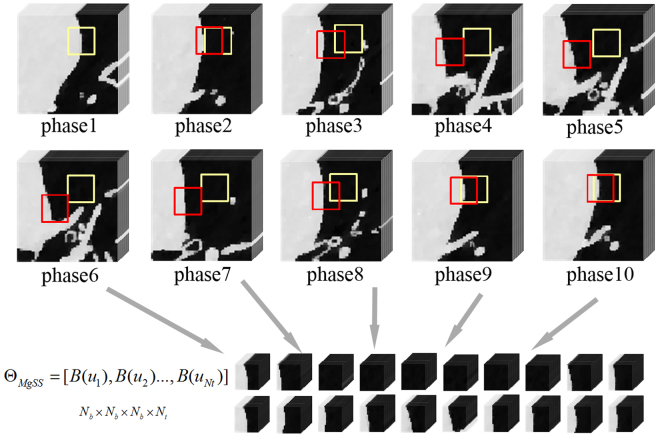
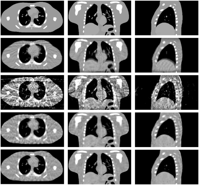
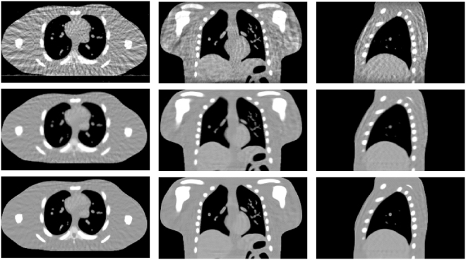
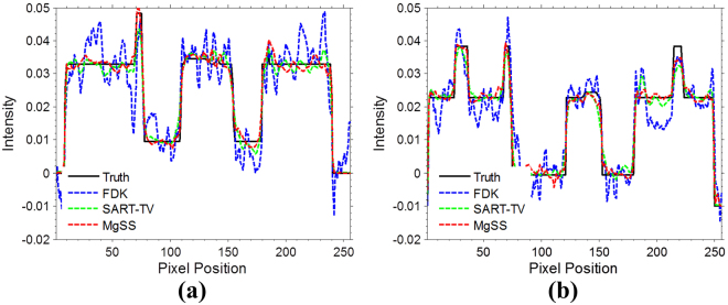
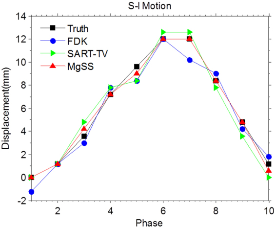
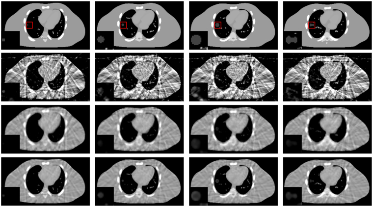
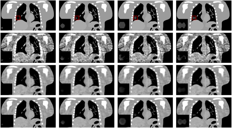
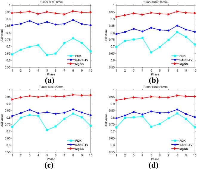
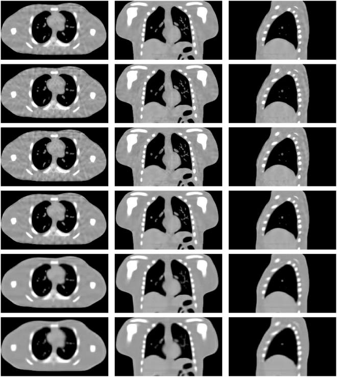
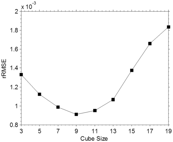
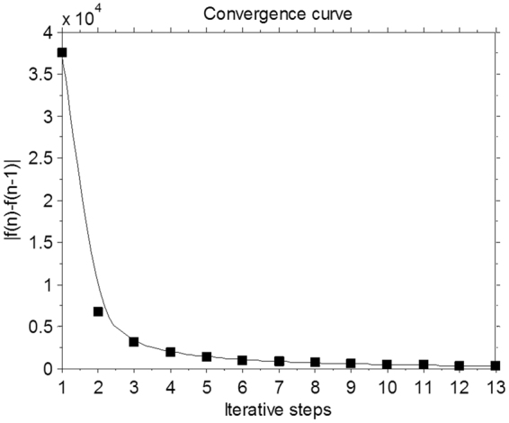
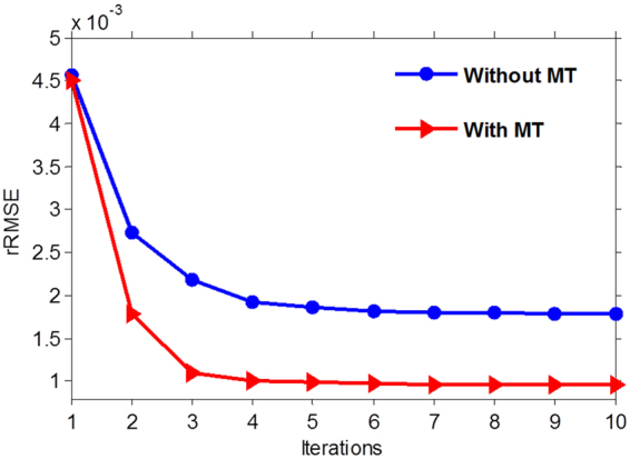
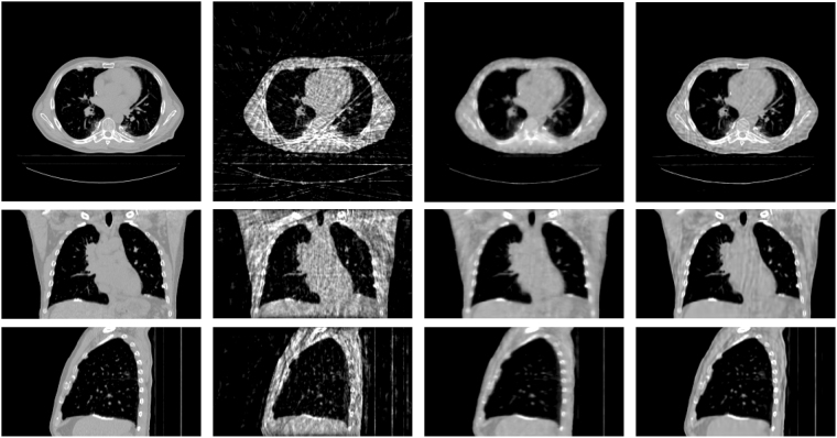
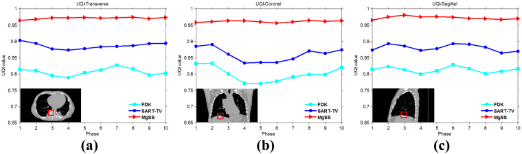
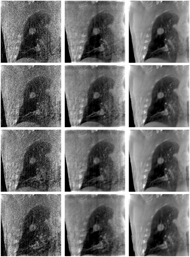
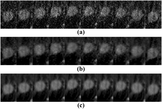
Conventional cone-beam computed tomography is often deteriorated by respiratory motion blur, which negatively affects target delineation. On the other side, the four dimensional cone-beam computed tomography (4D-CBCT) can be considered to describe tumor and organ motion. But for current on-board CBCT imaging system, the slow rotation speed limits the projection number at each phase, and the associated reconstructions are contaminated by noise and streak artifacts using the conventional algorithm. To address the problem, we propose a novel framework to reconstruct 4D-CBCT from the under-sampled measurements-Motion guided Spatiotemporal Sparsity (MgSS). In this algorithm, we try to divide the CBCT images at each phase into cubes (3D blocks) and track the cubes with estimated motion field vectors through phase, then apply regional spatiotemporal sparsity on the tracked cubes. Specifically, we recast the tracked cubes into four-dimensional matrix, and use the higher order singular value decomposition (HOSVD) technique to analyze the regional spatiotemporal sparsity. Subsequently, the blocky spatiotemporal sparsity is incorporated into a cost function for the image reconstruction. The phantom simulation and real patient data are used to evaluate this algorithm. Results show that the MgSS algorithm achieved improved 4D-CBCT image quality with less noise and artifacts compared to the conventional algorithms.


Conflict of interest statement

The authors declare that they have no competing interests.

Figures

Figure 1
Figure 1
An example of blocks tracked (red squares) and not tracked (yellow squares) through all the frames.
Figure 2
Figure 2
Results of 4D NCAT phantom with 21 projections for each phase. First row shows the begin-expiration phase of digital phantom. Second row shows the 3D-CBCT reconstructed from all projections by FDK. The third to last rows show 4D-CBCT images at the begin-expiration phase reconstructed by using FDK, SART-TV and proposed MgSS algorithm, respectively. The transverse, coronal, and sagittal planes have been shown in the first, second and third columns, respectively.
Figure 3
Figure 3
Reconstructions of 4D NCAT phantom with 31 projections for each phase. The first to third rows show 4D-CBCT at the begin-expiration phase reconstructed by FDK, SART-TV and proposed MgSS algorithms, respectively.
Figure 4
Figure 4
Reconstructions of 4D NCAT phantom with 51 projections for each phase. The first to third rows show 4D-CBCT at the begin-expiration phase reconstructed by FDK, SART-TV and proposed MgSS algorithms, respectively.
Figure 5
Figure 5
Horizontal profiles of reconstructions shown in Fig. 2. The profiles located at the pixel position x from 0 to 256 and y = 46 (a), y = 67 (b). The ‘black line’ generated from the NCAT phantom acts as the ground-truth for comparison.
Figure 6
Figure 6
Motion trajectories extracted from the result of 4D-CBCT along the superior–inferior direction.
Figure 7
Figure 7
Reconstructions of 4D NCAT phantom with 21 projections. The first to the fourth columns show the transverse planes of 4D NCAT phantom with tumors of diameters: 6 mm, 16 mm, 22 mm and 28 mm, respectively.
Figure 8
Figure 8
Reconstructions of 4D NCAT phantom with 21 projections. The first to the fourth columns show the coronal planes of 4D NCAT phantom with tumors of diameters: 6 mm, 16 mm, 22 mm and 28 mm, respectively.
Figure 9
Figure 9
The UQI measures on the ROIs in Fig. 8 for ten phase.
Figure 10
Figure 10
Ten phases reconstructions from -21 projections with block size set to be 5 × 5 × 5, 7 × 7 × 7, 9 × 9 × 9, 13 × 13 × 13, 17 × 17 × 17, 23 × 23 × 23.
Figure 11
Figure 11
The averaged rRMSEs of ten phases reconstructions with different cube size.
Figure 12
Figure 12
The convergence curve of the MgSS algorithm.
Figure 13
Figure 13
The rRMSE measures as a function of the number of iterations for the MgSS algorithm with and without motion tracking.
Figure 14
Figure 14
Results of realistic digital phantom with 21 projections for each phase. First column shows the begin-expiration phase of the 4DCT images. The second to last columns show 4D-CBCT images at the begin-expiration phase reconstructed by using FDK, SART-TV and proposed MgSS algorithms, respectively.
Figure 15
Figure 15
The UQI tests on ten phases reconstructions from -21views projection at the transverse, sagittal and coronal planes separately.
Figure 16
Figure 16
Results of patient data reconstructed by using different methods. The left to the right columns show the 4D-CBCT reconstructed by FDK, SART-TV and proposed MgSS algorithm, respectively.Each row shows images at the different phases in the breathing cycle: 20–30%, 40–50%, 60–70%, 80–90%.
Figure 17
Figure 17
The zoomed tumor areas in the reconstructions of ten phases. The first to the third rows show the tumor image which were reconstructed by FDK, SART-TV, and MgSS, respectively.

References

    1. Jaffray DA, Siewerdsen JH, Wong JW, Martinez AA. Flat-panel cone-beam computed tomography for image-guided radiation therapy. International Journal of Radiation Oncology* Biology* Physics. 2002;53:1337–1349. doi: 10.1016/S0360-3016(02)02884-5. - DOI - PubMed
    1. Oldham M, et al. Cone-beam-CT guided radiation therapy: A model for on-line application. Radiotherapy and oncology. 2005;75:271. E1–271. E8. doi: 10.1016/j.radonc.2005.03.026. - DOI - PubMed
    1. Pouliot J, et al. Low-dose megavoltage cone-beam CT for radiation therapy. International Journal of Radiation Oncology* Biology* Physics. 2005;61:552–560. doi: 10.1016/j.ijrobp.2004.10.011. - DOI - PubMed
    1. Chen GT, Kung JH, Beaudette KP. Artifacts in computed tomography scanning of moving objects. Semin Radiat Oncol. 2004;14:19–26. doi: 10.1053/j.semradonc.2003.10.004. - DOI - PubMed
    1. Sonke J, Zijp L, Remeijer P, van Herk M. Respiratory correlated cone beam CT. Medical physics. 2005;32:1176–1186. doi: 10.1118/1.1869074. - DOI - PubMed

Publication types

LinkOut - more resources